This website uses cookies to ensure you get the best experience on our website.
- Table of Contents
32 Citations 4 Q&As
11 Citations 9 Q&As
51 Citations 4 Q&As
71 Citations 6 Q&As
7 Citations 3 Q&As
7 Citations 4 Q&As
7 Citations 3 Q&As
15 Citations 4 Q&As
11 Citations 4 Q&As
10 Citations 5 Q&As
9 Citations 17 Q&As
17 Citations 16 Q&As
9 Citations 16 Q&As
18 Citations 16 Q&As
Facts about Cellular tumor antigen p53.
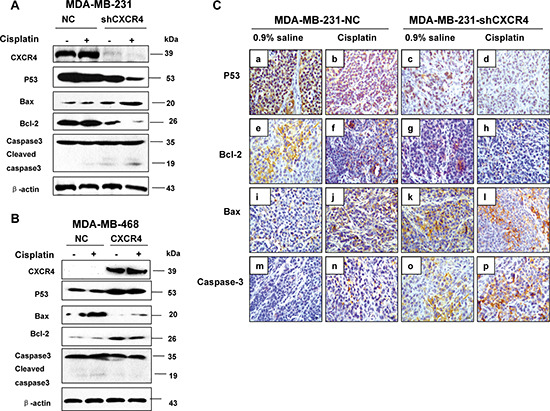
One of the activated genes is an inhibitor of cyclin-dependent kinases. Apoptosis induction seems to be mediated either by stimulation of BAX and FAS antigen expression, or by repression of Bcl-2 expression.
Human | |
---|---|
Gene Name: | TP53 |
Uniprot: | P04637 |
Entrez: | 7157 |
Belongs to: |
---|
p53 family |
Antigen NY-CO-13; BCC7; FLJ92943; LFS1; LFS1TRP53; p53 tumor suppressor; p53; P53cellular tumor antigen p53; Phosphoprotein p53; TP53; transformation-related protein 53; TRP53; tumor protein p53; Tumor suppressor p53
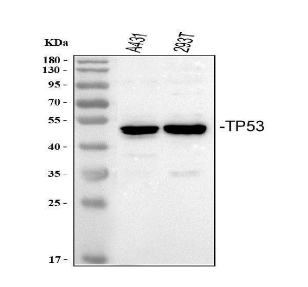
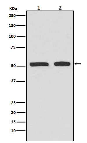
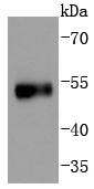
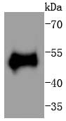
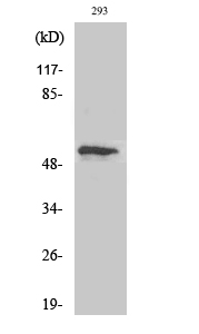
Mass (kDA):
43.653 kDA
Human | |
---|---|
Location: | 17p13.1 |
Sequence: | 17; NC_000017.11 (7668402..7687550, complement) |
Ubiquitous. Isoforms are expressed in a wide range of normal tissues but in a tissue-dependent manner. Isoform 2 is expressed in most normal tissues but is not detected in brain, lung, prostate, muscle, fetal brain, spinal cord and fetal liver. Isoform 3 is expressed in most normal tissues but is not detected in lung, spleen, testis, fetal brain, spinal cord and fetal liver. Isoform 7 is expressed in most normal tissues but is not detected in prostate, uterus, skeletal muscle and breast. Isoform 8 is detected only in colon, bone marrow, testis, fetal brain and intestine. Isoform 9 is expressed in most normal tissues but is not detected in brain, heart, lung, fetal liver, salivary gland, breast or intestine.
Cytoplasm. Nucleus. Nucleus, PML body. Endoplasmic reticulum. Mitochondrion matrix. Cytoplasm, cytoskeleton, microtubule organizing center, centrosome. Interaction with BANP promotes nuclear localization (PubMed:15701641). Recruited into PML bodies together with CHEK2 (PubMed:12810724). Translocates to mitochondria upon oxidative stress (PubMed:22726440). Translocates to mitochondria in response to mitomycin C treatment (PubMed:27323408).; [Isoform 1]: Nucleus. Cytoplasm. Predominantly nuclear but localizes to the cytoplasm when expressed with isoform 4.; [Isoform 2]: Nucleus. Cytoplasm. Local
In this article, we'll talk about the role played by the TP53 marker in progenitor-cell populations. We'll also go over the IHC process that is used to assess the expression of TP53. TP53 is required by cells in order to maintain a progenitor cells population. Mutations in TP53 could result in Li-Fraumeni syndrome.
The TP53 gene mutations cause a rare genetic disorder known as Li-Fraumeni syndrome. The TP53 gene is affected by mutations that hinder normal functioning of the protein, allowing damaged cells to continue to divide and multiply. These cells can grow into malignant tumors that spread throughout the body. In fact 60 to 80 percent of the classic LFS families have detectable germ-line TP53 mutations. The majority of mutations detected in the TP53 gene are missense mutations in the DNA-binding domain.
To understand if TP53 mutations are the cause of Li-Fraumeni syndrome, scientists need to learn how these genes operate. The TP53 gene encodes a nuclear protein of 53 kD transcription factor that regulates cell expansion and homeostasis. Because the gene is located in a variety of cells, TP53 mutations are vital to the development and growth of cancers. Extensive cell line research and transgenic animal experiments have revealed that TP53 mutations play an integral role in the malignant transformation of cells. A mutation may alter the function of the p53 gene. The protein can have dominant and recessive effects.
TP53 mutations are autosomal dominant. This condition can lead to multiple cancer types in patients which include lung, breast, and prostate. LFS may cause multiple primary tumors in one person while other hereditary diseases are limited to a single site. In addition to multiple primary tumors, people with LFS are prone to developing metachronous tumors throughout their organs. The most commonly-suspected types of cancers that are related to Li-Fraumeni syndrome comprise soft tissue sarcomas, adrenocortical cancer and breast cancer.
Women with the TP53 mutation in their germline are at risk of developing cancer. Prenatal genetic screenings of patients with TP53 mutations are a reliable way to identify the condition early. LFS patients can also receive prenatal diagnosis and genetic counseling. The results of the test may be used in clinical practice for treatment or monitoring of the disease. While there is no cure for Lifraumeni syndrome it can assist women who are pregnant or plan to become pregnant in order to minimize the risk of becoming pregnant.
Human hepatocellular carcinoma (HCC) is caused by TP53 mutations. This hotspot is linked to Aflatoxin B1 treatment. However, many HCCs do not have the mutation. This is due to the lack of information about the role of TP53 during HCC development. However, the current knowledge of TP53's role in HCC development offers new insights into the biology behind HCC.
Hematopoietic stem cell populations have a hierarchy of differentiated hematopoietic progenitors. They constantly replenish the blood system. However, p53-KO mice display no abnormal hematopoiesis. Further research has revealed that p53 is involved in the differentiation of blood hematopoietic cell lines. Reconstitution of mice with p53-KO wild-type p53 resulted in the maturation of B-cellsand the expression of M immunoglobulin heavy chain as well as B220.
Studies using mice with altered p53 have revealed that it can influence the development of cancer and aging independently of its tumor suppressor role. Interestingly, p53 suppresses the activity of mTORC1 in human and mouse cells and helps maintain naive T-cell populations. The findings suggest that p53 plays an impact on aging that is complex and the role of p53 in this process is dependent on the genetic background.
In contrast the mutant cells of p53 depart from their normal homeostatic behaviour and undergo exponential growth in response to UVB exposure. CP cells are epidermal precursor cells that maintain homeostasis through acquiring one of three stochastic fates following division. This CP cell behavior was also observed in the back skin of mice. In this study, the self-duplication of progenitor cells completely compensates for the loss of CP cells.
TP53 mutations can also be discovered in urinary tract and respiratory tract cancers. G/C to A/T transitions are the most common in brain tumors as well as colorectal. They are associated with environmental mutagens that create DNA adducts and alter codons. Tobacco smoke-compounds have been proven to target TP53 codons 157, 158. Other environmental pollutants such as AFB1 are believed to alter p53 as well.
It is still not clear whether it is clear if p53 IHC has any clinical significance in BE. The current surveillance strategy focuses on a small window of dysplasia as well as advanced cancer, and better methods for identifying high-risk BE patients could enhance the effectiveness of screening. offer valuable information for individualized treatment, and target the small number of BE patients who will develop. However, IHC of TP53 is not suggested for risk stratification for this disease.
To determine TP53 mutations Immunohistochemical staining is a reliable method to identify the mutations. The amount and the intensity of stained nuclei is a sign that positive cells are present. Sometimes aberrant nuclear staining may be observed in certain cases. However these rare cases are not always indicative that TP53 mutations. The presence of nuclei that are weakly positive or groups of positive cells can serve as internal positive control in IHC.
Patients with breast cancer who carry TP53 gene mutations have experienced poor clinical outcomes. Different TP53 mutation types lead to accumulated mutant p53 protein, which can be detected using immunohistochemistry. Therefore, null mutations should be considered when there is no detectable p53 protein in the samples. However, these results aren't conclusive and further studies are needed to better understand the role of p53 in breast cancer.
In the absence of mutations in TP53 The cytoplasmic staining of this gene can indicate non-HGSCa. It is uncommon to find wild type TP53 in tumor cells. In HGSCa tumor cells, tumor cells that have wild-type TP53 exhibit simple tubular structures with hobnail nuclei. Low-grade serous carcinoma however shows papillary structures with medusa-like projections.
This article outlines a thorough IHC procedure using the TP53 marker. Then, the fresh tissues were fixed in paraffin and formalin. They were then cut into sections of 4 mm. Finally glass slides were bonded to the sections. Sections were then incubated at 60°C for two hours. The sections were then incubated at a temperature of 60°C for two hours.
This IHC workflow used serum cfDNA obtained by two different techniques that include the Ion AmpliSeq(tm) TP53 community panel and the Oncomine breast tissue cfDNA assay. The detection limit for each method was 0.1%. The Oncomine panel requires a greater reading depth than AmpliSeq panels. Therefore, at least two samples were required to complete the IHC workflow with the TP53 marker.
The study showed that nine out of 17 patients diagnosed with cancer had TP53 mutations in their tumors or in their serum cfDNA. 5/6 patients experienced progression of their disease within six months after diagnosis. 3/4 of patients suffering from residual disease also had mutations. TP53 mutations were also discovered in 5/6 patients who failed chemotherapy but who didn't have TP53 tumors that were mutant.
This IHC workflow is based upon the highest-quality TP53 panel, which spans all exons. It also targets oncogenically relevant SNVs and indels, simplifying the analysis of TP53 mutations. Moreover, the CleanPlex(r) TP53 panel uses highly sensitive CleanPlex technology. The TP53 panel utilizes genomic data taken from formalin-fixed paraffin embedded samples.
The TP53 gene is involved in a variety of functions in the human body , and determining its status requires IHC tests. Its role in the diagnosis of cancer and prognosis is essential for accurate cancer treatment. The TP53 gene was discovered in a variety cell forms, with some previously undiscovered for their function. In this chapter, we'll explore the role of the TP53 gene in IHC diagnosis and troubleshooting.
The detection of biomarkers with IHC is a growing practice in the era of targeted therapy. Diagnostic pathology laboratories have been able to play a more important role in diagnosing illnesses using biomarkers. While many diagnostic pathology labs employ this technique, certain difficulties remain. For precise results, IHC requires that you have certain conditions. To ensure accuracy, employ appropriate control methods to determine the most effective concentration of the target antigen.
Although the TP53 gene isn't very specific and sensitive, IHC can be used as a marker to detect TP53 mutations. For therapeutic reasons however, it's important to have high specificity and sensitivity. A significant proportion of cancers with a TP53 mutation will have intermediate levels of the gene and the TP53 expression levels must be high enough to exclude them.
The TP53 gene contains a series of mutations. All missense mutations cause an increase in mutp53 expression. Non-missense mutations may have a variable staining level. One nonsense mutation in exon 7 resulted in high levels of staining. A single nonsense mutation resulted in intermediate staining, and the remaining four are categorized as 'Integrative', or 'Low'.
PMID: 4006916 by Zakut-Houri R., et al. Human p53 cellular tumor antigen: cDNA sequence and expression in COS cells.
PMID: 2946935 by Lamb P., et al. Characterization of the human p53 gene.
*Showing only the more recent 20. More publications can be found for each product on its corresponding product page