This website uses cookies to ensure you get the best experience on our website.
- Table of Contents
and ELISA kits, proteins related to Myocytes.
Myocytes, the fundamental building blocks of muscle tissue, are essential for generating the force and movement required for both voluntary actions like walking and involuntary functions such as heartbeat regulation. These specialized muscle cells come in various types, including skeletal, cardiac, and smooth myocytes, each playing a unique role in maintaining bodily functions and overall health. Comprehensive research into myocytes is crucial for understanding muscle development, repair mechanisms, and the underlying causes of muscular diseases. Our range of high-quality antibodies is specifically designed to target key proteins and biomarkers within myocytes, facilitating precise detection and analysis in your studies. Whether you’re investigating muscle regeneration, exploring cardiovascular health, or delving into neuromuscular disorders, our specialized antibodies provide the reliability and specificity needed to advance your myocyte research. Empower your scientific endeavors with our expertly curated antibody solutions tailored for myocyte-focused studies.

Anti-Connexin 43/GJA1 Antibody Picoband®, Figure 5. IF analysis of GJA1 using anti-GJA1 antibody (A00599).
GJA1 was detected in immunocytochemical section of U20...

Anti-Lamin A+C/LMNA Antibody Picoband®, Figure 6. IF analysis of Lamin A/C using anti-Lamin A/C antibody (PB9280).
Lamin A/C was detected in immunocytochemical section...
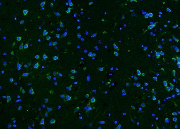
Anti-CaMKII alpha/CAMK2A Antibody Picoband®, Figure 6. IF analysis of CaMKII alpha/CAMK2A using anti-CaMKII alpha/CAMK2A antibody (A03241-2).
CaMKII alpha/...
| Protein Name | Gene Name | Function |
|---|---|---|
| Troponin I | TNNI3 | Regulates muscle contraction in cardiac myocytes. |
| Troponin T | TNNT2 | Part of the troponin complex, essential for cardiac muscle contraction. |
| Myosin Heavy Chain 7 | MYH7 | Major component of the cardiac sarcomere, involved in muscle contraction. |
| Myosin Binding Protein C | MYBPC3 | Regulates cardiac muscle contraction and sarcomere assembly. |
| Actin | ACTC1 | Forms thin filaments in the sarcomere, enabling muscle contraction. |
| Desmin | DES | Maintains the structural integrity of myocytes. |
| Titin | TTN | Contributes to sarcomere elasticity and stability. |
| Connexin 43 | GJA1 | Forms gap junctions for electrical coupling between myocytes. |
| SERCA2a | ATP2A2 | Pumps calcium into the sarcoplasmic reticulum, regulating muscle relaxation. |
| Ryanodine Receptor 2 | RYR2 | Releases calcium from the sarcoplasmic reticulum during muscle contraction. |
| Alpha-Actinin-2 | ACTN2 | Anchors actin filaments at the Z-disc in the sarcomere. |
| Lamin A/C | LMNA | Provides structural support to the nuclear envelope in myocytes. |
| Caveolin-3 | CAV3 | Involved in membrane organization and signal transduction in muscle cells. |
| Periostin | POSTN | Supports extracellular matrix organization and myocyte function. |
| Calmodulin | CALM1 | Regulates calcium signaling pathways in myocytes. |
| CAMKII | CAMK2A | Involved in calcium-dependent signaling and myocyte contraction regulation. |
| Natriuretic Peptide A | NPPA | Regulates blood pressure and fluid balance in cardiac function. |
| Natriuretic Peptide B | NPPB | Involved in cardiovascular homeostasis and cardiac hypertrophy regulation. |
| Tropomyosin | TPM1 | Stabilizes actin filaments and regulates access to myosin-binding sites. |
| Dystrophin | DMD | Links the cytoskeleton of myocytes to the extracellular matrix, providing structural stability. |
Excitation-contraction (E-C) coupling is a fundamental physiological process in myocytes, particularly in skeletal and cardiac muscle cells, that links electrical stimulation to mechanical contraction. This intricate mechanism begins with the generation of an action potential, an electrical impulse that travels along the sarcolemma and down the T-tubules of the myocyte. The action potential triggers the opening of voltage-sensitive dihydropyridine receptors (DHPRs) in the T-tubule membrane, which are mechanically coupled to ryanodine receptors (RyR) on the sarcoplasmic reticulum (SR). Activation of RyR channels leads to a rapid release of calcium ions (Ca²⁺) from the SR into the cytoplasm. The surge in intracellular Ca²⁺ binds to troponin C on the thin filaments of the myocyte, inducing a conformational change that allows actin and myosin interactions, ultimately resulting in muscle contraction. E-C coupling is tightly regulated to ensure precise control of muscle force and duration. Dysregulation of this process can lead to various muscular and cardiac disorders, highlighting its critical role in muscle physiology. Ongoing research in E-C coupling seeks to unravel the molecular intricacies and develop therapeutic strategies for related diseases.
Mitochondria are essential organelles within myocytes, serving as the powerhouse for energy production necessary for muscle contraction and overall cellular function. In myocytes, especially in cardiac and skeletal muscles, mitochondria are densely packed to meet the high energetic demands. They are primarily involved in oxidative phosphorylation, a metabolic pathway that generates adenosine triphosphate (ATP) through the electron transport chain and the citric acid cycle. Efficient mitochondrial function ensures a continuous supply of ATP, which is crucial for sustained muscle activity and rapid response to energy needs. Additionally, mitochondria play a pivotal role in calcium homeostasis by buffering intracellular Ca²⁺ levels, thereby contributing to the regulation of excitation-contraction coupling. Beyond energy production, mitochondria are involved in apoptotic signaling and reactive oxygen species (ROS) generation, which can impact muscle health and disease. Dysregulation of mitochondrial metabolism is implicated in various myopathies and cardiovascular diseases. Current research focuses on mitochondrial biogenesis, dynamics, and the interplay between metabolism and muscle function, aiming to develop interventions that enhance mitochondrial efficiency and mitigate metabolic dysfunctions in myocytes.