



Click image to see more details
-

-
-
-
-
+3
Product Info Summary
| SKU: | PA1484 |
|---|---|
| Size: | 100 μg/vial |
| Reactive Species: | Human |
| Host: | Rabbit |
| Application: | IHC, WB |
Customers Who Bought This Also Bought

Product info
Product Name
Anti-Toll-like receptor 4 TLR4 Antibody Picoband®
SKU/Catalog Number
PA1484
BA1717 is an alternative SKU for this antibody, used in previous lots.
Size
100 μg/vial
Form
Lyophilized
Description
Boster Bio Anti-Toll-like receptor 4 TLR4 Antibody catalog # PA1484. Tested in IHC, WB applications. This antibody reacts with Human. The brand Picoband indicates this is a premium antibody that guarantees superior quality, high affinity, and strong signals with minimal background in Western blot applications. Only our best-performing antibodies are designated as Picoband, ensuring unmatched performance.
Storage & Handling
Store at -20˚C for one year from date of receipt. After reconstitution, at 4˚C for one month. It can also be aliquotted and stored frozen at -20˚C for six months. Avoid repeated freeze-thaw cycles.
Cite This Product
Anti-Toll-like receptor 4 TLR4 Antibody Picoband® (Boster Biological Technology, Pleasanton CA, USA, Catalog # PA1484)
Host
Rabbit
Contents
Each vial contains 4 mg Trehalose, 0.9 mg NaCl, 0.2mg Na2HPO4, 0.05 mg NaN3.
Clonality
Polyclonal
Isotype
Rabbit IgG
Immunogen
A synthetic peptide corresponding to a sequence at the N-terminus of human TLR4.
Cross-reactivity
No cross-reactivity with other proteins
Reactive Species
PA1484 is reactive to TLR4 in Human
Observed Molecular Weight
80 kDa
Calculated molecular weight
95.7 kDa
Background of TLR4
TLR4, Toll-like receptor 4, is a protein that in humans is encoded by the TLR4 gene. TLR 4 is a toll-like receptor. TLR4, the human homolog of Drosophila Toll, is a type I transmembrane protein with an extracellular domain consisting of a leucine-rich repeat region and an intracellular domain homologous to that of human interleukin-1 receptor. The TLR4 gene is mapped to chromosome 9q32-q33 by fluorescence in situ hybridization.It detects lipopolysaccharide from Gram-negative bacteria and is thus important in the activation of the innate immune system.The protein encoded by this gene is a member of the Toll-like receptor (TLR) family, which plays a fundamental role in pathogen recognition and activation of innate immunity. TLRs are highly conserved from Drosophila to humans and share structural and functional similarities. They recognize pathogen-associated molecular patterns (PAMPs) that are expressed on infectious agents, and mediate the production of cytokines necessary for the development of effective immunity.
Antibody Validation
Boster validates all antibodies on WB, IHC, ICC, Immunofluorescence, and ELISA with known positive control and negative samples to ensure specificity and high affinity, including thorough antibody incubations.
Application & Images
Applications
PA1484 is guaranteed for IHC, WB Boster Guarantee
Recommend Dilution
| Application | Dilution | Species |
|---|---|---|
| Immunohistochemistry (Paraffin-embedded Section) | 0.5-1μg/ml | Human |
| Western blot | 0.1-0.5μg/ml | Human |
Tested application
Suggested blocking solution with 5% non-fat milk or BSA; (*)Recommended protein loading: 20-40 µg per lane
Use TE buffer pH 9.0 for antigen retrieval; (*) citrate buffer pH 6.0 is an alternative.
Validation Images & Assay Conditions
Click image to see more details
Anti-TLR4 antibody, PA1484, Western blotting
Lane 1: HELA Cell Lysate
Lane 2: SMMC Cell Lysate

Click image to see more details
Anti-TLR4 antibody, PA1484, IHC(P)
IHC(P): Human Lung Cancer Tissue
Click image to see more details
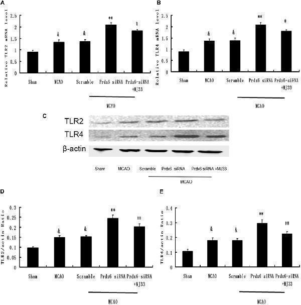
Expression of TLR2 and TLR4 in response to Prdx6 siRNA and MJ33. Real-time PCR showed that TLR2 (A) and TLR4 (B) expression increased after MCAO or giving Prdx6-siRNA. Combined treatment with Prdx6-siRNA and MJ33 downregulated TLR2 and TLR4 levels. Representative Western blot (C) and semi-quantitative analyses of the levels of TLR2 (D) and TLR4 (E) in the cortex after MCAO. Results are expressed as the mean ± SEM of three independent experiments. & p < 0.01 vs. Sham; ** p < 0.01 vs. Scramble; # p < 0.05, vs. Prdx6 siRNA; ## p < 0.01 vs. Prdx6 siRNA.
Index in PubMed under a CC BY license. PMID: 28424593

Click image to see more details
Effect of Prdx6-iPLA2 on Toll-like receptor 2 (TLR2) and TLR4 expression in microglia in response to OGD/R. TLR2 and TLR4 were activated during OGD/R exposure. Prdx6-iPLA2 siRNA or MJ33 treatment could inhibit TLR2 and TLR4 mRNA (A,B) and protein expression (C–E) . Results are expressed as the mean ± SEM of three independent experiments. ** p < 0.01 vs. Control; # p < 0.05 vs. Scramble; ## p < 0.01 vs. Scramble.
Index in PubMed under a CC BY license. PMID: 28424593
Click image to see more details
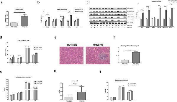
OTA-originated microbiota induces liver inflammation. a Liver LPS level in FMT (CON) and FMT (OTA) ducks. b Relative mRNA expressions of TLR4, MYD88, IKBα, IL-6, and TNF-α in the liver after FMT ( n = 6, mean with SEM). c Relative protein abundance of TLR4, MYD88, p-IKBα, p-IKBα/IKBα, and p-p65 in the liver after FMT ( n = 6, mean with SEM). d Effect of FMT on the liver levels of IL-1β, IL-6, TNF-α, and IL-10 ( n = 6, mean with SEM). e Representative H&E-stained liver sections. f Statistical analysis of the percentage of inflammatory cells in different groups shown in e ( n = 6, mean with SEM). g Serum levels of AST, ALT, ALP, and LDH in different groups ( n = 6, mean with SEM). h Serum LPS level in different FMT groups ( n = 6, mean with SEM). i Serum levels of IL-1β, IL-6, and TNF-α after FMT ( n = 6, mean with SEM). FMT (CON): ducks received the CON group fecal microbiota. FMT (OTA): ducks received the OTA group fecal microbiota. Data were analyzed with unpaired t test, n = 6. * P < 0.05; ** P < 0.01, *** P < 0.001, **** P <0.0001
Index in PubMed under a CC BY license. PMID: 31779704
Click image to see more details
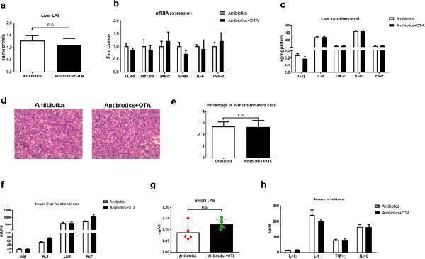
OTA fails to promote liver inflammation in antibiotics-treated ducks. a Liver LPS level in different groups ( n = 6, mean with SEM). b Relative mRNA expressions of TLR4, MYD88, IKBα, IL-6, and TNF-α in the liver of antibiotics-treated ducks with or without OTA ( n = 6, mean with SEM). c Levels of IL-1β, IL-6, TNF-α, IL-8, IL-10, and IFN-γ in the liver of antibiotics-treated ducks with or without OTA ( n = 6, mean with SEM). d Representative images of H&E-stained liver sections (magnification × 400; scale bar 100 μm; n = 6). e Statistical analysis of the percentage of inflammatory cells in different groups shown in d ( n = 6, mean with SEM). f Effect of OTA on serum levels of AST, ALT, ALP and LDH in antibiotics-treated ducks ( n = 6, mean with SEM). g Serum LPS level with or without OTA treatment in antibiotics-treated ducks ( n = 6, mean with SEM). h Effect of OTA on serum levels of IL-1β, IL-6, TNF-α, and IL-10 ( n = 6, mean with SEM). Data were analyzed with unpaired t test. n.s., not significant
Index in PubMed under a CC BY license. PMID: 31779704
Click image to see more details
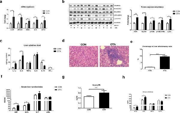
OTA promotes liver inflammation. a Relative mRNA expressions of TLR4, MYD88, IKBα, IL-6, and TNF-α in the liver after OTA oral gavage ( n = 6, mean with SEM). b Relative protein abundances of TLR4, MYD88, p-IKBα, p-IKBα/IKBα, and p-p65 in the liver after OTA oral gavage ( n = 6, mean with SEM). c Effect of OTA on levels of liver cytokines, including IL-1β, IL-6, TNF-α, IL-8, IL-10, and IFN-γ ( n = 6, mean with SEM). d Representative images of H&E-stained liver sections in CON and OTA group (magnification × 400, scale bar 100 μm, n = 6). e Statistical analysis of the percentage of inflammatory cells in different groups shown in d ( n = 6, mean with SEM). f Effect of OTA on serum levels of AST, ALT, ALP, and LDH ( n = 6, mean with SEM). g Serum LPS level with or without OTA treatment ( n = 6, mean with SEM). h Effect of OTA on serum levels of IL-1β, IL-6, TNF-α, and IL-10 ( n = 6, mean with SEM). Data in a , b , c , e , f , g , and h were analyzed with unpaired t test, * P < 0.05; ** P < 0.01, *** P < 0.001, P <0.0001
Index in PubMed under a CC BY license. PMID: 31779704
Specific Publications For Anti-Toll-like receptor 4 TLR4 Antibody Picoband® (PA1484)
Loading publications
Recommended Resources
Here are featured tools and databases that you might find useful.
- Boster's Pathways Library
- Protein Databases
- Bioscience Research Protocol Resources
- Data Processing & Analysis Software
- Photo Editing Software
- Scientific Literature Resources
- Research Paper Management Tools
- Molecular Biology Software
- Primer Design Tools
- Bioinformatics Tools
- Phylogenetic Tree Analysis
Customer Reviews
Have you used Anti-Toll-like receptor 4 TLR4 Antibody Picoband®?
Share your experimental results or join a short interview to earn up to $1,000 in product credits or other rewards.
0 Reviews For Anti-Toll-like receptor 4 TLR4 Antibody Picoband®
Customer Q&As
Have a question?
Find answers in Q&As, reviews.
Can't find your answer?
Submit your question
5 Customer Q&As for Anti-Toll-like receptor 4 TLR4 Antibody Picoband®
Question
We are currently using anti-TLR4 antibody PA1484 for human tissue, and we are content with the IHC results. The species of reactivity given in the datasheet says human. Is it possible that the antibody can work on feline tissues as well?
Verified Customer
Verified customer
Asked: 2020-05-07
Answer
The anti-TLR4 antibody (PA1484) has not been tested for cross reactivity specifically with feline tissues, but there is a good chance of cross reactivity. We have an innovator award program that if you test this antibody and show it works in feline you can get your next antibody for free. Please contact me if I can help you with anything.
Boster Scientific Support
Answered: 2020-05-07
Question
Our lab used your anti-TLR4 antibody for IHC on cerebellum last year. I am using human, and We intend to use the antibody for WB next. My lab would like examining cerebellum as well as fetal liver in our next experiment. Could give a recommendation on which antibody would work the best for WB?
P. Zhao
Verified customer
Asked: 2019-12-30
Answer
I looked at the website and datasheets of our anti-TLR4 antibody and I see that PA1484 has been tested on human in both IHC and WB. Thus PA1484 should work for your application. Our Boster satisfaction guarantee will cover this product for WB in human even if the specific tissue type has not been validated. We do have a comprehensive range of products for WB detection and you can check out our website bosterbio.com to find out more information about them.
Boster Scientific Support
Answered: 2019-12-30
Question
We have been able to see staining in human spleen. Any tips? Is anti-TLR4 antibody supposed to stain spleen positively?
Verified Customer
Verified customer
Asked: 2019-07-16
Answer
According to literature spleen does express TLR4. According to Uniprot.org, TLR4 is expressed in leukocyte, spleen, fetal liver, lung placenta, hippocampus, kidney uterus, cerebellum, monocyte, among other tissues. Regarding which tissues have TLR4 expression, here are a few articles citing expression in various tissues:
Cerebellum, Pubmed ID: 15489334
Fetal liver, Lung, and Placenta, Pubmed ID: 9435236
Hippocampus, Kidney, and Uterus, Pubmed ID: 14702039
Monocyte, Pubmed ID: 16622205
Spleen, Pubmed ID: 9237759
Boster Scientific Support
Answered: 2019-07-16
Question
My colleagues were happy with the WB result of your anti-TLR4 antibody. However we have seen positive staining in leukocyte cell membrane using this antibody. Is that expected? Could you tell me where is TLR4 supposed to be expressed?
K. Kulkarni
Verified customer
Asked: 2018-02-12
Answer
From literature, leukocyte does express TLR4. Generally TLR4 expresses in cell membrane. Regarding which tissues have TLR4 expression, here are a few articles citing expression in various tissues:
Cerebellum, Pubmed ID: 15489334
Fetal liver, Lung, and Placenta, Pubmed ID: 9435236
Hippocampus, Kidney, and Uterus, Pubmed ID: 14702039
Monocyte, Pubmed ID: 16622205
Spleen, Pubmed ID: 9237759
Boster Scientific Support
Answered: 2018-02-12
Question
My lab would like using your anti-TLR4 antibody for positive regulation of i-kappab kinase/nf-kappab signaling studies. Has this antibody been tested with western blotting on lung cancer tissue? We would like to see some validation images before ordering.
R. Krishna
Verified customer
Asked: 2016-10-04
Answer
I appreciate your inquiry. This PA1484 anti-TLR4 antibody is validated on hela cell lysate, smmc cell lysate, lung cancer tissue. It is guaranteed to work for IHC, WB in human. Our Boster guarantee will cover your intended experiment even if the sample type has not been be directly tested.
Boster Scientific Support
Answered: 2016-10-04

