Click image to see more details
Product Info Summary
| SKU: | M01551 |
|---|---|
| Size: | 100ug |
| Reactive Species: | Human, Mouse |
| Host: | Mouse |
| Application: | ELISA, Flow Cytometry, IP, IHC, WB |
Customers Who Bought This Also Bought

Product info
Product Name
Anti-BIN1/Amphiphysin Ii Monoclonal Antibody
SKU/Catalog Number
M01551
Size
100ug
Form
Liquid (sterile filtered)
Description
Boster Bio Anti-BIN1/Amphiphysin Ii Monoclonal Antibody (Catalog # M01551). Tested in ELISA, WB applications. This antibody reacts with Human, Mouse.
Storage & Handling
Store vial at -20°C prior to opening. Aliquot contents and freeze at -20°C or below for extended storage. Avoid cycles of freezing and thawing. Centrifuge product if not completely clear after standing at room temperature. This product is stable for several weeks at 4°C as an undiluted liquid. Dilute only prior to immediate use. Expiration date is one (1) year from date of opening. (Ship on dry ice.)
Cite This Product
Anti-BIN1/Amphiphysin Ii Monoclonal Antibody (Boster Biological Technology, Pleasanton CA, USA, Catalog # M01551)
Host
Mouse
Contents
0.02 M Potassium Phosphate, 0.15 M Sodium Chloride, pH 7.2, 0.01% (w/v) Sodium Azide
Clonality
Monoclonal
Clone Number
Clone: 99D IgG2b
Isotype
IgG2b
Immunogen
Anti-BIN1 (MOUSE) Monoclonal Antibody was produced in mouse by repeated immunizations with a fragment portion of recombinant human BIN1 protein followed by hybridoma development.
Reactive Species
M01551 is reactive to BIN1 in Human, Mouse
Calculated molecular weight
64.7 kDa
Background of BIN1
Bin1 is a conserved member of the BAR family of genes that have been implicated in diverse cellular processes including endocytosis, actin organization, programmed cell death, stress responses, and transcriptional control. The first mammalian BAR protein to be discovered, Amphiphysin I (AmphI), was identified in an immunoscreen for proteins associated with the plasma membranes of synaptic neurons, functions in the control of clathrin-dependent synaptic vesicle endocytosis. The mammalian Bin1 gene was first identified in a two hybrid screen for polypeptides that bind to the N-terminal Myc box 1 (MB1) portion of the c-Myc oncoprotein. Bin1 is similar to AmphI in overall structure, with an N-terminal BAR domain and a C-terminal SH3 domain. However, the Bin1 gene is more complex than the AmphI gene, encoding at least seven different splice variants that differ widely in subcellular localization, tissue distribution, and ascribed functions. Alternate splicing of the Bin1 gene results in ten transcript variants encoding different isoform. Bin1 is expressed ubiquitously in mammalian cells. Certain splice variants of Bin1 are expressed in the neurons, muscle cells or tumor cells. Bin1 may act as a cancer suppressor and inhibits malignant cell transformation. Studies in mouse suggest that this gene plays an important role in cardiac muscle development. Bin1 has also been implicated in Alzheimer disease and cardiac disease. Defects in Bin1 are the cause of centronuclear myopathy autosomal recessive; also known as autosomal recessive myotubular myopathy.
Antibody Validation
Boster validates all antibodies on WB, IHC, ICC, Immunofluorescence, and ELISA with known positive control and negative samples to ensure specificity and high affinity, including thorough antibody incubations.
Application & Images
Applications
M01551 is guaranteed for ELISA, Flow Cytometry, IP, IHC, WB Boster Guarantee
Recommend Dilution
| Application | Dilution | Species |
|---|---|---|
| Anti-BIN1 antibody has been tested for use in ELISA | IP | and Western Blot. This antibody is suitable for use in IHC and Flow Cytometry. Specific conditions for reactivity should be optimized by the end user. |
Validation Images & Assay Conditions
Click image to see more details
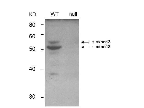
Western Blot of Anti-BIN1 Antibody. Lane 1: Keratinocyte derived from Bin1 wild type mice. Lane 2: Keratinocyte derived from Bin1 null mice. Load: 35 µg per lane. Primary antibody: BIN1 monoclonal Antibody. Secondary antibody: IRDye800™ mouse secondary antibody at 1:10,000 for 45 min at RT. Block: 1xPBS, 0.4% Tween-20. Other band(s): non-specific.

Click image to see more details
Flow Cytometry of Mouse Anti-BIN1 Antibody. Cells: C2C12 cells. Stimulation: none. Primary antibody: Anti-IgD (control), Anti-BIN-1 Antibody (99D clone). Secondary antibody: Biotin mouse secondary antibody at 1:10,000 for 45 min at RT and streptavidin PE at 1:5,000 for 30 min at RT.
Specific Publications For Anti-BIN1/Amphiphysin Ii Monoclonal Antibody (M01551)
Loading publications
Recommended Resources
Here are featured tools and databases that you might find useful.
- Boster's Pathways Library
- Protein Databases
- Bioscience Research Protocol Resources
- Data Processing & Analysis Software
- Photo Editing Software
- Scientific Literature Resources
- Research Paper Management Tools
- Molecular Biology Software
- Primer Design Tools
- Bioinformatics Tools
- Phylogenetic Tree Analysis
Customer Reviews
Have you used Anti-BIN1/Amphiphysin Ii Monoclonal Antibody?
Share your experimental results or join a short interview to earn up to $1,000 in product credits or other rewards.
0 Reviews For Anti-BIN1/Amphiphysin Ii Monoclonal Antibody
Customer Q&As
Have a question?
Find answers in Q&As, reviews.
Can't find your answer?
Submit your question
