Click image to see more details
Product Info Summary
| SKU: | M00555-6 |
|---|---|
| Size: | 100 μg/vial |
| Reactive Species: | Human |
| Host: | Mouse |
| Application: | Flow Cytometry, IF, IHC, WB |
Customers Who Bought This Also Bought

Product info
Product Name
Anti-CD45 Antibody Picoband® (monoclonal, 3H6)
SKU/Catalog Number
M00555-6
Size
100 μg/vial
Form
Lyophilized
Description
Boster Bio Anti-CD45 Antibody Picoband® (monoclonal, 3H6) catalog # M00555-6. Tested in Flow Cytometry, IF, IHC, WB applications. This antibody reacts with Human. The brand Picoband indicates this is a premium antibody that guarantees superior quality, high affinity, and strong signals with minimal background in Western blot applications. Only our best-performing antibodies are designated as Picoband, ensuring unmatched performance.
Storage & Handling
At -20°C for one year from date of receipt. After reconstitution, at 4°C for one month. It can also be aliquotted and stored frozen at -20°C for six months. Avoid repeated freezing and thawing.
Cite This Product
Anti-CD45 Antibody Picoband® (monoclonal, 3H6) (Boster Biological Technology, Pleasanton CA, USA, Catalog # M00555-6)
Host
Mouse
Contents
Each vial contains 4 mg Trehalose, 0.9 mg NaCl and 0.2 mg Na2HPO4.
Clonality
Monoclonal
Clone Number
3H6
Isotype
Mouse IgG1
Immunogen
A synthetic peptide corresponding to a sequence at the C-terminus of human CD45, different from the related mouse sequence by eight amino acids, and from the related rat sequence by ten amino acids.
Cross-reactivity
No cross-reactivity with other proteins.
Reactive Species
M00555-6 is reactive to PTPRC in Human
Observed Molecular Weight
220 kDa
Calculated molecular weight
147.5 kDa
Background of PTPRC
CD45 (Cluster of Differentiation 45), also known as PTPRC, LCA or CD45R, is an enzyme that, in humans, is encoded by the PTPRC gene. It is a member of the protein tyrosine phosphatase (PTP) family. CD45 is a major high molecular mass leukocyte cell surface molecule which is also an integral membrane protein tyrosine phosphatase. The cytogenetic location of CD45 is 1q31.3-q32.1. This gene is especially a prototype for transmembrane protein-tyrosine phosphatase (PTP). Targeted disruption of the CD45 gene leads to enhanced cytokine and interferon receptor-mediated activation of JAKs and STAT proteins. In vitro, CD45 directly dephosphorylates and binds to JAKs. Functionally, CD45 negatively regulates interleukin-3-mediated cellular proliferation, erythropoietin-dependent hematopoiesis, and antiviral responses in vitro and in vivo. In addition, CD45 has been best studied in T cells, where it determines T cell receptor signaling thresholds. CD45 is moved into or out of the immunological synapse (IS) membrane microdomain depending on the relative influence of interaction with the extracellular galectin lattice or the intracellular actin cytoskeleton. Galectin interaction can be finetuned by varying usage of the heavily Oglycosylated spliced regions and sialylation of Nlinked carbohydrates.
Antibody Validation
Boster validates all antibodies on WB, IHC, ICC, Immunofluorescence, and ELISA with known positive control and negative samples to ensure specificity and high affinity, including thorough antibody incubations.
Application & Images
Applications
M00555-6 is guaranteed for Flow Cytometry, IF, IHC, WB Boster Guarantee
Recommend Dilution
| Application | Dilution | Species |
|---|---|---|
| Western blot | 0.25-0.5 μg/ml | Human |
| Immunohistochemistry(Paraffin-embedded Section) | 2-5 μg/ml | Human |
| Immunofluorescence | 5 μg/ml | Human |
| Flow Cytometry (Fixed) | 1-3 μg/1x106 cells | Human |
Tested application
Suggested blocking solution with 5% non-fat milk or BSA; (*)Recommended protein loading: 20-40 µg per lane
Validation Images & Assay Conditions
Click image to see more details
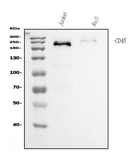
Western blot analysis of CD45 using anti-CD45 antibody (M00555-6).
Electrophoresis was performed on a 5-20% SDS-PAGE gel at 70V (Stacking gel) / 90V (Resolving gel) for 2-3 hours. The sample well of each lane was loaded with 30 ug of sample under reducing conditions.
Lane 1: human Jurkat whole cell lysates,
Lane 2: human Raji whole cell lysates.
After electrophoresis, proteins were transferred to a nitrocellulose membrane at 150 mA for 50-90 minutes. Blocked the membrane with 5% non-fat milk/TBS for 1.5 hour at RT. The membrane was incubated with mouse anti-CD45 antigen affinity purified monoclonal antibody (Catalog # M00555-6) at 0.5 μg/mL overnight at 4°C, then washed with TBS-0.1%Tween 3 times with 5 minutes each and probed with a goat anti-mouse IgG-HRP secondary antibody at a dilution of 1:10000 for 1.5 hour at RT. The signal is developed using an Enhanced Chemiluminescent detection (ECL) kit (Catalog # EK1001) with Tanon 5200 system. A specific band was detected for CD45 at approximately 220 kDa. The expected band size for CD45 is at 220 kDa.

Click image to see more details
IF analysis of CD45 using anti-CD45 antibody (M00555-6).
CD45 was detected in a paraffin-embedded section of human tonsli tissue. Heat mediated antigen retrieval was performed in EDTA buffer (pH 8.0, epitope retrieval solution). The tissue section was blocked with 10% goat serum. The tissue section was then incubated with 5 μg/mL mouse anti-CD45 Antibody (M00555-6) overnight at 4°C. Biotin conjugated goat anti-mouse IgG (BA1001) was used as secondary antibody and incubated for 30 minutes at 37°C. The tissue section was developed using DyLight®488 Conjugated Avidin (BA1128). The section was counterstained with DAPI. Visualize using a fluorescence microscope and filter sets appropriate for the label used.

Click image to see more details
Flow Cytometry analysis of Raji cells using anti-CD45 antibody (M00555-6).
Overlay histogram showing Raji cells stained with M00555-6 (Blue line). The cells were fixed with 4% paraformaldehyde and blocked with 10% normal goat serum. And then incubated with mouse anti-CD45 Antibody (M00555-6, 1 μg/1x106 cells) for 30 min at 20°C. DyLight®488 conjugated goat anti-mouse IgG (BA1126, 5-10 μg/1x106 cells) was used as secondary antibody for 30 minutes at 20°C. Isotype control antibody (Green line) was mouse IgG (1 μg/1x106) used under the same conditions. Unlabelled sample without incubation with primary antibody and secondary antibody (Red line) was used as a blank control.
Specific Publications For Anti-CD45 Antibody Picoband® (monoclonal, 3H6) (M00555-6)
Loading publications
Recommended Resources
Here are featured tools and databases that you might find useful.
- Boster's Pathways Library
- Protein Databases
- Bioscience Research Protocol Resources
- Data Processing & Analysis Software
- Photo Editing Software
- Scientific Literature Resources
- Research Paper Management Tools
- Molecular Biology Software
- Primer Design Tools
- Bioinformatics Tools
- Phylogenetic Tree Analysis
Customer Reviews
Have you used Anti-CD45 Antibody Picoband® (monoclonal, 3H6)?
Share your experimental results or join a short interview to earn up to $1,000 in product credits or other rewards.
0 Reviews For Anti-CD45 Antibody Picoband® (monoclonal, 3H6)
Customer Q&As
Have a question?
Find answers in Q&As, reviews.
Can't find your answer?
Submit your question
