This website uses cookies to ensure you get the best experience on our website.
- Table of Contents
2 Citations 6 Q&As
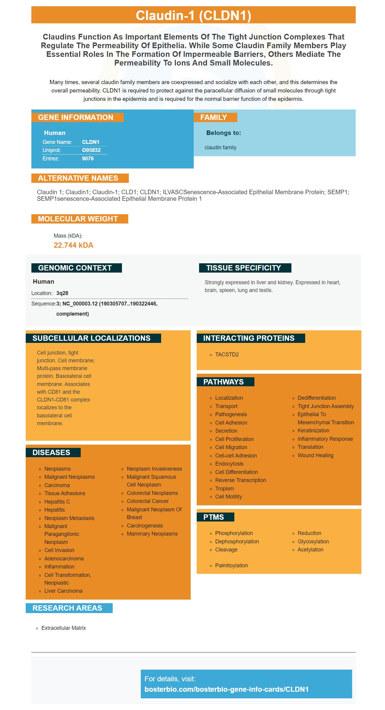
Facts about Claudin-1.
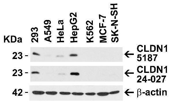
Many times, several claudin family members are coexpressed and socialize with each other, and this determines the overall permeability. CLDN1 is required to protect against the paracellular diffusion of small molecules through tight junctions in the epidermis and is required for the normal barrier function of the epidermis.
Human | |
---|---|
Gene Name: | CLDN1 |
Uniprot: | O95832 |
Entrez: | 9076 |
Belongs to: |
---|
claudin family |
claudin 1; Claudin1; Claudin-1; CLD1; CLDN1; ILVASCSenescence-associated epithelial membrane protein; SEMP1; SEMP1senescence-associated epithelial membrane protein 1
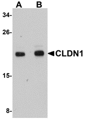
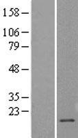
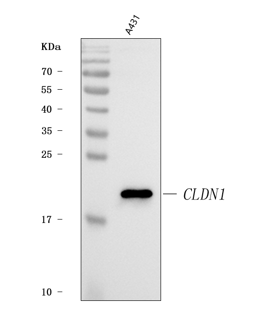
Mass (kDA):
22.744 kDA
Human | |
---|---|
Location: | 3q28 |
Sequence: | 3; NC_000003.12 (190305707..190322446, complement) |
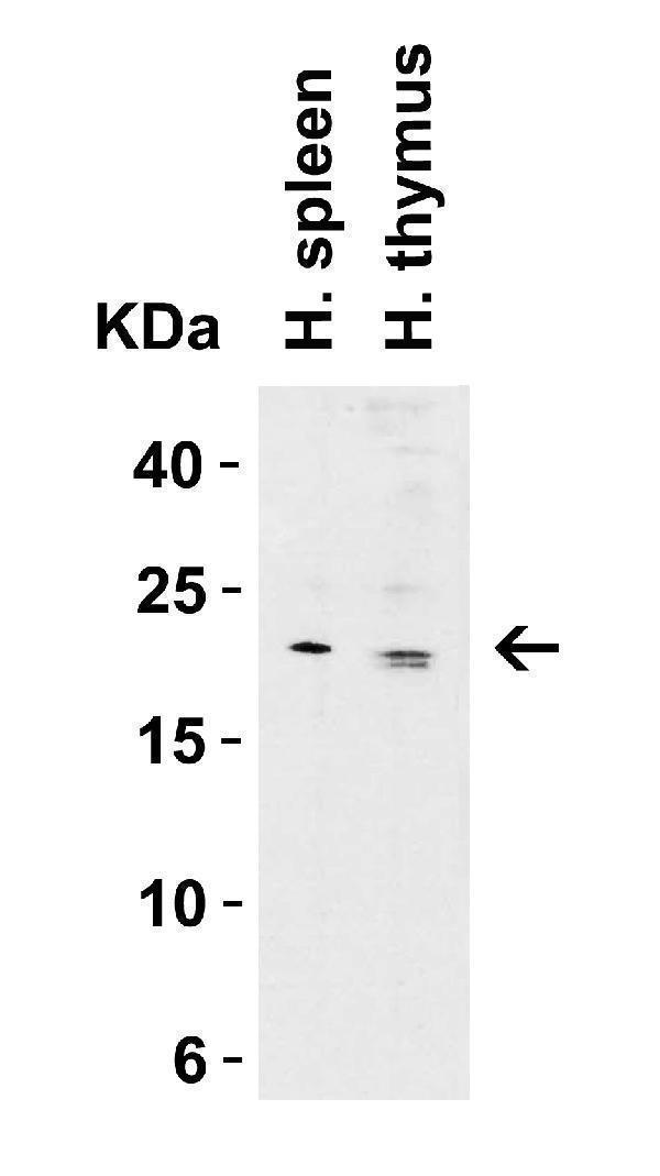
Strongly expressed in liver and kidney. Expressed in heart, brain, spleen, lung and testis.
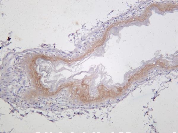
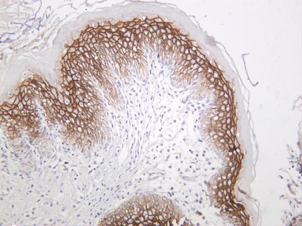
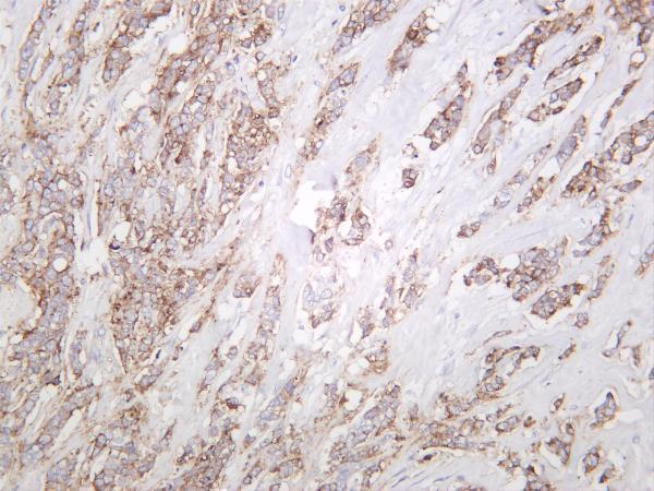
Cell junction, tight junction. Cell membrane; Multi-pass membrane protein. Basolateral cell membrane. Associates with CD81 and the CLDN1-CD81 complex localizes to the basolateral cell membrane.
The Claudin protein family includes many proteins that play key roles in the formation impermeable barriers. Others are responsible for permeability to small molecules. Claudin-1 antibodies are used to detect the protein in biological assays. Boster Bio develops its antibodies for Claudin-1 using rabbit and mouse. Many members of the family can be found in cells, tissues, immune system, and cells.
Boster Bio's Anti-Claudin 1 CLDN1 monoclonal antibody targets human Claudin-1. It is a transmembrane-protein. It is composed two extracellular layers, one comprising 53 amino acids and one which contains 27 amino acids. As with other Claudin family members, human Claudin-1 is composed of naturally occurring variants that are found in HCV-permissive human populations.
The Human version of the anti-Claudin 1 CLDN1 mono-clonal antibody is highly specific for this protein. It reacts well with both CLDN1 & Human. The antibody is available in PBS as a liquid and separately as a blocking peptide. The company also validates its antibody across a variety platforms, including cell lines derived from human lymphomas.
Boster researchers tested the antiCLDN1 MAb against HEK293T cells as well as a mouse CLDN1-expressing line of cells. The results showed that antiCLDN1 monoclonal anti-CLDN1 antibody bound to Huh7.5.1 cells at high affinity and reduced HCV infection. The antibody also inhibited potential cytotoxicity.
CLDN1 has a structure that was recently determined. It's a symmetrical protein that contains two extracellular loops. CLDN1 plays an important role in the physiological barriers and regulates solute movement in the paracellular area. In addition, hepatocytes are highly polarized cells, with TJs separating their plasma membranes into an apical-canalicular domain and a basolateral-sinusoidal domain. HCV can enter the body if CLDN1 is broken. This is why the anti-Claudin1 MAb must be used to target this domain. This protein has no or minimal adverse effects on the human liver function.
Researchers also found that Anti-Claudin 1 CLDN1 monoclonal antibody, Anti-Claudin 1, inhibited HCV infection of human liver-chimeric mice. This is promising for the development of novel anti-HCV agents. These findings indicate that anti-CLDN1 antiviral antibodies play an important part in preventing HCV infection in vivo.
Boster Bio's ACLDN1 monoclonal antibody has clinical potential as an immunotherapy for tumors. It has been shown to be expressed in normal colon mucosa and neoplastic tissues, making it a great candidate for immunotherapy. This anti-CLDN1 antitumor polyclonal antibody is capable, in addition to its antitumor effects, of inhibiting the growth and spread of tumor cells.
There are many uses for the CLDN1 marker. It is associated to increased copy number and protein expression in cervical cancer. CLDN1 expression in SiHa cells can increase invasive and antipoptotic activity. CLDN1 is also known to decrease the epithelial marker Ecadherin as well as increase the expression of mesenchymal marker VImentin. It is also known for promoting the growth and metastasis o athymic nude mouse.
In a study, CLDN1 expression was linked to increased CIN metastasis risk and subcutaneous tumor development in mice. This finding has important implications for cancer research. Although further research is required to understand the role of this marker in cancer, it has been shown to be therapeutic. Additionally, CLDN1 overexpression in cancer cells leads to decreased apoptosis as well as increased proliferation.
Molecular cloning is possible using a CLDN1 plasmid containing RNA. The CLDN1 gene was cloned using pcDNA3.1 (+), which contains Hind III, and BamHI. The primer sequences were GCTAAGCTTGCCCAAACTCTCTTC, and the antisense primer was 5' CGGGATCCTTTCTCTCACTTTCT. As a label, small interferingRNA was used.
This gene has been linked to the development of cervical cancer. Increased CLDN1 levels have been linked to the increase in 3q. These findings suggest that cervical carcinoma cells contain more CLDN1 copy than normal cervical tissue. The CLDN1 gene could have therapeutic potential in colorectal carcinoma. This gene may be useful in developing cervical cancer treatments if it is identified as a risk factor.
CLDN1 belongs the family of claudin-genes, which includes 24 members. These proteins are integral, transmembrane proteins that are involved in the formation and maintenance of tight junctions within vertebrates. Tight junctions are cellular structures which help maintain polarity, electro-osmotic gradients, and other cellular functions. They also act semi-permeable as paracellular endothelial dissemination barriers.
The CLDN1 gene is an important regulator of the transcription of many genes, including several that are involved in OC. Although there is limited evidence to suggest that CLDN1 has an effect on the expression of these genes in any way, it is known this gene can impact the recurrence rate of OC. Its involvement with OC aggressiveness is important because of its therapeutic potential. Targeting this gene with targeted therapies could increase chemosensitivity and decrease tumor metastasis.
Researchers used in vitro and in-vivo studies to test the expression of CDh2 (and other EMT markers) to investigate the role of CLDN1 for liver metastasis. These studies showed that OVCAR5 and SKOV8 cells had a slight increase in CDh2 expression after CLDN1 was knocked down. CLDN1 knockdown in CAOV2 cells did not result in significant changes to CDh2 expression.
A further study investigated the expression levels of CLDN1 in 18 SSA/P and eight MVHP samples. The expression levels CLDN1 were measured using qRTPCR and normalized to GAPDH. There were ten male and eight women SSA/P, whose ages ranged between 66 and 82 years. The MVHP samples, however, were between 54-78 and had a mean age 56.
Using Affymetrix microarray data from 16 matched primary and recurrent OC tissues, we found an inverse correlation between CLDN1 and CD44 in primary tumors, with no significant association between the two markers in recurrent cases. This finding may have implications for the role of CLDNs as it relates to the progression and treatment of ovarian cancer. In addition, the CLDN1 marker was found to be significantly associated with CD44 and CD133 in primary OCs, although this association was weaker in recurrent tumors.
YM201636 is another potential therapeutic candidate that isn't specific to CLDN1. Although it isn’t clear whether CLDN1 is a marker or ovarian tumor, it has been shown in animal models to reduce tumor growth. In fact, CLDN1 expression has a significant impact on cell-to-cell interactions and migration. Therefore, siRNA CLDN1 knockdown could have important clinical implications. It is a useful biomarker in the detection of ovarian cancer.
Many researchers believe that the CLDN1 marker, which is a key feature in metastasis, is responsible for the loss or cancer-cell junctions. It is not clear what genetic alterations are responsible for the absence of CLDN1. Researchers have found no genetic changes in CLDN1's promoter sequences or coding sequences in human carcinoma cell lines. They concluded that CLDN1 was not expressed in all cancer cells in humans, but that it plays an important role during metastasis.
CLDN1 also inhibits the expression of SLUG and regulates the activity EPHB6. This protein prevents breast cancer cells from migrating by suppressing ERK1/2 as well as SLUG. CLDN1 regulates SLUG transcription, which may play an important role in suppressing cancer cell metastasis. CLDN1 may also be a key regulator for EPHB-ephrin members.
Several studies have shown that CLDN1 enhances sensitivity of cancer cells to cisplatin and other chemotherapy drugs. CLDN1 and RUNX3 are essential in slowing the progression of lung cancer. These studies indicate that CLDN1 could be a prognostic sign for lung cancer. Further research is needed before it is considered a viable treatment option.
This marker is not of clinical value, but it is important to be mentioned for its role in tumorigenesis. It also represents CSC properties and is known to be a tumor-sensitizing factor. CLDN1 is highly expressed in tumor cells and has been associated with malignancy. The mechanism of action remains to be discovered. As with most molecular markers, its occurrence in cancer cells may be a sign of a metastatic disease.
PMID: 9931503 by Swisshelm K.L., et al. SEMP1, a senescence-associated cDNA isolated from human mammary epithelial cells, is a member of an epithelial membrane protein superfamily.
PMID: 10828592 by Halford S., et al. Assignment of claudin-1 (CLDN1) to human chromosome 3q28-->q29 with somatic cell hybrids.
*More publications can be found for each product on its corresponding product page