Click image to see more details
-

-
-
-
-
+14
Product Info Summary
| SKU: | PB9939 |
|---|---|
| Size: | 100 μg/vial |
| Reactive Species: | Mouse, Rat |
| Host: | Rabbit |
| Application: | IHC, WB |
Customers Who Bought This Also Bought

Product info
Product Name
Anti-Collagen I/COL1A1 Antibody Picoband®
SKU/Catalog Number
PB9939
PB0981 is an alternative SKU for this antibody, used in previous lots.
Size
100 μg/vial
Form
Lyophilized
Description
Boster Bio Anti-Collagen I/COL1A1 Antibody Picoband® catalog # PB9939. Tested in IHC, WB applications. This antibody reacts with Mouse, Rat. The brand Picoband indicates this is a premium antibody that guarantees superior quality, high affinity, and strong signals with minimal background in Western blot applications. Only our best-performing antibodies are designated as Picoband, ensuring unmatched performance.
Storage & Handling
Store at -20˚C for one year from date of receipt. After reconstitution, at 4˚C for one month. It can also be aliquotted and stored frozen at -20˚C for six months. Avoid repeated freeze-thaw cycles.
Cite This Product
Anti-Collagen I/COL1A1 Antibody Picoband® (Boster Biological Technology, Pleasanton CA, USA, Catalog # PB9939)
Host
Rabbit
Contents
Each vial contains 4 mg Trehalose, 0.9 mg NaCl and 0.2mg Na2HPO4.
Clonality
Polyclonal
Isotype
Rabbit IgG
Immunogen
A synthetic peptide corresponding to a sequence at the C-terminus of mouse Collagen I, different from the related human sequence by three amino acids, and identical to the related rat sequence.
Cross-reactivity
No cross-reactivity with other proteins
Reactive Species
PB9939 is reactive to Col1a1 in Mouse, Rat
Observed Molecular Weight
130,180-200 kDa
Calculated molecular weight
138.0 kDa
Background of Col1a1
Collagen, type I, alpha 1, also known as COL1A1, is a human gene that encodes the major component of type I collagen, the fibrillar collagen found in most connective tissues, including cartilage. This gene is mapped to 17q21.33. And this gene encodes the pro-alpha1 chains of type I collagen whose triple helix comprises two alpha1 chains and one alpha2 chain. Type I is a fibril-forming collagen found in most connective tissues and is abundant in bone, cornea, dermis and tendon. Mutations in this gene are associated with osteogenesis imperfecta types I-IV, Ehlers-Danlos syndrome type VIIA, Ehlers-Danlos syndrome Classical type, Caffey Disease and idiopathic osteoporosis.
Antibody Validation
Boster validates all antibodies on WB, IHC, ICC, Immunofluorescence, and ELISA with known positive control and negative samples to ensure specificity and high affinity, including thorough antibody incubations.
Application & Images
Applications
PB9939 is guaranteed for IHC, WB Boster Guarantee
Recommend Dilution
| Application | Dilution | Species |
|---|---|---|
| Western blot | 0.1-0.5μg/ml | Mouse, Rat |
| Immunohistochemistry (Paraffin-embedded Section) | 0.5-1μg/ml | Mouse, Rat |
Tested application
Suggested blocking solution with 5% non-fat milk or BSA; (*)Recommended protein loading: 20-40 µg per lane
Use TE buffer pH 9.0 for antigen retrieval; (*) citrate buffer pH 6.0 is an alternative.
Validation Images & Assay Conditions
Click image to see more details
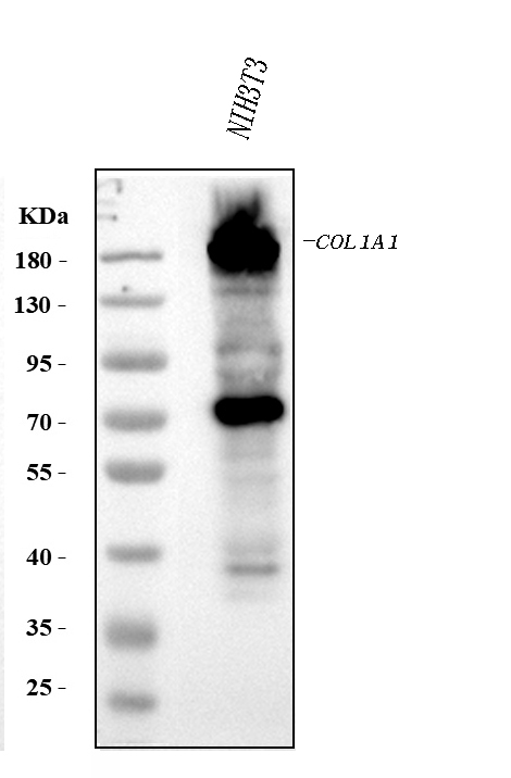
Western blot analysis of COL1A1 using anti-COL1A1 antibody (PB9939).
Electrophoresis was performed on a 8% SDS-PAGE gel at 80V (Stacking gel) / 120V (Resolving gel) for 2 hours. The sample well of each lane was loaded with 30 ug of sample under reducing conditions.
Lane 1: rat lung tissue lysates,
Lane 2: rat skin tissue lysates,
Lane 3: mouse lung tissue lysates,
Lane 4: mouse skin tissue lysates,
Lane 5: mouse NIH/3T3 whole cell lysates.
After electrophoresis, proteins were transferred to a nitrocellulose membrane at 150 mA for 50-90 minutes. Blocked the membrane with 5% non-fat milk/TBS for 1.5 hour at RT. The membrane was incubated with rabbit anti-COL1A1 antigen affinity purified polyclonal antibody (PB9939) at 0.5 μg/mL overnight at 4°C, then washed with TBS-0.1%Tween 3 times with 5 minutes each and probed with a goat anti-rabbit IgG-HRP secondary antibody (Catalog # BA1054) at a dilution of 1:5000 for 1.5 hour at RT. The signal is developed using an ECL Plus Western Blotting Substrate (Catalog # AR1196-200) with Tanon 5200 system. A specific band was detected for COL1A1 at approximately 130,180-200 kDa. The expected band size for COL1A1 is at 138 kDa.

Click image to see more details
OST treatment attenuated TiPs-induced osteogenesis inhibition in vivo. ( A ) Representative Masson’s trichrome staining images of calvarial sections. Scale bar, 200 μm (upper), 50 μm (lower). ( B ) Quantitative analysis of new bone area fraction (%) determined by Masson’s trichrome staining. ( C ) Representative images of calcein (green) and alizarin red (red) double labeling with a 10-day interval. Scale bar, 50 μm (upper), 20 μm (lower). ( D ) Quantitative analysis of average periosteum mineral apposition rates (MAR, µm/d). ( E-F ) Representative images and quantitative analysis of immunohistochemical (IHC) staining for osteocalcin (OCN). Scale bar, 100 μm (upper), 50 μm (lower). ( G-H ) Representative images and quantitative analysis of IHC staining for collagen type I alpha 1 (COL1α1). Scale bar, 100 μm (upper), 50 μm (lower). ( I-J ) Western blot analysis of the expression of osteogenic markers (OCN, COL1α1) in calvarial bone tissue samples from each group. n = 6. The selected images reflect typical examples from each group, closely representing the median degree as per statistical analysis. Data are presented as mean ± SD. One-way ANOVA with Tukey’s post hoc test. The relative COL1α1 protein levels were analyzed using Brown-Forsythe and Welch ANOVA tests followed by Tamhane T2 post hoc tests. ** P < 0.01 versus the Sham group. # P < 0.05 and ## P < 0.01 versus the TiPs group. ns , not statistically significant versus the TiPs group
Index in PubMed under a CC BY license. PMID: 39707212

Click image to see more details
Antifibrotic effect of microcystin (MC) in renal fibrosis mice. Mice were treated with MC-LR (20 μg/kg/day) or MC-RR (20 μg/kg/day) by intragastrical administration for 4 weeks in advance, and then unilateral ureteral ligation was performed to construct a mouse model of obstructive renal fibrosis. The operated mice were administrated with MC-LR or MC-RR for another week, and then were euthanized for further analysis. (A) Schematic diagram of the experimental design. Forty mice were divided into four groups, Sham, unilateral ureteral obstruction (UUO), UUO + MC-LR, and UUO + MC-RR groups, each with 10 mice. No mice died during the experiment. (B,C) Kidney tissue sections were employed for H&E and Masson staining (scale bar: 40 μm). (D) Fibrosis positive area in Masson staining were assessed by pathologist blind to this study ( n = 6). (E,F) The protein level of α -smooth muscle actin (α-SMA), fibronectin and collagen I were measured in the obstructive renal tissues of UUO mice by western blot. * p < 0.05, ** p < 0.01 determined by one-way ANOVA with S–N–K post hoc analysis. (G) Residual contents of MC were detected in the kidney tissues of model mice treatment with MC-LR or MC-RR using the ELISA method. Data were analyzed using independent Student’s t -test, * p < 0.05.
Index in PubMed under a CC BY license. PMID: 35754468

Click image to see more details
Renoprotection effect of microcystin (MC)-RR in different doses on unilateral ureteral obstruction (UUO) mice. The regimen of MC-RR was used in UUO mice. Twenty-five mice were divided into five groups, Sham, UUO, and UUO mice with treatment of MC-RR in three different doses (5, 10, and 20 μg/kg/day), each group with 5 mice. (A,B) Kidney tissue sections were employed for H&E and Masson staining (scale bar: 40 μm). (C) Fibrosis positive area in Masson staining were assessed by pathologist blind to this study ( n = 5). (D,E) Protein level of α -smooth muscle actin (α-SMA), fibronectin, and collagen I were measured in the renal tissues of UUO mice by western blot. (F,G) Protein expression of TGF-β, p-Smad3, and Smad3 in kidney tissue was measured by western blot. * p < 0.05, ** p < 0.01 determined by one-way ANOVA with S–N–K post hoc analysis.
Index in PubMed under a CC BY license. PMID: 35754468

Click image to see more details
Morphological features and identification of primary cultured tibial osteoblasts of broiler chicks. (A) Morphological image of P0 cells, day of 3 culture; (B) morphological image of P1 cells, day of 2 culture; (C) morphological image of P1 cells, day of 10 culture; (D) primary osteoblasts were positive in ALP straining; (E) immunofluorescence of COL1A1 in the primary tibial osteoblasts; (F) mineralized nodules were positive in ARS staining. ALP, alkaline phosphatase; COL1A1, collagen type I alpha 1; ARS, alizarin res S.
Index in PubMed under a CC BY license. PMID: 35392115

Click image to see more details
KD inhibits the expression of the fibrosis-related proteins in the rat model (A) The results of the IHC staining displayed the expression of α -SMA of the liver histology slices. KD significantly reduced the expression of brown stained α -SMA after irradiation (B) The IHC scores for grading the expression of α -SMA (C) Expression and localization of collagen in the liver tissue. The red stain represented collagen, which indicated that collagen expression in the IR + KD group was significantly lower than that in the IR group (D-F) The results of the western blots showed the expression of α -SMA and FN proteins in the liver tissue. The administration of KD after irradiation resulted in decreased expression of a -SMA and FN. For all results in this figure, original magnification, ×100 and ×200. Mean ± SEM. n. s. denotes not significant; * p < 0.05, ** p < 0.01, *** p < 0.001, **** p < 0.0001.
Index in PubMed under a CC BY license. PMID: 35126163

Click image to see more details
Fluorescence images of tenocytes after 5 days of culture on the surface of a culture plate (control group) and acellular amniotic membrane (amnion group). Tenocytes presented a clear cytoskeleton, good biocompatibility with acellular amniotic membrane, and even distribution on the surface of the materials, showing better growth activity than the control group (A,B) . Cell viability was measured by CCK-8, and the proliferation curve of tenocytes in the control and amniotic membrane groups was drawn (C) . Western blot assay for collagen I, fibronectin, TGF-β1, and bFGF expression in the tenocytes of the control and amnion groups for 1 week (D,E) . * p < 0.05; ** p < 0.01; and *** p < 0.001.
Index in PubMed under a CC BY license. PMID: 32478059

Click image to see more details
Representative immunofluorescence images of collagen I. Fluorescent micrographs of tenocytes after 2 and 5 days of culture on the surface of a culture plate and acellular amniotic membrane. Tenocyte nucleus shape observed under a fluorescence microscope (A,D,G,J) . Collagen I presented positive after the fluorescent FITC mark was observed under a fluorescence microscope (B,E,H,K) . Tenocyte nucleus and collagen I merging (C,F,I,L) . The corresponding semi quantitative analysis of collagen fluorescence intensity in panels (M,N) (scale bar = 50 um, n = 5, * P < 0.05 and ** P < 0.01).
Index in PubMed under a CC BY license. PMID: 32478059

Click image to see more details
Evaluation of the effect of CGA on liver histopathological and immunohistochemistry (IHC) in liver tissue. (A) Histological images of rat livers stained with H&E (original magnification, ×200). (B) The histopathologic detection of collagen in the liver by Masson’s trichrome stain (original magnification, ×100). (C,D) Effects of CGA on α-SMA and collagen I expression were examined with immunohistochemistry in liver tissue (original magnification, ×100).
Index in PubMed under a CC BY license. PMID: 29311932

Click image to see more details
VDR deficiency exacerbated hepatic fibrosis and steatosis in mice. A mRNA levels of fibrogenic markers ( α-Sma , Col1a1 , and Timp1 ). B mRNA levels of lipogenic enzymes ( Fasn , Acc-1 , and Acly ) and fatty acid desaturases ( Fads1/2 and Scd1 ) (n = 5). C Western blot analysis of VDR and TGFβ/Smad pathway (n = 3), D Serum levels of liver function markers (AST, ALT, ALP, and TBIL) (n = 5). * P < 0.05, ** P < 0.01 Full size image
Index in PubMed under a CC BY license. PMID: 40514735

Click image to see more details
VDR activation attenuated TGF-β1-induced fibrosteatotic changes in HSCs. A Western blot analysis of VDR and α-SMA (n = 3). B mRNA levels of fibrogenic markers ( α-Sma , Col1a1 , and Timp1 ). C mRNA levels of lipogenic enzymes ( Fasn , Acc-1 , and Acly ) and fatty acid desaturases ( Fads1/2 and Scd1 ) (n = 6). * P < 0.05, ** P < 0.01 Full size image
Index in PubMed under a CC BY license. PMID: 40514735

Click image to see more details
SI inhibited TGF-β1-induced activation of HSCs. A Chemical structure of the SI. B Cell viability of HSCs. C mRNA levels of fibrogenic markers ( α-Sma , Col1a1 , and Timp1 ). D Immunofluorescence analysis of α-SMA. scale bar = 20 μm. (n = 6). E Western blot analysis of α-SMA. F Molecular docking results of VDR and SI. G CETSA analysis. (n = 3) * P < 0.05, ** P < 0.01 Full size image
Index in PubMed under a CC BY license. PMID: 40514735

Click image to see more details
SI ameliorated CCl 4 -induced liver fibrosis in mice. A Serum levels of liver function markers (AST, ALT, ALP, and TBIL). B Liver tissue photograph and histopathological analysis (HE, Sirius Red, Masson), scale bar = 200 μm. C Serum calcium concentrations. D mRNA levels of fibrogenic markers ( α-Sma , Col1a1 , and Timp1 ). (n = 5) * P < 0.05, ** P < 0.01 Full size image
Index in PubMed under a CC BY license. PMID: 40514735

Click image to see more details
SI regulated VDR and fatty acid metabolism to suppress TGFβ/Smad pathway in CCl 4 -induced mice. A Immunohistochemistry analysis of TGF-β1, p-Smad3, α-SMA, COL1 A1. scale bar = 200 μm. (n = 5). B Western blot analysis of VDR, TGF-β1, and α-SMA. C mRNA levels of Cpt1a . D mRNA levels of lipogenic enzymes ( Fasn , Acc-1 , and Acly ) and fatty acid desaturases ( Fads1/2 and Scd1 ) (n = 6) * P < 0.05, ** P < 0.01 Full size image
Index in PubMed under a CC BY license. PMID: 40514735

Click image to see more details
VDR knockout abolished the SI-induced inhibition of HSCs activation. A Western blot analysis (n = 3). B Immunofluorescence analysis of α-SMA. scale bar = 20 μm. (n = 6). C mRNA levels of fibrogenic markers ( α-Sma , Col1a1 , and Timp1 ). (n = 6) *P < 0.05, **P < 0.01 compared with si-NC + TGF-β1; $ P < 0.05, $$ P < 0.01 compared with si-NC; # P < 0.05, ## P < 0.01 compared with si-VDR + TGF-β1 Full size image
Index in PubMed under a CC BY license. PMID: 40514735

Click image to see more details
IHC analysis of COL1A1 using anti COL1A1 antibody (PB9939).
COL1A1 was detected in a paraffin-embedded section of mouse lung tissue. Heat mediated antigen retrieval was performed in EDTA buffer (pH 8.0, epitope retrieval solution). The tissue section was blocked with 10% goat serum. The tissue section was then incubated with 2 μg/ml rabbit anti-COL1A1 Antibody (PB9939) overnight at 4°C. Peroxidase Conjugated Goat Anti-rabbit IgG was used as secondary antibody and incubated for 30 minutes at 37°C. The tissue section was developed using HRP Conjugated Rabbit IgG Super Vision Assay Kit (Catalog # SV0002) with DAB as the chromogen.

Click image to see more details
IHC analysis of COL1A1 using anti COL1A1 antibody (PB9939).
COL1A1 was detected in a paraffin-embedded section of rat lung tissue. Heat mediated antigen retrieval was performed in EDTA buffer (pH 8.0, epitope retrieval solution). The tissue section was blocked with 10% goat serum. The tissue section was then incubated with 2 μg/ml rabbit anti-COL1A1 Antibody (PB9939) overnight at 4°C. Peroxidase Conjugated Goat Anti-rabbit IgG was used as secondary antibody and incubated for 30 minutes at 37°C. The tissue section was developed using HRP Conjugated Rabbit IgG Super Vision Assay Kit (Catalog # SV0002) with DAB as the chromogen.

Click image to see more details
IHC analysis of COL1A1 using anti COL1A1 antibody (PB9939).
COL1A1 was detected in a paraffin-embedded section of rat kidney tissue. Heat mediated antigen retrieval was performed in EDTA buffer (pH 8.0, epitope retrieval solution). The tissue section was blocked with 10% goat serum. The tissue section was then incubated with 2 μg/ml rabbit anti-COL1A1 Antibody (PB9939) overnight at 4°C. Peroxidase Conjugated Goat Anti-rabbit IgG was used as secondary antibody and incubated for 30 minutes at 37°C. The tissue section was developed using HRP Conjugated Rabbit IgG Super Vision Assay Kit (Catalog # SV0002) with DAB as the chromogen.
Specific Publications For Anti-Collagen I/COL1A1 Antibody Picoband® (PB9939)
Loading publications
Recommended Resources
Here are featured tools and databases that you might find useful.
- Boster's Pathways Library
- Protein Databases
- Bioscience Research Protocol Resources
- Data Processing & Analysis Software
- Photo Editing Software
- Scientific Literature Resources
- Research Paper Management Tools
- Molecular Biology Software
- Primer Design Tools
- Bioinformatics Tools
- Phylogenetic Tree Analysis
Customer Reviews
Have you used Anti-Collagen I/COL1A1 Antibody Picoband®?
Share your experimental results or join a short interview to earn up to $1,000 in product credits or other rewards.
0 Reviews For Anti-Collagen I/COL1A1 Antibody Picoband®
Customer Q&As
Have a question?
Find answers in Q&As, reviews.
Can't find your answer?
Submit your question
16 Customer Q&As for Anti-Collagen I/COL1A1 Antibody Picoband®
Question
I would like using your anti-Collagen I/COL1A1 antibody for response to estradiol studies. Has this antibody been tested with western blotting on mouse lung? We would like to see some validation images before ordering.
Verified Customer
Verified customer
Asked: 2020-04-08
Answer
Thanks for your inquiry. This PB9939 anti-Collagen I/COL1A1 antibody is validated on mouse lung. It is guaranteed to work for IHC, WB in mouse, rat. Our Boster guarantee will cover your intended experiment even if the sample type has not been be directly tested.
Boster Scientific Support
Answered: 2020-04-08
Question
I have a question about product PB9939, anti-Collagen I/COL1A1 antibody. I was wondering if it would be possible to conjugate this antibody with biotin. I would need it to be without BSA or sodium azide. I am planning on using a buffer exchange of sodium azide with PBS only. Would there be problems for me to conjugate the antibody and store it in -20 degrees in small aliquots?
Verified Customer
Verified customer
Asked: 2020-03-27
Answer
We suggest not storing this antibody with PBS buffer only in -20 degrees. If you want to store it in -20 degrees it is best to add some cryoprotectant like glycerol. If you want carrier free PB9939 anti-Collagen I/COL1A1 antibody, we can provide it to you in a special formula with trehalose and/or glycerol. These molecules will not interfere with conjugation chemistry and provide a good level of protection for the antibody from degradation. Please be sure to specify this in your purchase order.
Boster Scientific Support
Answered: 2020-03-27
Question
Will anti-Collagen I/COL1A1 antibody PB9939 work for IHC with fetal brain cortex?
Verified Customer
Verified customer
Asked: 2019-09-02
Answer
According to the expression profile of fetal brain cortex, COL1A1 is highly expressed in fetal brain cortex. So, it is likely that anti-Collagen I/COL1A1 antibody PB9939 will work for IHC with fetal brain cortex.
Boster Scientific Support
Answered: 2019-09-02
Question
See below the WB image, lot number and protocol we used for fetal brain cortex using anti-Collagen I/COL1A1 antibody PB9939. Please let me know if you require anything else.
Verified Customer
Verified customer
Asked: 2019-08-07
Answer
Thank you very much for the data. Our lab team are working to resolve this as quickly as possible, and we appreciate your patience and understanding! You have provided everything we needed. Please let me know if there is anything you need in the meantime.
Boster Scientific Support
Answered: 2019-08-07
Question
Do you have a BSA free version of anti-Collagen I/COL1A1 antibody PB9939 available?
Verified Customer
Verified customer
Asked: 2019-07-03
Answer
Thank you for your recent telephone inquiry. I can confirm that some lots of this anti-Collagen I/COL1A1 antibody PB9939 are BSA free. For now, these lots are available and we can make a BSA free formula for you free of charge. It will take 3 extra days to prepare. If you require this antibody BSA free again in future, please do not hesitate to contact me and I will be pleased to check which lots we have in stock that are BSA free.
Boster Scientific Support
Answered: 2019-07-03
Question
We were happy with the WB result of your anti-Collagen I/COL1A1 antibody. However we have been able to see positive staining in fetal brain cortex extracellular using this antibody. Is that expected? Could you tell me where is COL1A1 supposed to be expressed?
Verified Customer
Verified customer
Asked: 2019-03-11
Answer
According to literature, fetal brain cortex does express COL1A1. Generally COL1A1 expresses in secreted, extracellular space, extracellular. Regarding which tissues have COL1A1 expression, here are a few articles citing expression in various tissues:
Bone, Pubmed ID: 3340531
Brain, Pubmed ID: 15489334
Liver, Pubmed ID: 24275569
Skin, Pubmed ID: 4319110, 5529814
Boster Scientific Support
Answered: 2019-03-11
Question
We are currently using anti-Collagen I/COL1A1 antibody PB9939 for rat tissue, and we are satisfied with the IHC results. The species of reactivity given in the datasheet says mouse, rat. Is it true that the antibody can work on zebrafish tissues as well?
Verified Customer
Verified customer
Asked: 2019-02-05
Answer
The anti-Collagen I/COL1A1 antibody (PB9939) has not been tested for cross reactivity specifically with zebrafish tissues, but there is a good chance of cross reactivity. We have an innovator award program that if you test this antibody and show it works in zebrafish you can get your next antibody for free. Please contact me if I can help you with anything.
Boster Scientific Support
Answered: 2019-02-05
Question
I see that the anti-Collagen I/COL1A1 antibody PB9939 works with IHC, what is the protocol used to produce the result images on the product page?
A. Krishna
Verified customer
Asked: 2019-01-11
Answer
You can find protocols for IHC on the "support/technical resources" section of our navigation menu. If you have any further questions, please send an email to support@bosterbio.com
Boster Scientific Support
Answered: 2019-01-11
Question
I was wanting to use your anti-Collagen I/COL1A1 antibody for IHC for rat fetal brain cortex on frozen tissues, but I want to know if it has been validated for this particular application. Has this antibody been validated and is this antibody a good choice for rat fetal brain cortex identification?
Verified Customer
Verified customer
Asked: 2018-09-12
Answer
It shows on the product datasheet, PB9939 anti-Collagen I/COL1A1 antibody has been validated for IHC, WB on mouse, rat tissues. We have an innovator award program that if you test this antibody and show it works in rat fetal brain cortex in IHC-frozen, you can get your next antibody for free.
Boster Scientific Support
Answered: 2018-09-12
Question
Is this PB9939 anti-Collagen I/COL1A1 antibody reactive to the isotypes of COL1A1?
G. Collins
Verified customer
Asked: 2018-04-03
Answer
The immunogen of PB9939 anti-Collagen I/COL1A1 antibody is A synthetic peptide corresponding to a sequence at the C-terminus of mouse Collagen I (1185-1207aa YDFSFLPQPPQEKSQDGGRYYRA), different from the related human sequence by three amino acids, and identical to the related rat sequence. Could you tell me which isotype you are interested in so I can help see if the immunogen is part of this isotype?
Boster Scientific Support
Answered: 2018-04-03
Question
We are interested in to test anti-Collagen I/COL1A1 antibody PB9939 on rat fetal brain cortex for research purposes, then I may be interested in using anti-Collagen I/COL1A1 antibody PB9939 for diagnostic purposes as well. Is the antibody suitable for diagnostic purposes?
Verified Customer
Verified customer
Asked: 2017-11-06
Answer
The products we sell, including anti-Collagen I/COL1A1 antibody PB9939, are only intended for research use. They would not be suitable for use in diagnostic work. If you have the means to develop a product into diagnostic use, and are interested in collaborating with us and develop our product into an IVD product, please contact us for more discussions.
Boster Scientific Support
Answered: 2017-11-06
Question
Is a blocking peptide available for product anti-Collagen I/COL1A1 antibody (PB9939)?
Verified Customer
Verified customer
Asked: 2017-05-15
Answer
We do provide the blocking peptide for product anti-Collagen I/COL1A1 antibody (PB9939). If you would like to place an order for it please contact support@bosterbio.com and make a special request.
Boster Scientific Support
Answered: 2017-05-15
Question
We appreciate helping with my inquiry over the phone. Here are the WB image, lot number and protocol we used for fetal brain cortex using anti-Collagen I/COL1A1 antibody PB9939. Let me know if you need anything else.
D. Edwards
Verified customer
Asked: 2016-06-29
Answer
I appreciate the data. You have provided everything we needed. Our lab team are working to resolve your inquiry as quickly as possible, and we appreciate your patience and understanding! Please let me know if there is anything you need in the meantime.
Boster Scientific Support
Answered: 2016-06-29
Question
We have been able to see staining in rat bone. What should we do? Is anti-Collagen I/COL1A1 antibody supposed to stain bone positively?
D. Johnson
Verified customer
Asked: 2014-04-01
Answer
According to literature bone does express COL1A1. According to Uniprot.org, COL1A1 is expressed in spinal cord, spleen, brain, skin, fetal brain cortex, bone, liver, among other tissues. Regarding which tissues have COL1A1 expression, here are a few articles citing expression in various tissues:
Bone, Pubmed ID: 3340531
Brain, Pubmed ID: 15489334
Liver, Pubmed ID: 24275569
Skin, Pubmed ID: 4319110, 5529814
Boster Scientific Support
Answered: 2014-04-01
Question
We ordered your anti-Collagen I/COL1A1 antibody for WB on skin last year. I am using mouse, and We want to use the antibody for IHC next. My question regards examining skin as well as spinal cord in our next experiment. Do you have any suggestion on which antibody would work the best for IHC?
H. Zhao
Verified customer
Asked: 2013-10-28
Answer
I took a look at the website and datasheets of our anti-Collagen I/COL1A1 antibody and it appears that PB9939 has been validated on mouse in both WB and IHC. Thus PB9939 should work for your application. Our Boster satisfaction guarantee will cover this product for IHC in mouse even if the specific tissue type has not been validated. We do have a comprehensive range of products for IHC detection and you can check out our website bosterbio.com to find out more information about them.
Boster Scientific Support
Answered: 2013-10-28
Question
Will PB9939 anti-Collagen I/COL1A1 antibody work on parafin embedded sections? If so, which fixation method do you recommend we use (PFA, paraformaldehyde, other)?
R. Moore
Verified customer
Asked: 2013-06-07
Answer
You can see on the product datasheet, PB9939 anti-Collagen I/COL1A1 antibody as been tested on IHC. It is best to use PFA for fixation because it has better tissue penetration ability. PFA needs to be prepared fresh before use. Long term stored PFA turns into formalin, as the PFA molecules congregate and become formalin.
Boster Scientific Support
Answered: 2013-06-07
