Click image to see more details
-

-
-
-
-
+1
Product Info Summary
| SKU: | A00455-2 |
|---|---|
| Size: | 100 μg/vial |
| Reactive Species: | Human |
| Host: | Rabbit |
| Application: | ELISA, Flow Cytometry, IHC, WB |
Customers Who Bought This Also Bought

Product info
Product Name
Anti-CXCR2 Antibody Picoband®
SKU/Catalog Number
A00455-2
Size
100 μg/vial
Form
Lyophilized
Description
Boster Bio Anti-CXCR2 Antibody Picoband® catalog # A00455-2. Tested in ELISA, Flow Cytometry, IHC, WB applications. This antibody reacts with Human. The brand Picoband indicates this is a premium antibody that guarantees superior quality, high affinity, and strong signals with minimal background in Western blot applications. Only our best-performing antibodies are designated as Picoband, ensuring unmatched performance.
Storage & Handling
Store at -20˚C for one year from date of receipt. After reconstitution, at 4˚C for one month. It can also be aliquotted and stored frozen at -20˚C for six months. Avoid repeated freeze-thaw cycles.
Cite This Product
Anti-CXCR2 Antibody Picoband® (Boster Biological Technology, Pleasanton CA, USA, Catalog # A00455-2)
Host
Rabbit
Contents
Each vial contains 4mg Trehalose, 0.9mg NaCl, 0.2mg Na2HPO4, 0.01mg NaN3.
Clonality
Polyclonal
Isotype
Rabbit IgG
Immunogen
E.coli-derived human CXCR2 recombinant protein (Position: F32-I54;A177-R212).
Cross-reactivity
No cross-reactivity with other proteins
Reactive Species
A00455-2 is reactive to CXCR2 in Human
Observed Molecular Weight
41 kDa
Calculated molecular weight
40.8 kDa
Background of CXCR2
CXCR2 is a receptor for Interleukin 8, which is a powerful neutrophil chemotactic factor. It is a member of the GPCR family (subfamily, chemokine). Binding of IL8 to the receptor causes activation of neutrophils. This response is mediated via a G-protein that activate a phosphatidylinositol-calcium second messenger system. This receptor binds to IL8 with a high affinity and to GRO/MGSA and NAP2 also with a high affinity. It has been reported to be expressed in a wide variety of tissues. ESTs have been isolated from human placenta and thymus libraries.
Antibody Validation
Boster validates all antibodies on WB, IHC, ICC, Immunofluorescence, and ELISA with known positive control and negative samples to ensure specificity and high affinity, including thorough antibody incubations.
Application & Images
Applications
A00455-2 is guaranteed for ELISA, Flow Cytometry, IHC, WB Boster Guarantee
Recommend Dilution
| Application | Dilution | Species |
|---|---|---|
| Western blot | 0.25-0.5μg/ml | Human |
| Immunohistochemistry (Paraffin-embedded Section) | 2-5μg/ml | Human |
| Flow Cytometry (Fixed) | 1-3μg/1x106 cells | Human |
| ELISA | 0.1-0.5μg/ml | - |
Tested application
Suggested blocking solution with 5% non-fat milk or BSA; (*)Recommended protein loading: 20-40 µg per lane
Use TE buffer pH 9.0 for antigen retrieval; (*) citrate buffer pH 6.0 is an alternative.
Validation Images & Assay Conditions
Click image to see more details
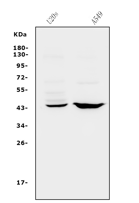
Western blot analysis of CXCR2 using anti-CXCR2 antibody (A00455-2).
Electrophoresis was performed on a 5-20% SDS-PAGE gel at 70V (Stacking gel) / 90V (Resolving gel) for 2-3 hours. The sample well of each lane was loaded with 30ug of sample under reducing conditions.
Lane 1: human U20S whole cell lysates,
Lane 2: human A549 whole cell lysates.
After Electrophoresis, proteins were transferred to a Nitrocellulose membrane at 150mA for 50-90 minutes. Blocked the membrane with 5% Non-fat Milk/ TBS for 1.5 hour at RT. The membrane was incubated with rabbit anti-CXCR2 antigen affinity purified polyclonal antibody (Catalog # A00455-2) at 0.5 μg/mL overnight at 4°C, then washed with TBS-0.1%Tween 3 times with 5 minutes each and probed with a goat anti-rabbit IgG-HRP secondary antibody at a dilution of 1:5000 for 1.5 hour at RT. The signal is developed using an Enhanced Chemiluminescent detection (ECL) kit (Catalog # EK1002) with Tanon 5200 system. A specific band was detected for CXCR2 at approximately 41KD. The expected band size for CXCR2 is at 41KD.

Click image to see more details
RM-1 cell inoculation increases CXCR2 mRNA and protein expression in the spinal cord. (A) Real-time PCR results show the increase of CXCR2 mRNA expression in the spinal cord. CXCR2 mRNA was increased from 7 days to 21 days after inoculation. *P <0.05 vs . sham. n = 4 mice per group. (B) Western blot shows time course of CXCR2 protein expression in the spinal cord after inoculation (B) . n = 3 mice per group. (C, D) Immunostaining shows the CXCR2 expression in the spinal cord in sham (C) and inoculated (D) animals. CXCR2-IR was increased at 7 days after inoculation. (E-G) Double staining shows CXCR2 was colocalized with neuronal marker NeuN. H , I . SB2205002 attenuated RM-1 cell inoculation-induced mechanical allodynia (H) and heat hyperalgesia (I) . * P <0.05; ** P <0.01; *** P <0.001 vs . vehicle.
Index in PubMed under a CC BY license. PMID: 24580964

Click image to see more details
IHC analysis of CXCR2 using anti-CXCR2 antibody (A00455-2).
CXCR2 was detected in paraffin-embedded section of human appendicitis tissue. Heat mediated antigen retrieval was performed in EDTA buffer (pH8.0, epitope retrieval solution). The tissue section was blocked with 10% goat serum. The tissue section was then incubated with 2μg/ml rabbit anti-CXCR2 Antibody (A00455-2) overnight at 4°C. Biotinylated goat anti-rabbit IgG was used as secondary antibody and incubated for 30 minutes at 37°C. The tissue section was developed using Strepavidin-Biotin-Complex (SABC) (Catalog # SA1022) with DAB as the chromogen.

Click image to see more details
IHC analysis of CXCR2 using anti-CXCR2 antibody (A00455-2).
CXCR2 was detected in paraffin-embedded section of human tonsil tissue. Heat mediated antigen retrieval was performed in EDTA buffer (pH8.0, epitope retrieval solution). The tissue section was blocked with 10% goat serum. The tissue section was then incubated with 2μg/ml rabbit anti-CXCR2 Antibody (A00455-2) overnight at 4°C. Biotinylated goat anti-rabbit IgG was used as secondary antibody and incubated for 30 minutes at 37°C. The tissue section was developed using Strepavidin-Biotin-Complex (SABC) (Catalog # SA1022) with DAB as the chromogen.

Click image to see more details
Flow Cytometry analysis of THP-1 cells using anti-CXCR2 antibody (A00455-2).
Overlay histogram showing THP-1 cells stained with A00455-2 (Blue line). To facilitate intracellular staining, cells were fixed with 4% paraformaldehyde and permeabilized with permeabilization buffer. The cells were blocked with 10% normal goat serum. And then incubated with rabbit anti-CXCR2 Antibody (A00455-2, 1μg/1x106 cells) for 30 min at 20°C. DyLight®488 conjugated goat anti-rabbit IgG (BA1127, 5-10μg/1x106 cells) was used as secondary antibody for 30 minutes at 20°C. Isotype control antibody (Green line) was rabbit IgG (1μg/1x106) used under the same conditions. Unlabelled sample without incubation with primary antibody and secondary antibody (Red line) was used as a blank control.
Specific Publications For Anti-CXCR2 Antibody Picoband® (A00455-2)
Loading publications
Recommended Resources
Here are featured tools and databases that you might find useful.
- Boster's Pathways Library
- Protein Databases
- Bioscience Research Protocol Resources
- Data Processing & Analysis Software
- Photo Editing Software
- Scientific Literature Resources
- Research Paper Management Tools
- Molecular Biology Software
- Primer Design Tools
- Bioinformatics Tools
- Phylogenetic Tree Analysis
Customer Reviews
Have you used Anti-CXCR2 Antibody Picoband®?
Share your experimental results or join a short interview to earn up to $1,000 in product credits or other rewards.
0 Reviews For Anti-CXCR2 Antibody Picoband®
Customer Q&As
Have a question?
Find answers in Q&As, reviews.
Can't find your answer?
Submit your question
