Click image to see more details
Product Info Summary
| SKU: | A00101-2 |
|---|---|
| Size: | 1mg |
| Reactive Species: | Human |
| Host: | Rabbit |
| Application: | ELISA, Flow Cytometry, IP, IHC, WB |
Customers Who Bought This Also Bought

Product info
Product Name
Anti-IL-1 beta Antibody
SKU/Catalog Number
A00101-2
Size
1mg
Form
Liquid (sterile filtered)
Description
Boster Bio Anti-IL-1 beta Antibody (Catalog # A00101-2). Tested in ELISA, IHC, WB applications. This antibody reacts with Human.
Storage & Handling
Store vial at -20°C prior to opening. Aliquot contents and freeze at -20°C or below for extended storage. Avoid cycles of freezing and thawing. Centrifuge product if not completely clear after standing at room temperature. This product is stable for several weeks at 4°C as an undiluted liquid. Dilute only prior to immediate use. Expiration date is one (1) year from date of opening. (Ship on dry ice.)
Cite This Product
Anti-IL-1 beta Antibody (Boster Biological Technology, Pleasanton CA, USA, Catalog # A00101-2)
Host
Rabbit
Contents
0.02 M Potassium Phosphate, 0.15 M Sodium Chloride, pH 7.2
Clonality
Polyclonal
Isotype
IgG
Immunogen
This antibody was prepared by repeated immunizations with recombinant human IL-1ß produced in E.coli. The MW of the recombinant 153 aa IL-1ß was 17 kDa with the N-terminal amino acid at position alanine 117. This cleavage site is generated by the IL-1ß converting enzyme (ICE, capase-1).
Cross-reactivity
Detects ~20kDa. Does not cross-react with αB-crystallin, βL-crystallin, ΒH- crystallin, γ-crystallin, HSP25, HSP27 or HSP47 proteins.
Reactive Species
A00101-2 is reactive to IL1B in Human
Observed Molecular Weight
68 kDa
Calculated molecular weight
30.7 kDa
Background of IL1B
IL-1 beta (also known as Interleukin-1 beta, IL-1ß and catabolin) is produced by activated macrophages. IL-1 stimulates thymocyte proliferation by inducing IL-2 release, B-cell maturation and proliferation, and fibroblast growth factor activity. IL-1 proteins are involved in the inflammatory response, being identified as endogenous pyrogens, and are reported to stimulate the release of prostaglandin and collagenase from synovial cells. IL-1ß is a monomeric secreted protein that may be released by damaged cells or is secreted by a mechanism differing from that used for other secretory proteins.
Antibody Validation
Boster validates all antibodies on WB, IHC, ICC, Immunofluorescence, and ELISA with known positive control and negative samples to ensure specificity and high affinity, including thorough antibody incubations.
Application & Images
Applications
A00101-2 is guaranteed for ELISA, Flow Cytometry, IP, IHC, WB Boster Guarantee
Recommend Dilution
| Application | Dilution | Species |
|---|---|---|
| ELISA: 1:500 - 1:2 | 000 | |
| WB: 1:1 | 000 | |
| Anti-Human IL-1ß has been tested for use in ELISA | immunohistochemistry | immunoblotting. This antibody is suitable for neutralizations, radioimmunoassays, flow cytometry, and immunoprecipitation. It recognizes the 17,000 MW mature IL-1ß. For immunoblots, typically, IL-1ß is detected from supernatants or lysates of 2 x 10E6 endotoxin-stimulated peripheral blood mononuclear cells (PBMC). PBMC are stimulated for 24 hours with 1% (v/v) serum plus 10 ng/mL E.coli LPS. For immunoprecipitation pre-clearing the preparation with a non-specific Rabbit IgG (p/n 011-001-297) to reduce background is suggested. For immunohistochemistry either paraffin fixation or cryofixation can be used for sample preparation to stain intracellular IL-1ß. For ELISA use HRP Conjugated Anti-Rabbit IgG [H&L] (Goat) (611-1302) for detection. In ELISA formats this antibody is best used as the second antibody in combination with a monoclonal antibody as a capture antibody. This antibody is also useful for neutralization of human and primate IL-1ß activity in bioassays. It does not neutralize the biological activity IL-1α. It does not neutralize the biological activity of murine, rat or rabbit IL-1ß. For neutralization, it is recommended to incubate the sample with a dilution of the antibody for at least 4 hours before being tested. A control of similarly diluted normal rabbit IgG is recommended. This antibody can be used for FACS analysis. Caution should be exhibited as the F(c) domain of the rabbit IgG molecule may interact with cells non-specifically. |
Validation Images & Assay Conditions
Click image to see more details
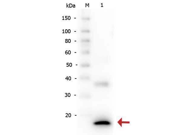
Western Blot of Rabbit anti-Human IL-1ß antibody. Lane 1: Human IL-1ß. Load: 5 ng per lane. Primary antibody: Human IL-1ß antibody at 1:2,000 for overnight at 4°C. Secondary antibody: Peroxidase rabbit secondary antibody at 1:40,000 for 30 min at RT. Block: Blocking Buffer for Fluorescent Western Blotting for 30 min at RT. Predicted/Observed size: 17 kDa, 17 kDa for Human IL-1ß. Other band(s): Unspecific band at ~35 kDa.

Click image to see more details
Immunohistochemistry of Human IL1 beta antibody. Tissue: human medullary lymph node. Fixation: formalin fixed paraffin embedded. Antigen retrieval: user optimized. Primary antibody: Human IL1 beta antibody. Secondary antibody: Peroxidase goat anti-rabbit at 1:10,000 for 45 min at RT. Localization: cytoplasm. Staining: Close up of medullary lymph node: positive staining in the cytoplasm of circulating macrophages. Neg Ctr (far right) of normal rabbit IgG with pH 6.2 at 40X.
Specific Publications For Anti-IL-1 beta Antibody (A00101-2)
Loading publications
Recommended Resources
Here are featured tools and databases that you might find useful.
- Boster's Pathways Library
- Protein Databases
- Bioscience Research Protocol Resources
- Data Processing & Analysis Software
- Photo Editing Software
- Scientific Literature Resources
- Research Paper Management Tools
- Molecular Biology Software
- Primer Design Tools
- Bioinformatics Tools
- Phylogenetic Tree Analysis
Customer Reviews
Have you used Anti-IL-1 beta Antibody?
Share your experimental results or join a short interview to earn up to $1,000 in product credits or other rewards.
0 Reviews For Anti-IL-1 beta Antibody
Customer Q&As
Have a question?
Find answers in Q&As, reviews.
Can't find your answer?
Submit your question
14 Customer Q&As for Anti-IL-1 beta Antibody
Question
We appreciate helping with my inquiry over the phone. Here are the WB image, lot number and protocol we used for macrophage using anti-IL-1 beta antibody A00101-2. Let me know if you need anything else.
Verified Customer
Verified customer
Asked: 2020-04-02
Answer
Thank you for the data. You have provided everything we needed. Our lab team are working to resolve your inquiry as quickly as possible, and we appreciate your patience and understanding! Please let me know if there is anything you need in the meantime.
Boster Scientific Support
Answered: 2020-04-02
Question
We bought anti-IL-1 beta antibody for IP on monocyte in the past. I am using human, and I plan to use the antibody for WB next. I am interested in examining monocyte as well as skin in our next experiment. Could you please give me some suggestion on which antibody would work the best for WB?
K. Dhar
Verified customer
Asked: 2019-11-11
Answer
I took a look at the website and datasheets of our anti-IL-1 beta antibody and it appears that A00101-2 has been tested on human in both IP and WB. Thus A00101-2 should work for your application. Our Boster satisfaction guarantee will cover this product for WB in human even if the specific tissue type has not been validated. We do have a comprehensive range of products for WB detection and you can check out our website bosterbio.com to find out more information about them.
Boster Scientific Support
Answered: 2019-11-11
Question
My question regarding product A00101-2, anti-IL-1 beta antibody. I was wondering if it would be possible to conjugate this antibody with biotin. I would need it to be without BSA or sodium azide. I am planning on using a buffer exchange of sodium azide with PBS only. Would there be problems for me to conjugate the antibody and store it in -20 degrees in small aliquots?
Verified Customer
Verified customer
Asked: 2019-10-18
Answer
We do not advise storing this antibody with PBS buffer only in -20 degrees. If you want to store it in -20 degrees it is best to add some cryoprotectant like glycerol. If you want carrier free A00101-2 anti-IL-1 beta antibody, we can provide it to you in a special formula with trehalose and/or glycerol. These molecules will not interfere with conjugation chemistry and provide a good level of protection for the antibody from degradation. Please be sure to specify this in your purchase order.
Boster Scientific Support
Answered: 2019-10-18
Question
Would anti-IL-1 beta antibody A00101-2 work for IP with macrophage?
Verified Customer
Verified customer
Asked: 2019-09-18
Answer
According to the expression profile of macrophage, IL1B is highly expressed in macrophage. So, it is likely that anti-IL-1 beta antibody A00101-2 will work for IP with macrophage.
Boster Scientific Support
Answered: 2019-09-18
Question
Do you have a BSA free version of anti-IL-1 beta antibody A00101-2 available?
Verified Customer
Verified customer
Asked: 2019-06-20
Answer
Thanks for your recent telephone inquiry. I can confirm that some lots of this anti-IL-1 beta antibody A00101-2 are BSA free. For now, these lots are available and we can make a BSA free formula for you free of charge. It will take 3 extra days to prepare. If you require this antibody BSA free again in future, please do not hesitate to contact me and I will be pleased to check which lots we have in stock that are BSA free.
Boster Scientific Support
Answered: 2019-06-20
Question
I was wanting to use your anti-IL-1 beta antibody for IP for human macrophage on frozen tissues, but I want to know if it has been validated for this particular application. Has this antibody been validated and is this antibody a good choice for human macrophage identification?
G. Edwards
Verified customer
Asked: 2018-09-03
Answer
You can see on the product datasheet, A00101-2 anti-IL-1 beta antibody has been tested for IP, IHC, WB on human tissues. We have an innovator award program that if you test this antibody and show it works in human macrophage in IHC-frozen, you can get your next antibody for free.
Boster Scientific Support
Answered: 2018-09-03
Question
Is a blocking peptide available for product anti-IL-1 beta antibody (A00101-2)?
Verified Customer
Verified customer
Asked: 2018-06-04
Answer
We do provide the blocking peptide for product anti-IL-1 beta antibody (A00101-2). If you would like to place an order for it please contact support@bosterbio.com and make a special request.
Boster Scientific Support
Answered: 2018-06-04
Question
We are currently using anti-IL-1 beta antibody A00101-2 for human tissue, and we are satisfied with the IHC results. The species of reactivity given in the datasheet says human. Is it possible that the antibody can work on monkey tissues as well?
Verified Customer
Verified customer
Asked: 2017-10-12
Answer
The anti-IL-1 beta antibody (A00101-2) has not been validated for cross reactivity specifically with monkey tissues, though there is a good chance of cross reactivity. We have an innovator award program that if you test this antibody and show it works in monkey you can get your next antibody for free. Please contact me if I can help you with anything.
Boster Scientific Support
Answered: 2017-10-12
Question
I see that the anti-IL-1 beta antibody A00101-2 works with IP, what is the protocol used to produce the result images on the product page?
Verified Customer
Verified customer
Asked: 2017-07-10
Answer
You can find protocols for IP on the "support/technical resources" section of our navigation menu. If you have any further questions, please send an email to support@bosterbio.com
Boster Scientific Support
Answered: 2017-07-10
Question
We have seen staining in human macrophage. What should we do? Is anti-IL-1 beta antibody supposed to stain macrophage positively?
Verified Customer
Verified customer
Asked: 2017-05-31
Answer
According to literature macrophage does express IL1B. According to Uniprot.org, IL1B is expressed in smooth muscle tissue, leukocyte, histiocytic lymphoma, monocyte, lung, skin, macrophage, among other tissues. Regarding which tissues have IL1B expression, here are a few articles citing expression in various tissues:
Histiocytic lymphoma, Pubmed ID: 3493774
Leukocyte, Pubmed ID: 3490654
Lung, Pubmed ID: 15489334
Macrophage, Pubmed ID: 20148899
Monocyte, Pubmed ID: 2635664, 11991722
Skin, Pubmed ID: 1919436
Boster Scientific Support
Answered: 2017-05-31
Question
I am interested in to test anti-IL-1 beta antibody A00101-2 on human macrophage for research purposes, then I may be interested in using anti-IL-1 beta antibody A00101-2 for diagnostic purposes as well. Is the antibody suitable for diagnostic purposes?
S. Li
Verified customer
Asked: 2016-09-01
Answer
The products we sell, including anti-IL-1 beta antibody A00101-2, are only intended for research use. They would not be suitable for use in diagnostic work. If you have the means to develop a product into diagnostic use, and are interested in collaborating with us and develop our product into an IVD product, please contact us for more discussions.
Boster Scientific Support
Answered: 2016-09-01
Question
My team were satisfied with the WB result of your anti-IL-1 beta antibody. However we have observed positive staining in skin cytosol using this antibody. Is that expected? Could you tell me where is IL1B supposed to be expressed?
M. Zhao
Verified customer
Asked: 2014-10-29
Answer
According to literature, skin does express IL1B. Generally IL1B expresses in cytoplasm, cytosol. Regarding which tissues have IL1B expression, here are a few articles citing expression in various tissues:
Histiocytic lymphoma, Pubmed ID: 3493774
Leukocyte, Pubmed ID: 3490654
Lung, Pubmed ID: 15489334
Macrophage, Pubmed ID: 20148899
Monocyte, Pubmed ID: 2635664, 11991722
Skin, Pubmed ID: 1919436
Boster Scientific Support
Answered: 2014-10-29
Question
I have attached the WB image, lot number and protocol we used for macrophage using anti-IL-1 beta antibody A00101-2. Please let me know if you require anything else.
K. Anderson
Verified customer
Asked: 2014-06-10
Answer
Thank you very much for the data. Our lab team are working to resolve this as quickly as possible, and we appreciate your patience and understanding! You have provided everything we needed. Please let me know if there is anything you need in the meantime.
Boster Scientific Support
Answered: 2014-06-10
Question
Does A00101-2 anti-IL-1 beta antibody work on parafin embedded sections? If so, which fixation method do you recommend we use (PFA, paraformaldehyde, other)?
D. Kulkarni
Verified customer
Asked: 2014-03-18
Answer
You can see on the product datasheet, A00101-2 anti-IL-1 beta antibody as been validated on IP. It is best to use PFA for fixation because it has better tissue penetration ability. PFA needs to be prepared fresh before use. Long term stored PFA turns into formalin, as the PFA molecules congregate and become formalin.
Boster Scientific Support
Answered: 2014-03-18
