This website uses cookies to ensure you get the best experience on our website.


- Table of Contents
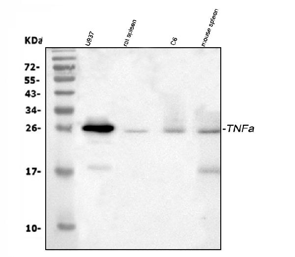
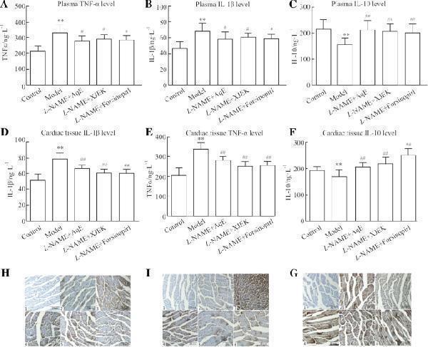
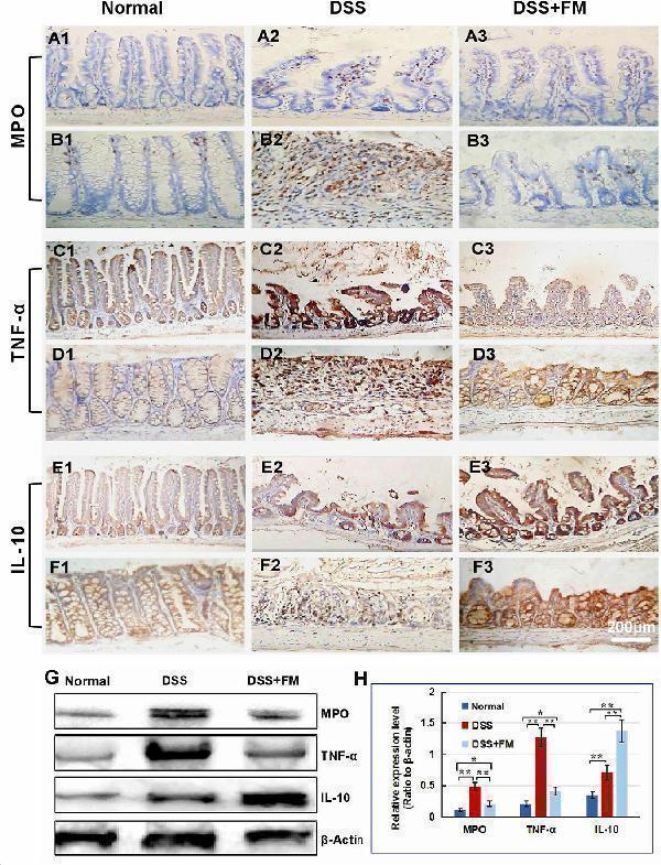
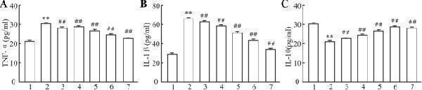
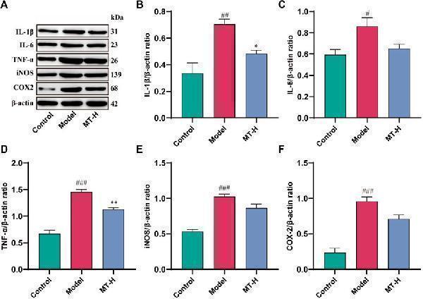
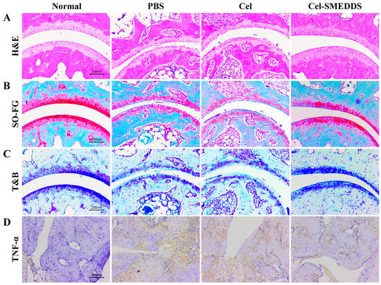
TNF antibody (PA1079) performs well in Flow Cytometry, IHC, WB and shows reactivity with Human, Mouse, Rat.



| Reactive Species | Host | Application |
|---|---|---|
| Human, Mouse, Rat | Rabbit | Flow Cytometry, IHC, WB |
TNF antibody was positively detected by WB in:
human U937 whole cell, rat spleen tissue, rat C6 whole cell, mouse spleen tissue
Suggested blocking solution with 5% non-fat milk or BSA; (*)Recommended protein loading: 20-40 ug per lane
TNF antibody was positively detected by IHC in:
human B lymphocytic tumor tissue
Use TE buffer pH 9.0 for antigen retrieval; (*) citrate buffer pH 6.0 is an alternative.
TNF antibody was positively detected by FC in:
CACO-2 cell
* It is recommended to optimize the amount of this reagent in each experimental system to achieve the best results.
Note: For flow cytometry (FCM), validated for fixed cells only. Not validated for FACS or surface flow cytometry using live cells.
25.6 kDa
The results generated by this homology analysis tool for commonly used animal species are shown below. For unlisted species, please refer to the homology analysis between the immunogen sequence and the corresponding species.
A homology of ≥90% is recommended. Generally, higher homology indicates a greater potential for cross-species reactivity. However, protein expression levels may vary among different species, tissues, and cell types. Therefore, the homology analysis results are for reference only and do not guarantee antibody cross-reactivity or experimental performance.







Submit a review and receive $370 in product credits, a $185 Amazon gift card, or our carefully curated gift bundles.
| SKU | PA1079 |
|---|---|
| Application | Western Blot |
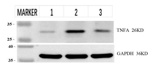
| Sample | PC-12 (rat adrenal pheochromocytoma cells, highly differentiated) |
| Sample Processing Description | ① Normal PC12 cells (rat adrenal pheochromocytoma cells, highly differentiated)② PC12 cells are treated with β-amyloid (Aβ) to establish an in vitro Alzheimer's disease (AD) model.③ The AD in vitro model is treated with the therapeutic drug. |
| Other Reagents | RIPA lysis buffer, Protease inhibitor, Electrophoresis buffer, Transfer buffer, Blocking buffer |
| Primary Antibody | TNF alpha Antibody Picoband® |
| Primary Incubation | 1:2000, overnight at 4 ℃ |
| Secondary Antibody | HRP-conjugated goat anti-rabbit IgG |
| Secondary Incubation | 1:10000, 1 hour in RT |
| Detection | Substrate: ECL substrate; Image system: ChemiDoc MP |
| Results Summary | ① Normal PC-12 cells (rat adrenal pheochromocytoma cells (highly differentiated)) ② PC12 cells were induced with β-amyloid protein (Aβ) to establish an in vitro model of Alzheimer's disease (AD) ③ The AD in vitro model was treated with therapeutic drugs. TNFα is a typical pro-inflammatory factor. The experimental results showed a clear target band. After modeling, the total TNFα protein expression level was upregulated, and after drug treatment, the expression level decreased, which was consistent with theory and expectations. |
Yue Zhang, Chengdu University of Traditional Chinese Medicine
Verified customer
Submitted 2026-08-26
| SKU | PA1079 |
|---|---|
| Application | Western Blot |
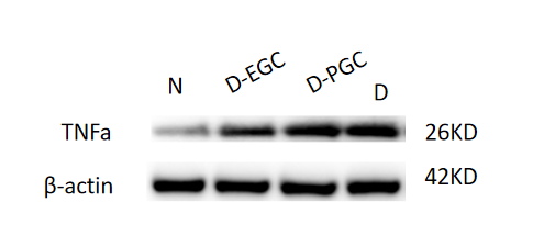
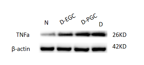
| Sample | rat alveolar bone |
| Sample Processing Description | ① Normal mouse hippocampal tissue, ② Hippocampal tissue from Alzheimer’s disease model mouse, ③ Hippocampal tissue from Alzheimer’s disease model mouse treated with a self-developed drug. Total protein was extracted from all samples. |
| Other Reagents | RIPA lysis buffer, Protease inhibitor, Running buffer, Transfer buffer, Blocking buffer |
| Primary Antibody | TNF alpha Antibody Picoband® |
| Primary Incubation | 1:1000, overnight at 4 ℃ |
| Secondary Antibody | HRP Conjugated AffiniPure Goat Anti-Rabbit IgG (H+L) (BA1054) |
| Secondary Incubation | 1:10000, 1 h in RT |
| Detection | Substrate: ECL substrate, Image system: ChemiDoc MP |
| Results Summary | TNFα is a typical pro-inflammatory factor that enhances inflammation, inhibits osteogenesis, and promotes bone resorption in bone defect models. The experimental results show that in rat alveolar bone tissue, TNFα levels increase in the following order: ① N: normal rats’ alveolar bone, ② D-EGC: EGCG-treated diabetic bone defect model, ③ D-PGC: PGC-1α-treated diabetic bone defect model, ④ D: diabetic bone defect model, indicating that both EGCG and PGC-1α can suppress TNFα expression. |
Yurou Chen, Shanxi Medical University, Stomatology Hospital
Verified customer
Submitted 2026-03-30
| SKU | PA1079 |
|---|---|
| Application | Western Blot |
| Sample | mouse brain tissue |
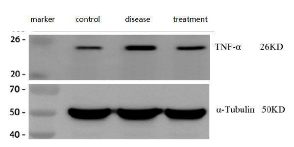
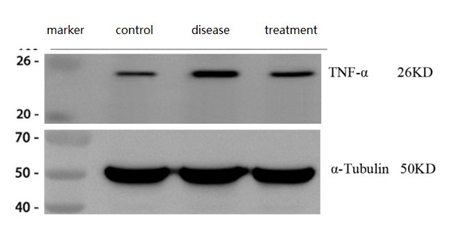
| Sample Processing Description | ① Normal mouse hippocampal tissue, ② Hippocampal tissue from Alzheimer’s disease model mouse, ③ Hippocampal tissue from Alzheimer’s disease model mouse treated with a self-developed drug. Total protein was extracted from all samples. |
| Other Reagents | RIPA lysis buffer, Protease inhibitor, Running buffer, Transfer buffer, Blocking buffer |
| Primary Antibody | TNF alpha Antibody Picoband® |
| Primary Incubation | 1:1000, overnight at 4 ℃ |
| Secondary Antibody | HRP Conjugated AffiniPure Goat Anti-Rabbit IgG (H+L) (BA1054) |
| Secondary Incubation | 1:10000, 1 h in RT |
| Detection | Substrate: ECL substrate, Image system: ChemiDoc MP |
| Results Summary | TNFα (tumor necrosis factor-alpha) is a key pro-inflammatory cytokine in the immune system, playing an essential role in normal immune defense and tissue homeostasis, with levels typically rising rapidly after inflammation. The results showed that TNFα expression was significantly increased in the brains of Alzheimer’s disease model mice and decreased after treatment. |
Huili Yin, Shandong First Medical University
Verified customer
Submitted 2026-03-27
| SKU | PA1079 |
|---|---|
| Application | Western Blot |
| Sample | Mouse lung tissue |
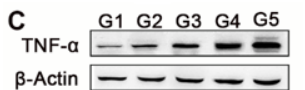
| Sample Processing Description | Tissue samples were directly lysed in RIPA buffer, mixed with loading buffer at the appropriate ratio, and denatured by heating at 98 °C. Load 20 µL of protein sample per lane onto SDS-PAGE. |
| Primary Incubation | The membrane was incubated with TNF-α primary antibodies (1:1000) overnight at 4 °C. |
| Secondary Antibody | HRP-conjugated Goat Anti-Rabbit IgG Secondary Antibody |
| Secondary Incubation | Incubate at room temperature for 1 hour |
| Other Reagents used | 5% Non-fat milk |
| Detection | Signal was developed using ECL substrate on an ChemiDoc MP system. |
| Results Summary | This antibody shows excellent consistency and reproducibility across batches, ensuring reliable and stable experimental results. Its performance remains consistent across different experiments, providing strong technical support for our research. |
Jiahui Yan, College of Chemistry and Chemical Engineering, Ocean University of China
Verified customer
Submitted 2025-09-25
We promise Anti-TNF alpha Antibody Picoband® performs excellently in relevant applications for Flow Cytometry, IHC, WB. If it doesn't perform as promised, we will refund the product. Learn more


We promise all of our products perform as described in datasheets.
This product is for Research Use Only. Not for use in diagnostic procedures.