Click image to see more details
-

-
-
-
-
+4
Product Info Summary
| SKU: | PB9010 |
|---|---|
| Size: | 100 μg/vial |
| Reactive Species: | Rat |
| Host: | Rabbit |
| Application: | WB |
Customers Who Bought This Also Bought

Product info
Product Name
Anti-TNF alpha Antibody Picoband®
SKU/Catalog Number
PB9010
PB0082 is an alternative SKU for this antibody, used in previous lots.
Size
100 μg/vial
Form
Lyophilized
Description
Boster Bio Anti-TNF alpha Antibody Picoband® catalog # PB9010. Tested in WB applications. This antibody reacts with Rat. The brand Picoband indicates this is a premium antibody that guarantees superior quality, high affinity, and strong signals with minimal background in Western blot applications. Only our best-performing antibodies are designated as Picoband, ensuring unmatched performance.
Storage & Handling
Store at -20˚C for one year from date of receipt. After reconstitution, at 4˚C for one month. It can also be aliquotted and stored frozen at -20˚C for six months. Avoid repeated freeze-thaw cycles.
Cite This Product
Anti-TNF alpha Antibody Picoband® (Boster Biological Technology, Pleasanton CA, USA, Catalog # PB9010)
Host
Rabbit
Contents
Each vial contains 4 mg Trehalose, 0.9 mg NaCl and 0.2 mg Na2HPO4.
Clonality
Polyclonal
Isotype
Rabbit IgG
Immunogen
E.coli-derived rat TNF alpha recombinant protein (Position: D89-L235). Rat TNF alpha shares 95% amino acid (aa) sequence identity with mouse TNF alpha.
Cross-reactivity
No cross-reactivity with other proteins
Reactive Species
PB9010 is reactive to Tnf in Rat
Observed Molecular Weight
26 kDa
Calculated molecular weight
25.8 kDa
Background of Tnf
TNF alpha (Tumor Necrosis Factor alpha) gene encodes a multifunctional proinflammatory cytokine that belongs to the tumor necrosis factor (TNF) superfamily. This cytokine is mainly secreted by macrophages. It can bind to, and thus functions through its receptors TNFRSF1A/TNFR1 and TNFRSF1B/TNFBR. This cytokine is involved in the regulation of a wide spectrum of biological processes including cell proliferation, differentiation, apoptosis, lipid metabolism, and coagulation. This cytokine has been implicated in a variety of diseases, including autoimmune diseases, insulin resistance, and cancer. Knockout studies in mice also suggested the neuroprotective function of this cytokine.
Antibody Validation
Boster validates all antibodies on WB, IHC, ICC, Immunofluorescence, and ELISA with known positive control and negative samples to ensure specificity and high affinity, including thorough antibody incubations.
Application & Images
Applications
PB9010 is guaranteed for WB Boster Guarantee
Recommend Dilution
| Application | Dilution | Species |
|---|---|---|
| Western blot | 0.1-0.5μg/ml | Rat |
Tested application
Suggested blocking solution with 5% non-fat milk or BSA; (*)Recommended protein loading: 20-40 µg per lane
Validation Images & Assay Conditions
Click image to see more details
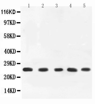
Western blot analysis of TNF alpha using anti-TNF alpha antibody (PB9010).
Electrophoresis was performed on a 5-20% SDS-PAGE gel at 70V (Stacking gel) / 90V (Resolving gel) for 2-3 hours. The sample well of each lane was loaded with 50ug of sample under reducing conditions.
Lane 1: PC-12 Whole Cell Lysate,
Lane 2: Rat Spleen Tissue Lysate,
Lane 3: Rat Brain Tissue Lysate,
Lane 4: Rat Kidney Tissue Lysate,
Lane 5: Rat Liver Tissue Lysate.
After Electrophoresis, proteins were transferred to a Nitrocellulose membrane at 150mA for 50-90 minutes. Blocked the membrane with 5% Non-fat Milk/ TBS for 1.5 hour at RT. The membrane was incubated with rabbit anti-TNF alpha antigen affinity purified polyclonal antibody (Catalog # PB9010) at 0.5 μg/mL overnight at 4°C, then washed with TBS-0.1%Tween 3 times with 5 minutes each and probed with a goat anti-rabbit IgG-HRP secondary antibody at a dilution of 1:10000 for 1.5 hour at RT. The signal is developed using an Enhanced Chemiluminescent detection (ECL) kit (Catalog # EK1002) with Tanon 5200 system. A specific band was detected for TNF alpha at approximately 26KD. The expected band size for TNF alpha is at 26KD.

Click image to see more details
Red nucleus IL-33 facilitates the early development of mononeuropathic pain by inducing TNF-α through activating ERK, p38 MAPK and JAK2/STAT3 signaling pathways. A Western blotting showed that red nucleus TNF-α was increased at 1 week post-SNI, intrarubral injection of anti-IL-33 antibody at 1 week post-SNI suppressed the overexpression of TNF-α ( n = 6 per group, F = 14.302, P < 0.001). B Western blotting displayed that intrarubral administration of PD98059, SB203580, or AG490 at 1 week post-SNI inhibited the production of TNF-α ( n = 6 per group, F = 13.157, P < 0.001). C Immunohistochemistry showed that red nucleus TNF-α was upregulated at 1 week post-SNI, intrarubral injection of anti-IL-33 antibody, PD98059, SB203580, or AG490 at 1 week post-SNI suppressed the production of TNF-α ( n = 4 per group, F = 33.029, P < 0.001). ** P < 0.01 and *** P < 0.001. Scale bars = 50 μm
Index in PubMed under a CC BY license. PMID: 34225736

Click image to see more details
Red nucleus IL-33 evokes mechanical hypersensitivity by inducing TNF-α through activating ERK, p38 MAPK, and JAK2/STAT3 signaling pathways. A Western blotting indicated that intrarubral administration of IL-33 stimulated the secretion of TNF-α in naive rats ( n = 6 per group, F = 15.143, P < 0.001). B Western blotting showed that intrarubral pre-injection of PD98059, SB203580, or AG490, 30 min before IL-33 administration, restrained IL-33-induced overexpression of TNF-α in naive rats ( n = 6 per group, F = 9.812, P < 0.001). C Immunohistochemistry showed that intrarubral injection of IL-33 potentiated the secretion of TNF-α in naive rats, intrarubral pre-injection of PD98059, SB203580, or AG490, 30 min prior to IL-33 administration, inhibited IL-33-induced overexpression of TNF-α in naive rats ( n = 4 per group, F = 44.310, P < 0.001). *** P < 0.001. Scale bars = 50 μm
Index in PubMed under a CC BY license. PMID: 34225736

Click image to see more details
DJ-1 interference increased the expression of TNF-α, IL-1β, and IL-6 after cerebral I/R injury. a and c Western blot detecting DJ-1 and the cytokines TNF-α, IL-1β, and IL-6 in rats. b , d Western blot detecting DJ-1 and the cytokines TNF-α, IL-1β, and IL-6 in astrocytes. e–g Quantification of TNF-α, IL-1β, and IL-6 in rats by ELISA. h‑j Quantification of TNF-α, IL-1β, and IL-6 in astrocytes by ELISA. The data are expressed as the mean ± SEM. * p < 0.05, ** p < 0.01, *** p < 0.001, **** p < 0.0001. n = 6 per group
Index in PubMed under a CC BY license. PMID: 32151250

Click image to see more details
DJ-1 inhibited the expression of TNF-α, IL-1β, and IL-6 after cerebral I/R injury via SHP-1. a , c After virus and TPI-1 were used to overexpress DJ-1 and inhibit SHP-1, respectively, Western blotting was used to detect the cytokines IL-1β, IL-6, and TNF-α in rats. b , d After virus and TPI-1 were used to overexpress DJ-1 and inhibit SHP-1, respectively, Western blotting was used to detect the cytokines IL-1β, IL-6, and TNF-α in astrocytes. e , g After treatment with an SHP-1 inhibitor, Western blotting was used to detect the cytokines IL-1β, IL-6, and TNF-α in rats. f , h After treatment with an SHP-1 inhibitor, Western blotting was used to detect the cytokines IL-1β, IL-6, and TNF-α in astrocytes. i , k , m Quantification of TNF-α, IL-1β, and IL-6 in rats by ELISA. j , l , n Quantification of TNF-α, IL-1β, and IL-6 in astrocytes by ELISA. The data are expressed as the mean ± SEM. * p < 0.05 vs. the sham group; # p < 0.05 vs. the MCAO group; & p < 0.05 vs. the overexpression group; ★ p < 0.05 vs. the DMSO group; △ p < 0.05 vs. the DMSO group; ◆ p < 0.05 vs. the TPI-1 group. n = 6 per group. The data are expressed as the mean ± SEM. * p < 0.05 vs. the control group; # p < 0.05 vs. the OGD/R group; & p < 0.05 vs. the overexpression group; ★ p < 0.05 vs. the DMSO group; △ p < 0.05 vs. the DMSO group; ◆ p < 0.05 vs. the TPI-1 group. n = 6 per group. The groups in a and b and their corresponding groups in the statistical graphs are as follows: sham (−−−), MCAO or OGD/R (+−−), scramble (+−−), overexpression (++−), overexpression + DMSO (++−), and overexpression + TPI-1 (+++). The groups in e and f and the corresponding groups in the statistical graphs are as follows: DMSO (+−−), TPI-1 (+−+), overexpression + DMSO (++−), overexpression + TPI-1 (+++)
Index in PubMed under a CC BY license. PMID: 32151250

Click image to see more details
DJ-1 regulated the disassociation of NLRX1 from TRAF6 after cerebral I/R injury via SHP-1. a , g After virus and TPI-1 were used to overexpress DJ-1 and inhibit SHP-1, respectively, Western blotting was used to detect NLRX1, TRAF6, and SHP-1 in rats. b , h After virus and TPI-1 were used to overexpress DJ-1 and inhibit SHP-1, respectively, Western blotting was used to detect NLRX1, TRAF6, and SHP-1 in astrocytes. c , i After treatment with an SHP-1 inhibitor, Western blotting was used to detect the cytokines IL-1β, IL-6, and TNF-α in rats. d , j After treatment with an SHP-1 inhibitor, Western blotting was used to detect the cytokines IL-1β, IL-6, and TNF-α in astrocytes. e Immunoprecipitation and immunoblot analyses of NLRX1-TRAF6 in rats. f Immunoprecipitation and immunoblot analyses of SHP-1-TRAF6 in rats. The data are expressed as the mean ± SEM. * p < 0.05 vs. the sham group; # p < 0.05 vs. the MCAO group; & p < 0.05 vs. the overexpression group; ★ p < 0.05 vs. the DMSO group; △ p < 0.05 vs. the DMSO group. n = 6 per group. The data are expressed as the mean ± SEM. * p < 0.05 vs. the control group; # p < 0.05 vs. the OGD/R group; & p < 0.05 vs. the overexpression group; ★ p < 0.05 vs. the DMSO group; △ p < 0.05 vs. the DMSO group. n = 6 per group
Index in PubMed under a CC BY license. PMID: 32151250

Click image to see more details
Fecal microbiota transplantation (FMT) reduced systemic or local inflammatory response. (A) Protein levels of the pro-inflammatory cytokines and chemokines in the blood. (B) mRNA levels of the pro-inflammatory cytokines and chemokines in kidney tissue using real-time PCR. (C) Western blot analysis of the pro-inflammatory cytokines in kidney tissue. (D) Protein band intensities quantified using optical densitometry and normalized to β-actin levels within the respective experimental groups. n = 6. Representative images from six independent experiments. The p -values above the data denote statistical comparisons on the groups—the control (Ctrl), ischemia–reperfusion injury plus normal saline (IRI+NS), and IRI+FMT groups—which were calculated using ANOVA with Tukey’s post-hoc test. * p < 0.05; *** p < 0.001.
Index in PubMed under a CC BY license. PMID: 41078364

Click image to see more details
Propionic acid reduced the systemic or local inflammatory response. (A) Protein levels of the pro-inflammatory cytokines and chemokines in the blood. (B) mRNA levels of the pro-inflammatory cytokines and chemokines in kidney tissue using real-time PCR. (C) Western blot analysis of the pro-inflammatory cytokines in kidney tissue. (D) Protein band intensities quantified using optical densitometry and normalized to β-actin levels within the respective experimental groups. n = 6. Representative images from six independent experiments. The p -values above the data denote statistical comparisons of the groups—the control (Ctrl), ischemia–reperfusion injury plus normal saline (IRI+NS), and IRI plus propionic acid (IRI+Prop) groups—which were calculated using ANOVA with Tukey’s post-hoc test. ** p < 0.01; *** p < 0.001.
Index in PubMed under a CC BY license. PMID: 41078364
Specific Publications For Anti-TNF alpha Antibody Picoband® (PB9010)
Loading publications
Recommended Resources
Here are featured tools and databases that you might find useful.
- Boster's Pathways Library
- Protein Databases
- Bioscience Research Protocol Resources
- Data Processing & Analysis Software
- Photo Editing Software
- Scientific Literature Resources
- Research Paper Management Tools
- Molecular Biology Software
- Primer Design Tools
- Bioinformatics Tools
- Phylogenetic Tree Analysis
Customer Reviews
Have you used Anti-TNF alpha Antibody Picoband®?
Share your experimental results or join a short interview to earn up to $1,000 in product credits or other rewards.
0 Reviews For Anti-TNF alpha Antibody Picoband®
Customer Q&As
Have a question?
Find answers in Q&As, reviews.
Can't find your answer?
Submit your question
15 Customer Q&As for Anti-TNF alpha Antibody Picoband®
Question
I am looking for using your anti-TNF alpha antibody for response to drug studies. Has this antibody been tested with western blotting on spleen tissue? We would like to see some validation images before ordering.
Verified Customer
Verified customer
Asked: 2020-05-08
Answer
We appreciate your inquiry. This PB9010 anti-TNF alpha antibody is tested on rat liver tissue, spleen tissue, tissue lysate, brain tissue, kidney tissue. It is guaranteed to work for WB in rat. Our Boster guarantee will cover your intended experiment even if the sample type has not been be directly tested.
Boster Scientific Support
Answered: 2020-05-08
Question
I was wanting to use to test anti-TNF alpha antibody PB9010 on rat leukocyte for research purposes, then I may be interested in using anti-TNF alpha antibody PB9010 for diagnostic purposes as well. Is the antibody suitable for diagnostic purposes?
Verified Customer
Verified customer
Asked: 2020-04-24
Answer
The products we sell, including anti-TNF alpha antibody PB9010, are only intended for research use. They would not be suitable for use in diagnostic work. If you have the means to develop a product into diagnostic use, and are interested in collaborating with us and develop our product into an IVD product, please contact us for more discussions.
Boster Scientific Support
Answered: 2020-04-24
Question
We have been able to see staining in rat prostatic carcinoma. Are there any suggestions? Is anti-TNF alpha antibody supposed to stain prostatic carcinoma positively?
Verified Customer
Verified customer
Asked: 2019-10-14
Answer
According to literature prostatic carcinoma does express TNF. According to Uniprot.org, TNF is expressed in leukocyte, blood, prostatic carcinoma, among other tissues. Regarding which tissues have TNF expression, here are a few articles citing expression in various tissues:
Blood, Pubmed ID: 15489334
Prostatic carcinoma, Pubmed ID: 8597870, 10205166
Boster Scientific Support
Answered: 2019-10-14
Question
Does anti-TNF alpha antibody PB9010 work for WB with leukocyte?
Verified Customer
Verified customer
Asked: 2019-09-18
Answer
According to the expression profile of leukocyte, TNF is highly expressed in leukocyte. So, it is likely that anti-TNF alpha antibody PB9010 will work for WB with leukocyte.
Boster Scientific Support
Answered: 2019-09-18
Question
I was wanting to use your anti-TNF alpha antibody for WB for rat leukocyte on frozen tissues, but I want to know if it has been tested for this particular application. Has this antibody been tested and is this antibody a good choice for rat leukocyte identification?
Verified Customer
Verified customer
Asked: 2019-02-06
Answer
It shows on the product datasheet, PB9010 anti-TNF alpha antibody has been validated for WB on rat tissues. We have an innovator award program that if you test this antibody and show it works in rat leukocyte in IHC-frozen, you can get your next antibody for free.
Boster Scientific Support
Answered: 2019-02-06
Question
Our team were satisfied with the WB result of your anti-TNF alpha antibody. However we have seen positive staining in prostatic carcinoma cell membrane using this antibody. Is that expected? Could you tell me where is TNF supposed to be expressed?
Verified Customer
Verified customer
Asked: 2019-01-24
Answer
From literature, prostatic carcinoma does express TNF. Generally TNF expresses in cell membrane. Regarding which tissues have TNF expression, here are a few articles citing expression in various tissues:
Blood, Pubmed ID: 15489334
Prostatic carcinoma, Pubmed ID: 8597870, 10205166
Boster Scientific Support
Answered: 2019-01-24
Question
I see that the anti-TNF alpha antibody PB9010 works with WB, what is the protocol used to produce the result images on the product page?
Verified Customer
Verified customer
Asked: 2018-12-25
Answer
You can find protocols for WB on the "support/technical resources" section of our navigation menu. If you have any further questions, please send an email to support@bosterbio.com
Boster Scientific Support
Answered: 2018-12-25
Question
See attached the WB image, lot number and protocol we used for leukocyte using anti-TNF alpha antibody PB9010. Please let me know if you require anything else.
Verified Customer
Verified customer
Asked: 2018-12-14
Answer
Thank you very much for the data. Our lab team are working to resolve this as quickly as possible, and we appreciate your patience and understanding! You have provided everything we needed. Please let me know if there is anything you need in the meantime.
Boster Scientific Support
Answered: 2018-12-14
Question
Is this PB9010 anti-TNF alpha antibody reactive to the isotypes of TNF?
Verified Customer
Verified customer
Asked: 2018-12-10
Answer
The immunogen of PB9010 anti-TNF alpha antibody is E.coli-derived rat TNF alpha recombinant protein (Position: D89-L235). Rat TNF alpha shares 95% amino acid (aa) sequence identity with mouse TNF alpha. Could you tell me which isotype you are interested in so I can help see if the immunogen is part of this isotype?
Boster Scientific Support
Answered: 2018-12-10
Question
Is a blocking peptide available for product anti-TNF alpha antibody (PB9010)?
W. Zhang
Verified customer
Asked: 2017-12-22
Answer
We do provide the blocking peptide for product anti-TNF alpha antibody (PB9010). If you would like to place an order for it please contact support@bosterbio.com and make a special request.
Boster Scientific Support
Answered: 2017-12-22
Question
Will PB9010 anti-TNF alpha antibody work on parafin embedded sections? If so, which fixation method do you recommend we use (PFA, paraformaldehyde, other)?
Verified Customer
Verified customer
Asked: 2017-07-25
Answer
As indicated on the product datasheet, PB9010 anti-TNF alpha antibody as been validated on WB. It is best to use PFA for fixation because it has better tissue penetration ability. PFA needs to be prepared fresh before use. Long term stored PFA turns into formalin, as the PFA molecules congregate and become formalin.
Boster Scientific Support
Answered: 2017-07-25
Question
Can you help my question with product PB9010, anti-TNF alpha antibody. I was wondering if it would be possible to conjugate this antibody with biotin. I would need it to be without BSA or sodium azide. I am planning on using a buffer exchange of sodium azide with PBS only. Would there be problems for me to conjugate the antibody and store it in -20 degrees in small aliquots?
D. Li
Verified customer
Asked: 2016-11-11
Answer
It is not recommended storing this antibody with PBS buffer only in -20 degrees. If you want to store it in -20 degrees it is best to add some cryoprotectant like glycerol. If you want carrier free PB9010 anti-TNF alpha antibody, we can provide it to you in a special formula with trehalose and/or glycerol. These molecules will not interfere with conjugation chemistry and provide a good level of protection for the antibody from degradation. Please be sure to specify this in your purchase order.
Boster Scientific Support
Answered: 2016-11-11
Question
We are currently using anti-TNF alpha antibody PB9010 for rat tissue, and we are content with the WB results. The species of reactivity given in the datasheet says rat. Is it possible that the antibody can work on dog tissues as well?
C. Taylor
Verified customer
Asked: 2014-09-04
Answer
The anti-TNF alpha antibody (PB9010) has not been tested for cross reactivity specifically with dog tissues, but there is a good chance of cross reactivity. We have an innovator award program that if you test this antibody and show it works in dog you can get your next antibody for free. Please contact me if I can help you with anything.
Boster Scientific Support
Answered: 2014-09-04
Question
Thanks for helping with my inquiry over the phone. Here are the WB image, lot number and protocol we used for leukocyte using anti-TNF alpha antibody PB9010. Let me know if you need anything else.
N. Li
Verified customer
Asked: 2014-07-16
Answer
We appreciate the data. You have provided everything we needed. Our lab team are working to resolve your inquiry as quickly as possible, and we appreciate your patience and understanding! Please let me know if there is anything you need in the meantime.
Boster Scientific Support
Answered: 2014-07-16
Question
Do you have a BSA free version of anti-TNF alpha antibody PB9010 available?
R. Mitchell
Verified customer
Asked: 2014-01-10
Answer
Thank you for your recent telephone inquiry. I can confirm that some lots of this anti-TNF alpha antibody PB9010 are BSA free. For now, these lots are available and we can make a BSA free formula for you free of charge. It will take 3 extra days to prepare. If you require this antibody BSA free again in future, please do not hesitate to contact me and I will be pleased to check which lots we have in stock that are BSA free.
Boster Scientific Support
Answered: 2014-01-10
