This website uses cookies to ensure you get the best experience on our website.
- Table of Contents

438 Citations 18 Q&As
609 Citations 11 Q&As
1585 Citations 12 Q&As
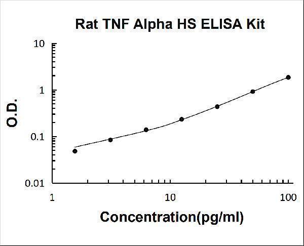
11 Citations 21 Q&As
1 Citations 18 Q&As
170 Citations 16 Q&As
1 Citations


4 Citations


Facts about Tumor necrosis factor.

Impairs regulatory T- cells (Treg) function in people with rheumatoid arthritis through FOXP3 dephosphorylation. Upregulates the expression of protein phosphatase 1 (PP1), which dephosphorylates the key'Ser-418' residue of FOXP3, thereby inactivating FOXP3 and producing Treg cells functionally defective (PubMed:23396208).
| Human | |
|---|---|
| Gene Name: | TNF |
| Uniprot: | P01375 |
| Entrez: | 7124 |

| Belongs to: |
|---|
| Tumor necrosis factor family |

APC1 protein; Cachectin; Cachetin; DIF; TNF; TNF, monocyte-derived; TNFA; TNF-A; TNFalpha; TNF-alpha; TNF-alphacachectin; TNFATNF, macrophage-derived; TNFG1F; TNFSF1A; TNFSF2; TNFSF2TNF superfamily, member 2; tumor necrosis factor (TNF superfamily, member 2); tumor necrosis factor alpha; Tumor necrosis factor ligand superfamily member 2; tumor necrosis factor; tumor necrosis factor-alpha
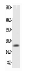
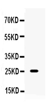
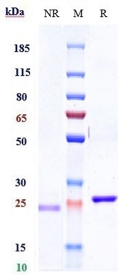
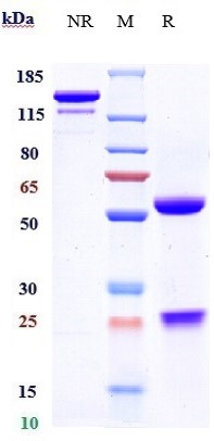
Mass (kDA):
25.644 kDA

| Human | |
|---|---|
| Location: | 6p21.33 |
| Sequence: | Chromosome 6; NC_000006.12 (31575565..31578336) |
Cell membrane; Single-pass type II membrane protein.; [Tumor necrosis factor, membrane form]: Membrane; Single-pass type II membrane protein.; [Tumor necrosis factor, soluble form]: Secreted.; [C-domain 1]: Secreted.; [C-domain 2]: Secreted.


Aggarwal, B.B. (2003). Signalling pathways of the TNF superfamily: a double-edged sword. Nature Reviews Immunology, 3, 745-756. doi: 10.1038/nri1184
Aggarwal, B.B., Moffat, B., & Harkins, R.N. (1984). Human lymphotoxin. Production by a lymphoblastoid cell line, purification, and initial characterization. Journal of Biological Chemistry, 259(1), 686-691.
Balkwill, F. (2006). TNF-alpha in promotion and progression of cancer. Cancer Metastasis Reviews, 25. doi: 10.1007/s10555-006-9005-3
Carswell, E.A., Old, L.J., Kassel, R.L., Green, S., Fiore, N., & Williamson, B. (1975). An endotoxin-induced serum factor that causes necrosis of tumors. Proceedings of the National Academy of Sciences of the United States of America, 72(9), 3666-3670. doi: 10.1073/pnas.72.9.3666
Diwan, A., Tran, T., Misra, A., & Mann, D.L. (2003). Inflammatory mediators and the failing heart: a translational approach. Current Molecular Medicine, 3(2), 161-182. doi: 10.2174/1566524033361537
Dowlati, Y., Herrmann, N., Swardfager, W., Liu, H., Sham, L., Reim, E.K., & Lanctot, K.L. (2010). A meta-analysis of cytokines in major depression. Biological Psychiatry, 67(5), 446-457. doi: 10.1016/j.biopsych.2009.09.033
Hehlgans, T., & Pfeffer, K. (2005). The intriguing biology of the tumour necrosis factor/tumour necrosis factor receptor superfamily: players, rules and the games. Immunology, 115(1), 1-20. doi: 10.1111/j.1365-2567.2005.02143.x
Horiuchi, T., Mitoma, H., Harashima, S., Tsukamoto, H., & Shimoda, T. (2010). Transmembrane TNF-alpha: structure, function and interaction with anti-TNF agents. Rheumatology (Oxford), 49(7), 1215-1228. doi: 10.1093/rheumatology/keq031
Lee, S.Y., Ju, M.K., Jeon, H.M., Jeong, E.K., Lee, Y.J., Kim, C.H..,?, Kang, H.S. (2018). Regulation of Tumor Progression by Programmed Necrosis. Oxidative Medicine and Cellular Longevity, 2018. doi: 10.1155/2018/3537471
Levine, B., Kalman, J., Mayer, L., Fillit, H.M., & Packer, M. (1990). Elevated circulating levels of tumor necrosis factor in severe chronic heart failure. New England Journal of Medicine, 323(4), 236-241. doi: 10.1056/NEJM199007263230405
Nagatsu, T., & Sawada, M. (2005). Inflammatory process in Parkinson's disease: role for cytokines. Current Pharmaceutical Design, 11(8), 999-1016. doi: 10.2174/1381612053381620
Parameswaran, N., & Patial, S. (2010). Tumor necrosis factor-alpha signaling in macrophages. Critical Reviews in Eukaryotic Gene Expression, 20(2), 87-103. doi: 10.1615/critreveukargeneexpr.v20.i2.10
Shrestha, B., Zhang, B., Purtha, W.E., Klein, R.S., & Diamond, M.S. (2008). Tumor necrosis factor alpha protects against lethal West Nile virus infection by promoting trafficking of mononuclear leukocytes into the central nervous system. Journal of Virology, 82(18), 8956-8964. doi: 10.1128/JVI.01118-08
Swardfager, W., Lanctot, K., Rothenburg, L., Wong, A., Cappell, J., & Herrmann, N. (2010). A meta-analysis of cytokines in Alzheimer's disease. Biology Psychiatry, 68(10), 930-941. doi: 10.1016/j.biopsych.2010.06.012
Swaroop, J.J., Rajarajeswari, D., & Naidu, J.N. (2012). Association of TNF-alpha with insulin resistance in type 2 diabetes mellitus. Indian Journal of Medical Research, 135(1), 127-130. doi: 10.4103/0971-5916.93435
Tang, P., Hung, M.C., & Klostergaard, J. (1996). Human pro-tumor necrosis factor is a homotrimer. Biochemistry, 35, 8216-8225. doi: 10.1021/bi952182t
Wajant, H., Pfizenmaier, K., & Scheurich, P. (2003). Tumor necrosis factor signaling. Cell Death & Differentiation, 10, 45-65. doi: 10.1038/sj.cdd.4401189
*Showing only the more recent 20. More publications can be found for each product on its corresponding product page