This website uses cookies to ensure you get the best experience on our website.


- Table of Contents
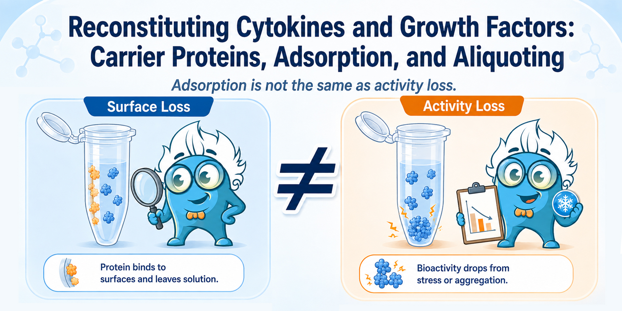
A recombinant cytokine can be prepared at the correct calculated concentration and still produce a weaker-than-expected response after dilution or frozen storage. The protein may have lost biological integrity, but another explanation is often overlooked: part of the cytokine may no longer be in the liquid phase because it has adsorbed to tubes, pipette tips, or assay plates. This handling issue matters across many recombinant proteins, particularly when they are used at low concentrations in cell culture or functional assays.
Expression system, purification, and final formulation can influence how a recombinant protein behaves after reconstitution. Boster’s guide to recombinant protein production provides background on these upstream variables without replacing the handling instructions for the finished product.
In practice, a reduced experimental response after reconstitution can reflect two different problems:
A carrier protein can help with the first problem when it is compatible with the assay.
It cannot reliably repair a cytokine that has already aggregated or denatured.
Proteins can adsorb non-specifically to both glass and plastic. This becomes especially important at low concentration: a small absolute loss to the container may represent a substantial fraction of the total protein in the sample. [1]
The type of plastic also matters. Studies have found differences in protein or peptide recovery among polypropylene materials and among products marketed as low binding. [2–5] The effect is protein-specific, so a tube that performs well for one analyte may not be the best choice for another.
Surface loss is more likely to affect the experiment when the cytokine is:
The calculated concentration can therefore be correct even when the amount that remains in solution and reaches the cells is lower.
Adsorption removes protein from the liquid phase. The molecules still in solution may retain their normal specific activity, but fewer molecules are delivered to the assay. A true loss of activity involves a change in the protein itself, such as aggregation, precipitation, partial unfolding, chemical modification, or proteolytic degradation.
Both mechanisms can occur in one workflow. A dilute cytokine may be lost to tube surfaces during preparation and then experience additional stress during storage. Distinguishing reduced recovery from true inactivation is useful because the corrective steps are different.
Albumin-based carrier proteins, including BSA and HSA, are commonly used to reduce the handling loss of dilute proteins. They can occupy potential binding sites on a vessel surface, leaving fewer sites available for the target cytokine. BSA-treated surfaces have improved recovery in several experimental protein systems. [1,4,5] For a broader overview of BSA’s laboratory roles and possible sources of interference, see Boster’s guide to BSA in laboratory buffers.
A carrier protein does not increase the intrinsic potency of a cytokine. Its role is to improve the amount of target protein that remains available during dilution, transfer, incubation, and storage. Once a cytokine has aggregated or denatured, adding albumin will not restore the original preparation. When BSA is compatible with the assay, Boster’s BSA Bovine Serum Albumin protein is one possible reagent source; the required grade and concentration should still be selected for the downstream application.
Use the solvent and minimum stock concentration specified for the exact product. Depending on the cytokine or growth factor, the recommended solution may be water, a buffered solution, dilute acid, or a formulation that already contains a stabilizer. Do not copy the reconstitution method from another protein simply because both are described as cytokines.
Product pages make this product-specific approach concrete. The following examples show why one recombinant-protein protocol should not be copied to another:
Product instructions are not interchangeable
These examples are not a shared protocol. They demonstrate why the exact product page and datasheet should be reviewed before reconstitution.
A concentrated stock contains more target protein relative to the available vessel surface than a highly dilute working solution. Storing the protein as a concentrated stock can therefore reduce the proportion lost to surfaces, although it does not eliminate adsorption or other stability risks. The stock concentration should still follow the product instructions.
Carrier protein is often most useful when the concentrated stock is diluted to the low concentration required by the experiment. This stage can introduce several new contact surfaces through serial dilution, transfer, and dispensing. When the downstream assay permits it, a carrier-containing dilution buffer can reduce surface-related loss. [1,3–5]
This is why the reconstitution buffer and the final dilution buffer do not always have the same composition.
Carrier proteins should not be treated as invisible ingredients. BSA, HSA, serum, and other stabilizers become part of the experimental system and may be unsuitable when:
A study of T-helper-cell polarization found that surface-bound albumin from recombinant cytokine preparations amplified the measured response under the tested culture conditions. [8] The result is not a general reason to avoid albumin, but it shows why a carrier-matched control may be necessary.
Control design
When a cytokine preparation adds BSA or another carrier to the culture, include the same carrier type and final concentration in the corresponding vehicle control whenever the experimental design permits.
Before opening the vial, confirm the recommended solvent, stock concentration, dissolution procedure, carrier or stabilizer requirements, storage temperature, and restrictions on freeze–thaw cycles. Product-specific instructions take priority over general laboratory habits.
If the datasheet instructs you to do so, briefly centrifuge the vial before opening so that displaced lyophilized material is collected at the bottom.
Reconstitution volume = protein amount ÷ desired stock concentration
Example: 10 µg ÷ 100 µg/mL = 0.1 mL = 100 µL. Check that the units are compatible before calculating the volume.
Add the recommended solvent using the handling method described in the datasheet. Avoid vigorous agitation or excessive foaming. Aggressive mixing increases exposure to air–liquid and solid–liquid interfaces, which may destabilize susceptible formulations.
Give the preparation the recommended time to hydrate and dissolve. Use gentle mixing when required, and avoid vortexing when the product instructions advise against it.
Before diluting the stock, decide whether the assay permits a carrier protein. For a low-concentration working solution, use the specified dilution buffer, limit transfer steps, choose suitable labware, and prepare the solution close to the time of use. Accurate dilution becomes more difficult when several serial steps or very small pipetting volumes are required. The practical principles in Boster’s ELISA standard preparation guide—including avoiding extremely small transfers and preparing standards close to use—can also reduce handling variation here. This is a dilution-technique reference, not a substitute for protein-specific stability data.
Once the protein is fully dissolved and gently mixed, divide it into practical portions. The aliquot should be large enough for accurate pipetting but small enough to avoid repeatedly thawing the same stock vial.
Low-binding tubes, tips, and plates can improve the recovery of some dilute proteins. However, “low binding” is not a guarantee of equal performance for every target. In published comparisons, recovery depended on the protein and the specific material or treatment used. [3–5]
When recovery is critical, compare candidate labware using the actual cytokine and buffer rather than relying only on the product label.
Each transfer creates another surface on which a small amount of cytokine may remain. Use only the required dilution steps, select a vessel that matches the solution volume, and avoid prolonged intermediate storage.
A dilute working solution usually presents a less favorable surface-to-protein ratio than a concentrated stock. Unless product-specific stability data support storage at the final assay concentration, prepare the working dilution shortly before use.
An aliquot should contain approximately the amount required for one experiment or a short, defined series of experiments. Oversized aliquots encourage repeated thawing, while extremely small aliquots increase pipetting error, dead-volume loss, evaporation risk, and relative surface exposure.
After the stock has dissolved completely, use the approved gentle mixing method before dispensing the aliquots. Uneven mixing can produce concentration differences among tubes.
Freezing changes the local environment around a protein. Ice formation can concentrate salts, excipients, and protein in the remaining liquid phase, while ice–liquid interfaces and thawing conditions may contribute to aggregation in susceptible formulations. [7]
The effect is protein- and formulation-dependent. Recombinant MCP-1/CCL2, for example, showed concentration- and storage-dependent losses after frozen storage and repeated freeze–thaw exposure. [6] That result should not be converted into a universal number of “safe” cycles for other cytokines. Single-use aliquots are the more conservative option when protein-specific stability data are unavailable.
Use the thawing procedure specified for the product, avoid partial thawing followed by refreezing, mix gently when required, and keep the interval between thawing and use consistent across experiments.
| Observation | Possible explanation | What to check |
|---|---|---|
| Weak response immediately after reconstitution | Incorrect solvent, incomplete dissolution, concentration error, adsorption, or unsuitable assay conditions | Verify the datasheet, calculation, dissolution method, labware, and biological controls. |
| Response falls after preparing the working dilution | Reduced recovery at low concentration | Review carrier compatibility, dilution buffer, tube type, transfer count, and delay before use. |
| Response falls after frozen storage | Aggregation, precipitation, repeated thawing, or unsuitable formulation | Review storage conditions, freeze–thaw history, aliquot size, and visible changes in the solution. |
| Different aliquots give inconsistent results | Uneven mixing, pipetting error, very small aliquots, or inconsistent thawing | Standardize stock mixing, dispensing volume, thawing, and time to use. |
| Cytokine works in serum-containing medium but not protein-free buffer | Serum proteins may reduce surface loss, while the two media may also alter the biological assay | Compare matched buffer conditions and use appropriate vehicle controls. |
| Adding BSA changes the cellular response | The carrier is affecting the assay rather than acting only as a stabilizer | Include a carrier-matched control and reassess the carrier type and concentration. |
| Recovery varies among tubes or plates | Protein-specific adsorption to the selected material | Compare candidate labware with the actual cytokine and buffer system. |
A weaker response may reflect reduced recovery from adsorption, a true loss of biological activity, or both. Check the solvent, stock concentration, labware, dilution process, storage history, and assay controls before attributing the result to protein degradation.
No. Some product instructions specify BSA or another stabilizer, while others specify water, buffer, or dilute acid without a carrier. Use the instructions for the exact product and confirm that any carrier is compatible with the downstream experiment.
The answer is product-specific. Carrier protein is often most useful when a concentrated stock is diluted to a low working concentration, but some proteins are formulated or reconstituted with a carrier from the start. Follow the datasheet rather than applying one formulation to every cytokine.
Only when the product instructions and experimental design support it. Medium composition can affect solubility, stability, and the biological response. A concentrated stock prepared in the specified solvent is often easier to control than direct reconstitution at the final working concentration.
Use portions that match one experiment or a short series of experiments. Avoid aliquots so large that they require repeated thawing and so small that pipetting error, dead volume, or surface exposure becomes excessive.
Some proteins may tolerate refreezing better than others, but there is no universal safe number of cycles. When protein-specific stability data are unavailable, designate aliquots for single use and avoid partial thawing and refreezing.
They may still be useful. Carrier proteins and low-binding labware address the same general risk through different means, and neither guarantees complete recovery for every cytokine. The combination should be selected according to the protein, buffer, and assay.
Looking for cytokines, chemokines, or growth factors for cell-based research?
Browse Boster recombinant proteins and review the formulation, reconstitution, and storage instructions provided for the exact product before use.
For projects that require a specific expression system, purification strategy, buffer exchange, lyophilization, or packaging format, explore Boster’s custom recombinant protein expression service.







