This website uses cookies to ensure you get the best experience on our website.
- Table of Contents
Key Considerations When Collecting Samples
Serum:
Considering the anticoagulation mechanisms and downstream applications, choose the necessary anticoagulant. For example:
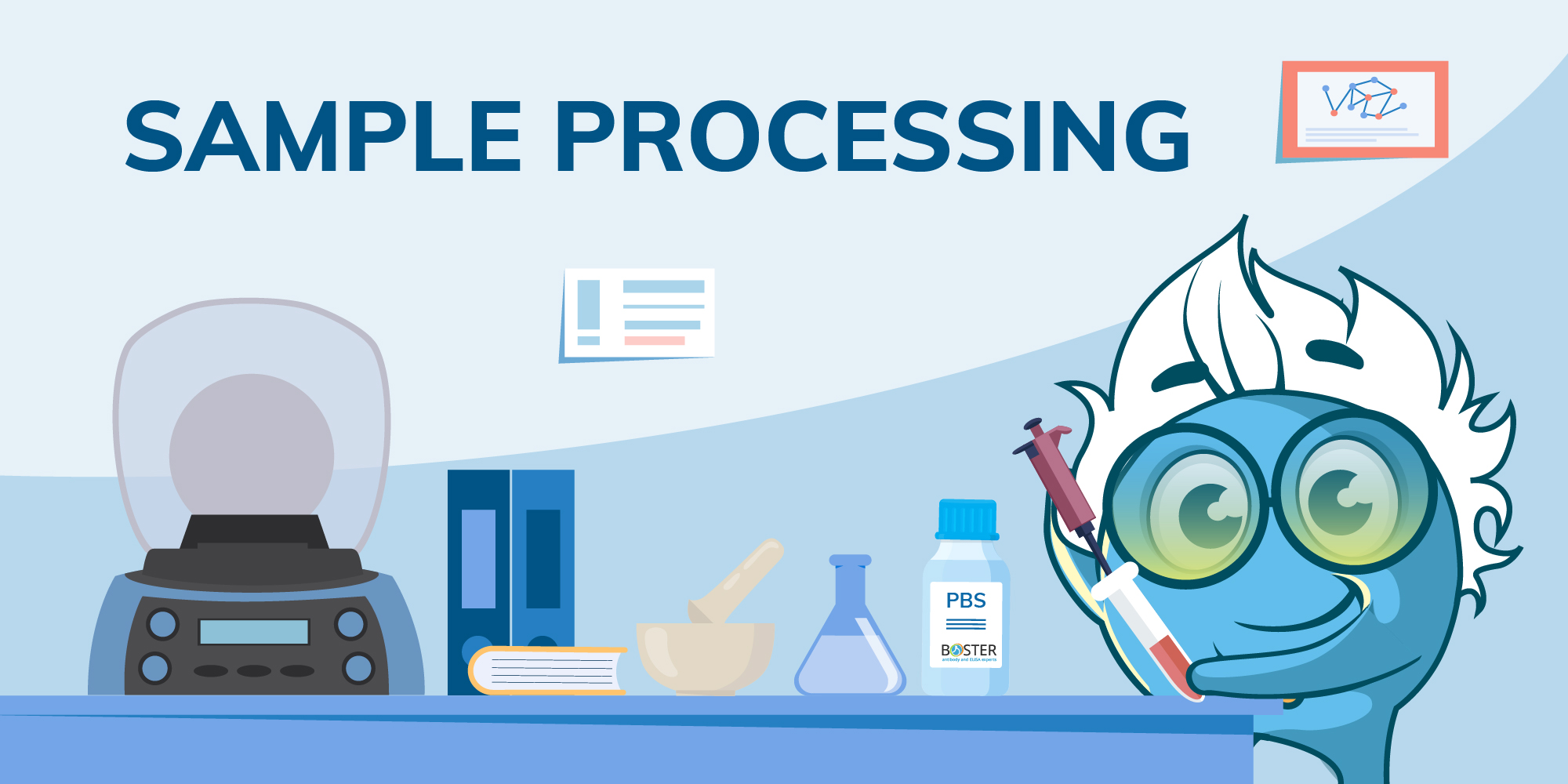
Rinse the tissue with pre-cooled PBS (0.01M, pH=7.4) to remove residual blood, weigh the tissue, and then mince it.
Add the minced tissue to a glass homogenizer with the appropriate volume of PBS (a 1:9 w/v ratio is typically used).
Homogenize the tissue on ice and further disrupt cells by freeze-thawing or ultrasonic processing.
Centrifuge the homogenate at 5000×g for 5-10 minutes at 2-8°C and collect the supernatant for testing.
Wash adherent cells with cold PBS, then digest them with trypsin. For downstream applications like western blotting, following a proper protein extraction protocol for western blot ensures that lysates are prepared correctly for reliable protein detection.
Collect the cells by centrifugation at 1000×g for 5 minutes and wash them 3 times with cold PBS.
For Every 10^6 cells, add 150-200 μL PBS (with protease inhibitors if necessary), lyse by freeze-thawing or sonication.
Centrifuge the lysate at 1500×g for 10 minutes at 2-8°C.
Collect the supernatant for testing.
Conditioned Media:
Growing cells to the desired confluence in a complete growth medium (with serum).
Remove the growth medium and wash the cells with warm PBS several times.
Replace with serum-free medium and incubate the cells for 1-2 days.
Collect the medium and centrifuge at 1500 rpm at 4°C for 10 minutes. The supernatant can be used for detection.
Collect plasma using EDTA, heparin, or citrate as an anticoagulant. Centrifuge for 15 minutes at 1000×g within 30 minutes of collection. Perform an additional centrifugation step of the plasma at 10,000×g for 10 minutes at 2-8°C to remove platelets. The supernatant can be used for detection.
Human Milk:
Centrifuge the sample at 1000×g for 15 minutes at 2-8°C. Collect the aqueous fraction and repeat this centrifugation step a total of three times. The supernatant can then be used for detection.
Following these guidelines can preserve sample integrity, ensuring accurate ELISA testing results. Always store samples at -20°C or lower to avoid degradation and minimize freeze-thaw cycles. For samples of human metabolites, special attention should be paid to their freshness.
1. Hemolysis should be avoided as much as possible when collecting blood samples.
2. After collecting anticoagulated whole blood, gently invert the tube to ensure full anticoagulation, preventing clotting caused by partial blood not coming into contact with the anticoagulant.
3. It is not recommended to use commercial lysis buffers when collecting tissue and cell extract samples. Surfactants in the lysis buffer may affect the native conformation of the analyte, potentially leading to significantly reduced measurements or false negatives. Additionally, since a new solvent is introduced, potential matrix interference is uncertain, which could increase background levels and affect measurement accuracy.
4. Avoid using hyperlipidemic samples. High-fat samples contain lipids and are not homogeneous solutions. If applied directly, they may interfere with antigen-antibody binding, thus affecting measurement accuracy.
5. If the samples are to be tested within a week, store them at 2-8°C. If testing cannot be performed promptly, aliquot the samples according to the usage amount, and store them at -20°C or -80°C to avoid repeated freeze-thaw cycles.
In sample processing, hemolysis refers to the rupture of red blood cells, releasing hemoglobin into the serum or plasma. Hemolysis can be caused by various physical, chemical, and toxic factors. In vitro, mechanical agitation or freezing at low temperatures can result in hemolysis.
After hemolysis occurs, endogenous HRP enzymes and hemoglobin's peroxidase-like activity can lead to uncontrolled nonspecific staining in ELISA, affecting the accuracy of the results. If the sample is slightly hemolyzed, it is recommended to wash the plate 1-3 times after the incubation with the standards to minimize interference. If severe hemolysis occurs, it is advisable to collect new samples.
To avoid hemolysis affecting test results, ensure that the venipuncture pressure is not too high, avoid collecting too small a volume of blood, handle collected blood samples gently to avoid vigorous shaking, do not centrifuge at excessively high speeds, process the collected whole blood into serum/plasma promptly, and avoid freeze-thawing whole blood. If anesthesia is required for blood collection, use an anesthetic that does not cause hemolysis.


