This website uses cookies to ensure you get the best experience on our website.
- Table of Contents
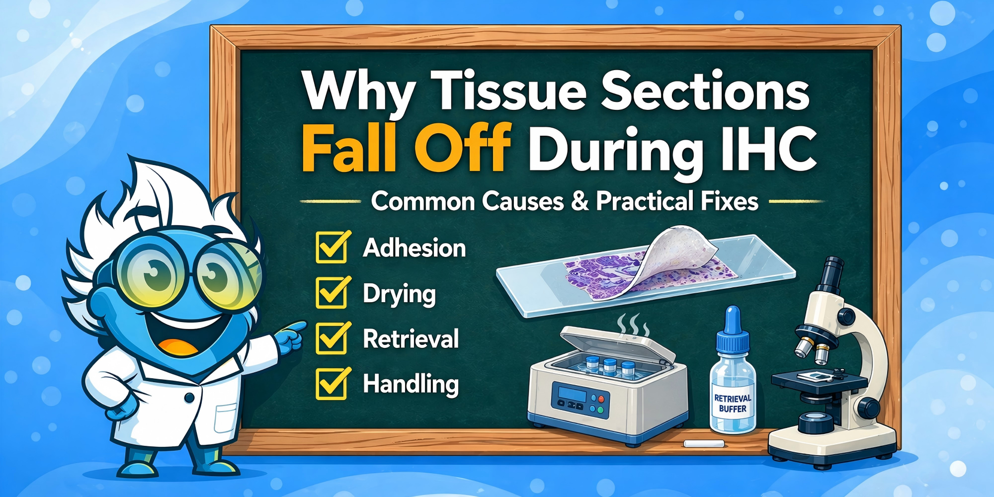
When tissue sections fall off during IHC staining, the fastest way to find the cause is to identify the step where detachment begins.
In most cases, tissue loss has more to do with slide adhesion, drying, antigen retrieval, sample preparation, or handling than with the antibody itself. A section that comes off early in the workflow, during retrieval, or late in the run is usually pointing to a different kind of problem.
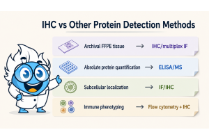
If you want to place this issue in the context of the broader workflow, it helps to start with the basics of IHC staining and the overall IHC protocol before narrowing in on section loss in a typical histology lab environment.
If detachment happens early in the workflow, the section may not have been fully secured to the slide before staining began. In addition to deparaffinization or rehydration, early section loss can also be related to poor section quality, folds, uneven section thickness, or incomplete drying after mounting.
In general, frozen sections prepared using OCT compound and liquid nitrogen are more likely to show detachment at these earlier stages, while formalin fixed paraffin embedded tissue sections and workflows more often remain intact until antigen retrieval.
This is one of the most common failure points, especially in FFPE IHC. Heat-induced epitope retrieval, also known as heat induced antigen retrieval or antigen unmasking, is essential in many workflows, but it also puts substantial physical stress on the section. If tissue loss starts here, the retrieval conditions may be too aggressive for that sample, or the tissue may already have been weakened earlier in processing, mounting, or drying. This can include use of a pressure cooker, water bath, or other heating systems, where thermal shock and rapid temperature shifts may weaken adhesion. Buffer choice also matters, including citrate buffer and high pH retrieval solutions, since pH matters in maintaining tissue integrity.
In some cases, switching to enzyme digestion or protease digestion may provide gentler alternatives to harsh heat-based retrieval.
When retrieval seems to be the breaking point, it is often worth reviewing antigen retrieval, the tradeoffs between HIER and PIER, and, where relevant, how sodium citrate buffer is being used.
If sections begin to come off during washes or incubation steps, the tissue may already have been weakened earlier in the run. At this stage, strong buffer flow, repeated agitation, or direct dispensing onto the tissue can make section loss more visible, but these are often not the only cause. Sometimes, improper handling using slide racks can contribute.
Using appropriate wash buffers such as PBS buffers prepared with deionized water helps maintain consistency, while avoiding waterbath contamination is critical when sections are floated before mounting.
If sections remain attached until counterstaining, dehydration, or mounting, the damage may have built up gradually through the run. Steps like immunoperoxidase staining, DAB reaction, and use of various detection reagents or signal amplification systems can stress already fragile tissue. In those cases, it can help to revisit how counterstains and later processing steps affect already stressed tissue.
If the section is not firmly attached to the slide, every later step becomes less forgiving. Detachment that starts at the edges or in thinner areas often points to weak initial adhesion.
For more demanding samples, positively charged or coated slides, a hydrophobic barrier or proper mounting techniques can help improve retention. This is especially useful for fragile paraffin sections or tissues that will go through more rigorous retrieval conditions.
A section can look attached and still not be fully set. Using a slide warmer ensures proper drying before staining begins and reduces the risk of tissue detachment during downstream steps. If drying is insufficient, residual moisture can weaken attachment once the tissue is exposed to solvents, heat, or retrieval buffer.
Common starting points include 60°C for about 1 hour or 56°C overnight, though exact conditions vary by tissue and lab workflow. The goal is not simply to warm the slide, but to make sure the section is genuinely secured before staining begins.
Antigen retrieval is necessary for many targets, but stronger is not always better. Excessive heat, extended retrieval time, or unsuitable buffer high-pH conditions can all increase the risk of section loss, especially in delicate samples. Fragile tissue samples may require milder retrieval approaches rather than standard protocols.
This pattern is especially common in FFPE workflows, where sections may remain intact through earlier steps and then fail under retrieval stress. It is also where upstream choices begin to matter. If tissue integrity was already weakened during fixation or processing, retrieval often becomes the step where that weakness shows up.
Fragile, fatty, decalcified, or otherwise compromised tissues are especially likely to detach under harsh retrieval conditions. In those cases, gentler retrieval conditions are often more useful than simply repeating the same standard protocol.
The problem does not always start on the staining bench. Weak fixation, over-fixation, harsh decalcification, or rough sectioning can all reduce tissue morphology integrity before IHC even begins.
Under-fixation can leave tissue cohesion too weak to withstand downstream handling, while over-fixation can make the section more brittle. If the issue seems to begin upstream of staining, it usually makes more sense to review sample preparation and the main fixative types used in IHC and ICC than to keep changing staining conditions alone.
Directly dispensing of buffers, onto the section, washing too forcefully, or handling slides too roughly can all increase tissue loss, especially after retrieval. These issues may also contribute to background staining or increased non-specific binding, especially if blocking and washing steps are not optimized.
At the same time, these steps often act on tissue that is already unstable. In practice, washing may be the point where section loss becomes obvious rather than the only reason it happens.
Some tissues are simply less tolerant of heat, agitation, and repeated solution changes. If the problem appears only in certain sample types while the rest of the workflow performs normally, tissue-specific fragility should be part of the troubleshooting logic.
For example, fatty tissues tend to have looser structural support, while decalcified tissues may lose some of the integrity they had before processing. In both cases, gentler handling and milder retrieval conditions may be necessary from the start.
When sections fall off during IHC, do not change everything at once. Start with the factor that best matches the step where failure occurs.
| If your result looks like this | Better next move |
|---|---|
| Routine FFPE tissue, first-round setup | Start with HIER |
| Weak signal, but tissue remains intact | Optimize HIER first |
| Heat damages morphology or section stability | Test enzymatic retrieval |
| Literature or antibody guidance supports protease digestion | Consider enzymatic retrieval earlier |
| Enzyme treatment over-digests tissue | Reduce digestion strength or reassess method fit |
A sensible troubleshooting order is:
A useful practical distinction is this: if sections begin to lift early in the workflow, adhesion, section quality, or drying are usually the first place to look. If they remain stable until retrieval and then start to fail, retrieval intensity or tissue fragility becomes a more likely explanation.
Once section retention is under control, broader assay quality questions are usually better addressed through IHC optimization, IHC troubleshooting, and well-designed IHC controls.
Many detachment problems begin long before antibody incubation starts. A few preventive checks can reduce failure later in the run:
Proper antibody application with the right antibody concentration, along with validated primary antibody, secondary antibody, or monoclonal antibody systems, also helps maintain consistency. Blocking steps targeting endogenous peroxidase and endogenous biotin can further improve staining quality.
These steps are simple, but they often make the difference between a stable section and one that begins to lift halfway through the protocol.
If the section is physically coming off the slide, start with section stability. Markers such as proliferating cell nuclear antigen are commonly used, but antibody performance may matter later, but it is usually not the first issue here.
Washing may be where the section comes off, but the underlying cause often started earlier.
If you change slide type, drying conditions, retrieval settings, and wash method all in one run, it becomes much harder to see what actually solved the problem.
Why do tissue sections often fall off during antigen retrieval?
Because retrieval combines heat and chemical stress. If adhesion or tissue integrity is already marginal, retrieval is often the step that exposes it. This is especially common in FFPE workflows.
When should I consider using coated or charged slides?
They are often worth considering for fragile tissues, difficult paraffin sections, or workflows that require more demanding retrieval conditions.
Can fixation affect whether sections stay on the slide?
Yes. Fixation and tissue processing both affect tissue integrity, which in turn affects how well the section holds up during staining.
Do all tissues need charged slides for IHC?
Not necessarily. Many tissues stain well without them, but charged or coated slides can be especially helpful when you are working with fragile samples or retrieval-heavy workflows.
How can I tell whether the problem is poor drying or harsh retrieval?
The timing often helps. If the section begins to lift early in the workflow, drying, slide adhesion, or section quality are more likely. If it stays stable until retrieval and then detaches, retrieval conditions or tissue fragility are usually better places to start.
When tissue sections fall off during IHC, the most useful question is simple: At what step did the section start to detach? In most cases, the answer points back to slide adhesion, section drying, retrieval intensity, tissue integrity, or physical handling.
Once section retention is under control, the rest of IHC optimization becomes much more straightforward. And in many cases, the most effective fix is not a dramatic protocol change, but a better match between the tissue, the slide, the retrieval conditions, and the way the section is handled from the start.