This website uses cookies to ensure you get the best experience on our website.
- Table of Contents
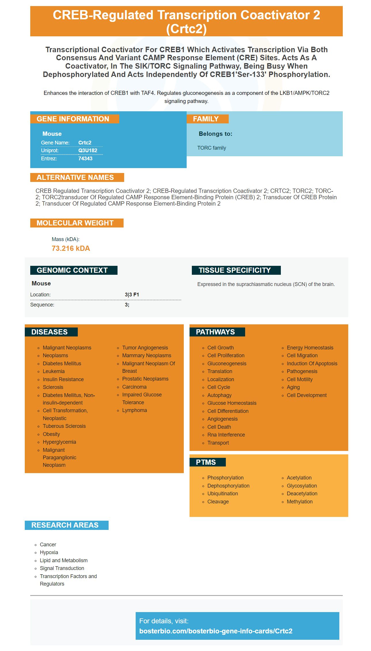
Facts about CREB-regulated transcription coactivator 2.
Enhances the interaction of CREB1 with TAF4. Regulates gluconeogenesis as a component of the LKB1/AMPK/TORC2 signaling pathway.
Mouse | |
---|---|
Gene Name: | Crtc2 |
Uniprot: | Q3U182 |
Entrez: | 74343 |
Belongs to: |
---|
TORC family |
CREB regulated transcription coactivator 2; CREB-regulated transcription coactivator 2; CRTC2; TORC2; TORC-2; TORC2transducer of regulated cAMP response element-binding protein (CREB) 2; Transducer of CREB protein 2; Transducer of regulated cAMP response element-binding protein 2
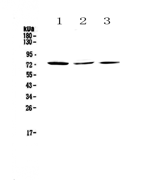
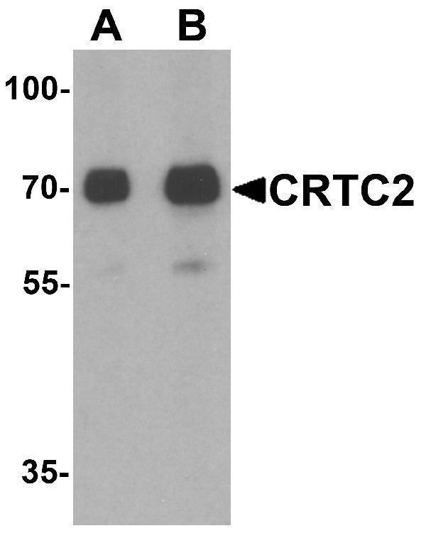
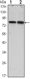
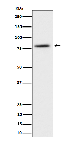
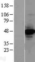
Mass (kDA):
73.216 kDA
Mouse | |
---|---|
Location: | 3|3 F1 |
Sequence: | 3; |
Expressed in the suprachiasmatic nucleus (SCN) of the brain.
Scientists are able to submit their results using the CRTC2Marker for species, applications, or special samples. Based on their results, scientists can also be eligible for product credits. This procedure is open to all scientists across the globe. The CRTC2 marker is shared by CRTC2 knockout mouse strains. This marker permits scientists to examine the expression of the CRTC2 gene in two different mouse strains and find the most effective combination for their experiments.
The CRTC2 gene encodes the fluorescent protein mTurquoise2. The gene is expressed in the mouse embryo, allowing scientists to track embryogenesis. This fluorescent protein is non-toxic, and has a high QY for monomeric fluorescent proteins. This feature allows researchers to use it to observe embryogenesis as well as for imaging in long-term studies. This fluorescent protein is also free of toxicities and photobleaching.
The CRTC2 marker is a fundamental fluorescent cyan fluorescent protein, derived from the blue algae Aequorea victoria. It has low acid sensitivity and is quickly produced as monomers. A structural analysis revealed that Ile146 was a poor residue. It was therefore targeted to further improve. This mutation increased QY to 0.93 and improved the packing of the chromophore.
It has been proven that CRTC2 can play a key role in the gluconeogenesis process of Hepatocytes. It was found to be abundant in the nuclei of hepatocytes after fasting as well as exposure to agents that increase cAMP. The overexpression of the CRTC2 gene led to an increase in glucoseconeogenesis while silencing it prevented the same phenomenon. The subcellular localization of CRTC2 was comparable to the B-cell receptor. A modification in the expression of SIKs was also found to affect the activity of CRTC2.
CRTC2 stimulates transcription of G6Pase, PEPCK. These transcription factors boost the rate of gluconeogenesis in the liver. The CRTC2 activity is inhibited by the Pgc1a transcriptional activator. Inhibition of CRTC2 by PKI partially blocked the effects of FSH on PEPCK and G6Pase.
Similarly, mTOR-CRTC2 signaling pathway contributes to insulin resistance development. Insulin resistance is a characteristic of CRTC2-/mice. Activated mTOR stimulates lipogenesis by blocking the browning of white adipocytes. The COX-2 inhibitor CRTC2 blocks prostaglandin synthesis through COX. It regulates expression of PGC-1a.
Expression by cloning cDNA is a very effective method to create new fluorescent proteins. The probability of cDNA insertion after the first start codon in pRSET is about 1/3, and transcriptional slippage is induced by the DNA sequence (dT) 14 that is located close to the first start codon. To confirm the frame-insensitive cloning process, eGFP was inserted in the various frames of the pRSETA. No significant differences were found in the expression levels of E.coli cells.
Recent research has shown improved CFP variants in mTurquoise. The mTurquoise2.1 variant contained the critical h248G mutation that was previously identified in mTurquoise-GL. Similar to mCerulean3, mCerulean3 also single mutation, called T65S, which is responsible for the high QY of mTurquoise. Both mCerulean2 as well as mCerulean3 had QYs significantly higher at 0.65 and 0.87 respectively.
The mTurquoise2.1 molecule has the same structure as of the original. The seventh strand is stabilized and pushed towards the bulk solvent. Tyr145 has a single conformation. It also shows an increase of interactions between the Tyr65 side chains and the Glu222 side chains that together make up half of the overall structure.
Recent studies have demonstrated that an mTurquoise2.1 mutation was created through site-directed mutation (SDM) using Cre-loxP vectors that contain the critical mutation h248G. The mutation was previously recognized in the mTurquoise-GL. Moreover, the improved mTurquoise2.1 contained only one mutation, T65S, which is the reason for its high QY. These mutants had QYs of 0.60 and 0.87 respectively.
mTurquoise2.1 does not show any significant increase in fluorescence if co-expressed with crTC2.1 and mCitrine shows the same increase in fluorescence. This suggests that mTurquoise2.1 has reached its highest levels of expression under constant conditions and is maturing rapidly. These results suggest that mTurquoise2.1 is a protein with a short maturation period and is a high-quality protein.
The expression of H2B is a crucial instrument in the field of cell biology. It is useful for studying cell behavior. pUCM-AAVS1-TO-H2B/mTurquoise2 vector was introduced into the PAX3-Venus iPSCs using CRISPR/Cas9 technology. Although the reporter gene isn't expressed in iPSCs but it is induced through doxycycline treatment is still expressed in iPSCs. It is possible to measure H2B-mTurquoise2 expression, which allows cell divisions and imaging of living cells.
We have looked at the structure of mTurquoise2.1 (purified version) and its spectral properties. The two spectral characteristics are nearly identical except for one residue, which is hydrophobic. However, the two structures were found to be indistinguishable. The simulations of molecular dynamics demonstrated that the two proteins interact through interactions with hydrophobic residues.
In mammalian cells, the mTurquoise2 gene emits over 1,600,000 photons before photobleaching and is superior to GFP17's estimated 400,000 photons. Additionally, mTurquoise2 exhibits higher photostability and performs better as an FRET donor. Therefore, mTurquoise2 is an attractive fluorescent protein.
We have measured the extinction coefficient of mTurquoise2.1 and other CFP variants. As we have previously reported, the Cerulean extinction factor is 43,000 M-1 cm-1. The mTurquoise's extinction coefficient is 40,000 M-1 cm-1. In addition the mTurquoise2 protein's extinction coefficient is similar to that of Cerulean.
PMID: 16148943 by Koo S.-H., et al. The CREB coactivator TORC2 is a key regulator of fasting glucose metabolism.
PMID: 16308421 by Shaw R.J., et al. The kinase LKB1 mediates glucose homeostasis in liver and therapeutic effects of metformin.