This website uses cookies to ensure you get the best experience on our website.
- Table of Contents
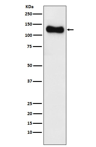
Facts about Disks large homolog 1.
May play a role in adherens junction assembly, signal transduction, cell proliferation, synaptogenesis and lymphocyte activation. Regulates the excitability of cardiac myocytes by regulating the functional expression of Kv4 channels.
Human | |
---|---|
Gene Name: | DLG1 |
Uniprot: | Q12959 |
Entrez: | 1739 |
Belongs to: |
---|
MAGUK family |
discs, large homolog 1 (Drosophila); disks large homolog 1; DKFZp761P0818; DKFZp781B0426; hDlg; large (Drosophila) homolog 1; presynaptic protein SAP97
Mass (kDA):
100.455 kDA
Human | |
---|---|
Location: | 3q29 |
Sequence: | 3; NC_000003.12 (197042560..197299321, complement) |
Abundantly expressed in atrial myocardium (at protein level). Expressed in lung fibroblasts, cervical epithelial and B-cells (at protein level). Widely expressed, with isoforms displaying different expression profiles.
Membrane; Peripheral membrane protein. Basolateral cell membrane. Endoplasmic reticulum membrane. Cell junction, synapse, postsynaptic density. Cell junction, synapse. Cell membrane, sarcolemma. Apical cell membrane. Cell junction. Cytoplasm. Colocalizes with EPB41 at regions of intercellular contacts. Basolateral in epithelial cells (PubMed:12807908). May also associate with endoplasmic reticulum membranes. Mainly found in neurons soma, moderately found at postsynaptic densities (By similarity).
In this article, we will review Steven Boster's bio as well as the clinical significance of DLG1 as a diagnostic and prognostic marker for colorectal cancer. We will also look at the interaction of DLG1 with other proteins, as well as the possibility of developing new therapies. We will end by discussing the most effective uses for the DLG1 marker in colorectal cancer.
CRC showed high levels of DLG1 mRNA expression. This was linked to several clinicopathologic characteristics that included an increase in tumor invasion or distant metastasis. However, high DLG1 mRNA levels were not related to age, gender, or lymph node metastasis. Therefore, DLG1 may serve as an effective biomarker to aid in the diagnosis and prognosis for CRC.
Prior research looked into the known causes of colorectal cancer in China in 2005. Drinking alcohol and a lack of physical activity were the main causes of 14.6% of deaths from colon cancer. Smoking and obesity are also responsible for a significant proportion of deaths, as was inactivity. However, the significance of DLG1 in colorectal cancer remains unclear. Further research is required to determine the molecular mechanisms through which DLG1 plays a role in colorectal tumor progression.
To evaluate the clinical relevance of DLG1 as a predictive and diagnostic marker in the field of colorectal cancer The first step was to examine the expression of tumor-derived genes in a group of 519 patients with cancer. DLG1 is a member of the MAGUK family of proteins, including Src homology 3 and PDZ. These proteins act as scaffolds for proteins, and are important in adhesion between cells. They also play a part in maintaining polarity and apical-basal.
The expression of miR-21 in colorectal cancer is negatively regulated by miR-124. The absence of miR-144 is associated with an increased liver metastasis and malignant capacity. MiR-155 negatively correlates with lymph node metastasis. However miR124 expression isn't directly linked to clinical pathological parameters.
The DLG1 gene is a mutable protein that has many functions in cellular processes. It has been found to regulate cell-cell junctions, and promote cell movement. Although it has been linked with cancer, its clinical significance isn't yet fully understood. Recent studies have suggested it might act as a tumor suppressor. Additionally, the knockdown of DLG1 in endometrial cancer cells resulted in an increase in cancer growth and invasion.
Recently, researchers have discovered that the DLG1 gene is an important marker for gastric cancer cells. The expression of this gene correlates with the expression of the marker CD33. Inhibiting the expression of the circDLG1 gene may prevent the cancer from growing. A higher number of MDSCs are also associated with the DLG1 gene. These cells are thought to contribute to gastric cancer progression. Therefore, targeting circDLG1 could be an appealing therapeutic option for this type of cancer.
In PTC, increased DLG1-AS1 expression is associated with a lower prognosis. In vitro, knockdown of DLG1-AS1 reduces tumor cell proliferation and invasion. This gene regulates the miR-497/YAP1 axis , and could be a therapeutic target. Although research is ongoing in PTC, the target of DLG1–AS1 inhibitor remains elusive.
DLG1 overexpression is associated with numerous pathological and clinical indicators in colorectal cancer. It can also be used as a diagnostic or prognostic marker. It could also be a potential therapeutic target for CRC. This study was funded by the Guangdong Planned Project for Science and Technology. In the near future the discovery of new therapeutic targets will allow patients to find the most effective treatment for cancer.
Disks large homolog 1, (DLG1), is a modular protein that plays a crucial role in cell-cell junctions. The clinical relevance of DLG1 expression in colorectal cancer remains largely unclear. Boster Bio has developed antibodies that can identify DLG1 and also its interactions with other proteins. To determine the clinical utility of these antibodies, the following procedures were employed.
Colorectal cancer is one of the most prevalent causes of death around the world. To prevent death it is essential to catch the disease early. Unfortunately, most patients are not given an accurate diagnosis until it is at an advanced stage that is accompanied by lymph nodes and distant metastases. The current treatments and surgical procedures have not made a difference in the survival rates of colorectal cancer patients, and the development of new prognostic biomarkers is necessary for creating targeted therapies.
Adult wings of DPPHR4/+ and ada3mutants are illustrated in Figure 1. DLG1 and aPKC staining of wing imaginal disc cells are shown in Figure 1A and D. These mutant clones demonstrate the location of the PCV and the presence of DLG1 and DAPI. The wing imaginal disc cells of ada3/+ mutants were constructed by MARCM, and DAPI was used to mark the nuclei.
A PDZ binding motif is a portion of a protein which functions like a ligand-binding site. The binding affinity of the PDZ domain could be a result of different conformations of its constituent residues. This article provides a brief overview of the structure and function of the Boster Bio PDZ Domain. The structure of a PDZ domain is an three-dimensional structure.
Many methods have been devised to study the genome-wide PDZ domain binding properties in recent years. The PDZ-containing proteins interact with a variety of other proteins in cells, so it is vital to comprehend the regulatory mechanisms that regulate the interactions between these protein complexes. This review examines recent advances in the field of structural biology, proteomic applications and the functions in cellular cells of PDZ-mediated interactions.
Various studies have shown that the C-terminal binding motif of PDZ is crucial for the synaptic distribution of dopaminergic neurons. The two mouse models are not dependent on PICK1 to maintain synaptic DAT levels as neither model displays significant phenotypes in mice that lack the PDZ-binding motif. It is crucial to continue to study the mechanism by the way this domain binds to DAT.
In addition to being an essential part of supramodule formation The PDZ-binding molecule in Boser Bio plays a role in controlling gene expression. The sequences of PDZ-binding motif of Boster Bio are identical to those of other proteins belonging to the same family. These peptides are present in numerous tissues and organs, and are essential to the normal development of all kinds of cells.
PMID: 7937897 by Lue R.A., et al. Cloning and characterization of hdlg: the human homologue of the Drosophila discs large tumor suppressor binds to protein 4.1.
PMID: 7477295 by Kim E., et al. Clustering of Shaker-type K+ channels by interaction with a family of membrane-associated guanylate kinases.