This website uses cookies to ensure you get the best experience on our website.
- Table of Contents
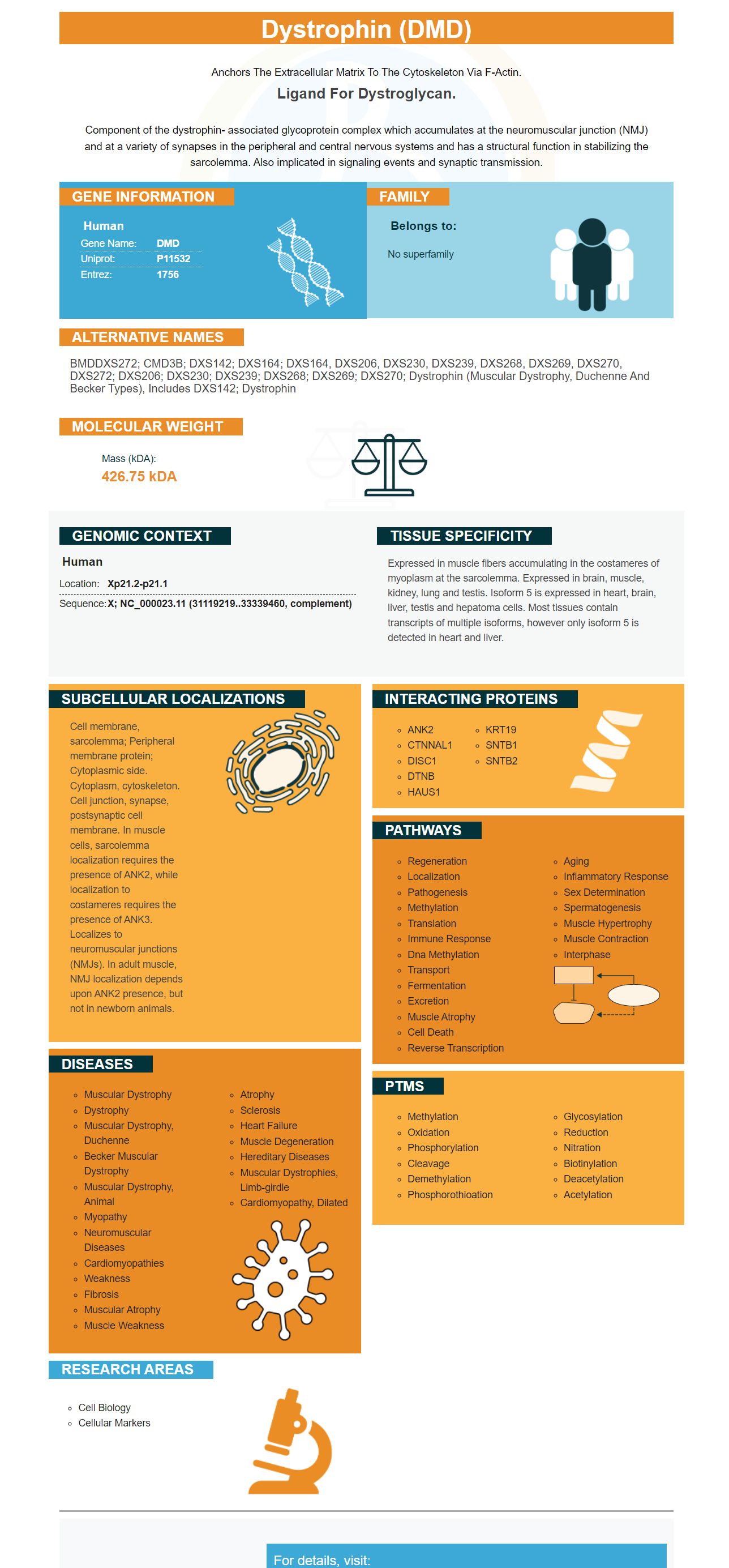
Facts about Dystrophin.
Anchors the extracellular matrix to the cytoskeleton via F-actin.
Ligand for dystroglycan.Component of the dystrophin- associated glycoprotein complex which accumulates at the neuromuscular junction (NMJ) and at a variety of synapses in the peripheral and central nervous systems and has a structural function in stabilizing the sarcolemma. Also implicated in signaling events and synaptic transmission.
| Human | |
|---|---|
| Gene Name: | DMD |
| Uniprot: | P11532 |
| Entrez: | 1756 |

| Belongs to: |
|---|
| No superfamily |

BMDDXS272; CMD3B; DXS142; DXS164; DXS164, DXS206, DXS230, DXS239, DXS268, DXS269, DXS270, DXS272; DXS206; DXS230; DXS239; DXS268; DXS269; DXS270; dystrophin (muscular dystrophy, Duchenne and Becker types), includes DXS142; dystrophin
Mass (kDA):
426.75 kDA

| Human | |
|---|---|
| Location: | Xp21.2-p21.1 |
| Sequence: | X; NC_000023.11 (31119219..33339460, complement) |
Expressed in muscle fibers accumulating in the costameres of myoplasm at the sarcolemma. Expressed in brain, muscle, kidney, lung and testis. Isoform 5 is expressed in heart, brain, liver, testis and hepatoma cells. Most tissues contain transcripts of multiple isoforms, however only isoform 5 is detected in heart and liver.
Cell membrane, sarcolemma; Peripheral membrane protein; Cytoplasmic side. Cytoplasm, cytoskeleton. Cell junction, synapse, postsynaptic cell membrane. In muscle cells, sarcolemma localization requires the presence of ANK2, while localization to costameres requires the presence of ANK3. Localizes to neuromuscular junctions (NMJs). In adult muscle, NMJ localization depends upon ANK2 presence, but not in newborn animals.


The Boster Bio is a great choice for your research, particularly when you're dealing with DMD. This marker is an excellent instrument to help you determine the health of a particular tissue or cell. This instrument can assist you in analysing your results. Here are some suggestions to maximize your research. Every experiment will experience some difficulties, but proper controls will eliminate the majority of the sources of error. Troubleshooting guides will help you quickly pinpoint the issue and get it fixed.
Although there are many benefits to the DMD marker, it isn't always a reliable surrogate marker for clinical studies. It is crucial to locate the surrogate marker that has the ability to detect changes in the concept of interest and is reliable. A single biomarker should be utilized for every trial intervention. A multi-modality analysis is essential due to the fact that the DMD population is very complex.
The DMD gene is the only gene which mutations can cause BMD and DMD-associated DCM. However the amount of information is not enough to determine how many patients carry a specific mutation. These mutations can be either duplications, point mutations, or deletions. These numbers are approximate. The DMD gene is present in around 1percent of people who suffer from DMD.
Additionally, research has shown that high DMD expression does not significantly predict survival in patients suffering from grade IV glioma. These results suggest that the DMD gene may be vital in the early stages of the glioma's development. It is essential to take the DMD gene into consideration when developing strategies for early detection and treatment. These types of cancer are generally less aggressive than the ones associated with DMD expression.
The DMD gene markers may also aid in monitoring the progress of DMD. Researchers have found that high levels DMD can reduce the biogenesis of ribosomes. These findings are particularly helpful in identifying patients suffering from DMD receiving treatment. They can be used to monitor the treatment's progress. If you're wondering if the DMD gene markers could aid in the early diagnosis and treatment, then look no further. Soon, you'll discover if the DMD gene marker offers other benefits.
There are limitations to DMD gene marker technology, despite the numerous benefits. DMD gene markers are not a reliable method of diagnosing all LGG patients however, they can be used as a starting point. In addition to identifying patients with LGG that are IDH mutant there are studies that indicate that dystrophin-related expression can enhance the risk stratification process. In this manner, the DMD gene markers could play a crucial role in the early diagnosis of the disease.
A series of international conferences and workshops highlighted the need for a reliable molecular marker that can keep track of DMD development and treatment response. This is especially important as promising therapeutic strategies are entering clinical trials. Monitoring the effectiveness of therapies has been proven to be a challenging task because outcomes are different at different stages of the disease. In addition, they are usually subjective and lack sensitivity. The goal of this paper is to outline guidelines for implementing a reliable DMD marker for use in clinical trials.
The first studies on biomarkers of proteins in DMD focused on a muscle-specific protein, called creatine kinase. This protein is a sign of damage to the sarcolemma. This marker is used to detect newborns for DMD however, it decreases with age. Because it is affected by muscle injury like trauma and exercise, it is not effective in determining progression of the disease.
Mutation detection techniques have been shown to detect mutations in 75 percent of DMD probands. This highlights the importance of fast and accurate methods to detect mutations. In fact, the MLPA assay is suggested as the first screening test for clinically suspected DMD patients and women with a DMD/BMD family history. Medical genetics laboratories should also utilize at minimum two different methods to determine the risk of DMD.
Noninvasive quantitative techniques such as ultrasound are used in the DMD clinical trial as a method for the assessment of disease progression and response to treatment. Because these methods are less invasive than conventional functional tests they can be used in clinical trials. This information can help physicians determine promising therapeutic targets. These techniques could be added to the existing functional measures and could help speed up the development of novel treatments for DMD.
A recent study was conducted to evaluate the carrier status of DMD families and to confirm multiplex ligation-dependent probe amplifying. These findings were compared with those obtained by other methods utilized in the detection of carriers. The authors also assessed the effectiveness of MLPA as a carrier analysis technique. The results of this study are comparable to the results obtained with MLPA. The technique could be used to detect DMD carriers among Indians , if further studies are conducted.
PMID: 3282674 by Koenig M., et al. The complete sequence of dystrophin predicts a rod-shaped cytoskeletal protein.
PMID: 2668885 by Rosenthal A., et al. Two human cDNA molecules coding for the Duchenne muscular dystrophy (DMD) locus are highly homologous.