This website uses cookies to ensure you get the best experience on our website.
- Table of Contents
2 Citations
Facts about Golgi membrane protein 1.

Unknown.
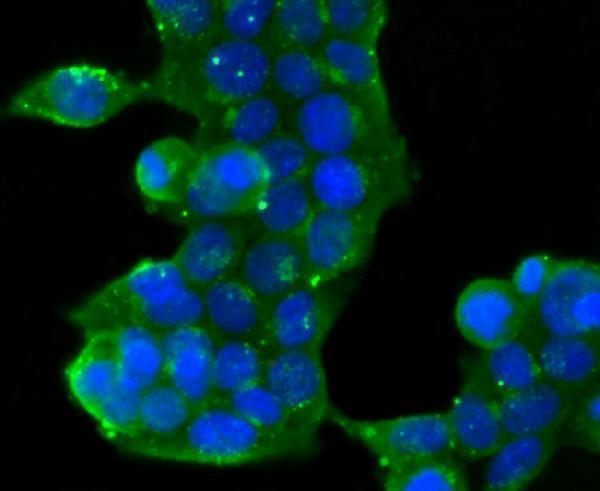
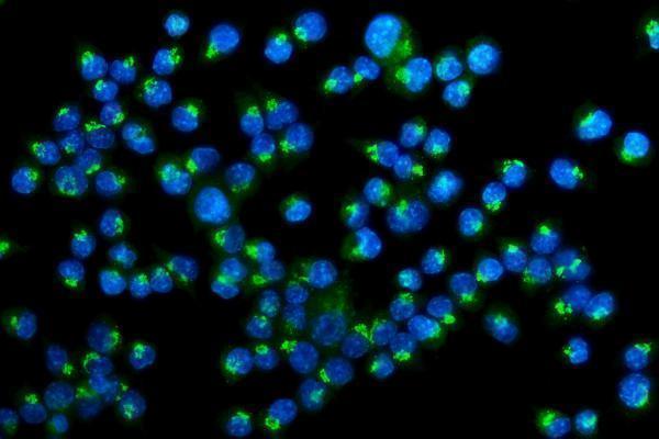
Cellular response protein to viral infection..
| Human | |
|---|---|
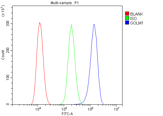
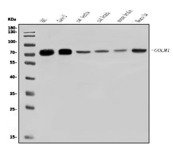
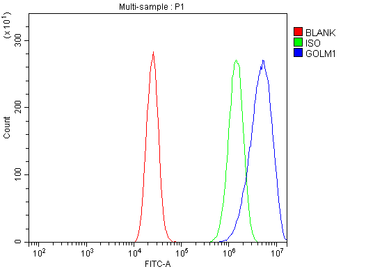
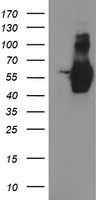
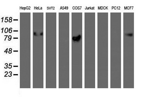
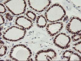
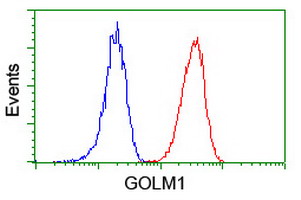
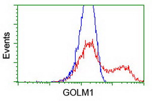
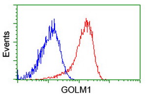
| Gene Name: | GOLM1 |
| Uniprot: | Q8NBJ4 |
| Entrez: | 51280 |

| Belongs to: |
|---|
| GOLM1/CASC4 family |

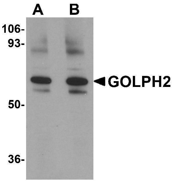
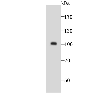
bA379P1.3; FLJ23608; golgi membrane protein 1; Golgi membrane protein GP73; Golgi phosphoprotein 2C9orf155; golgi protein, 73-kD; GOLPH2 FLJ22634; GP73 chromosome 9 open reading frame 155; GP73; PSEC0257
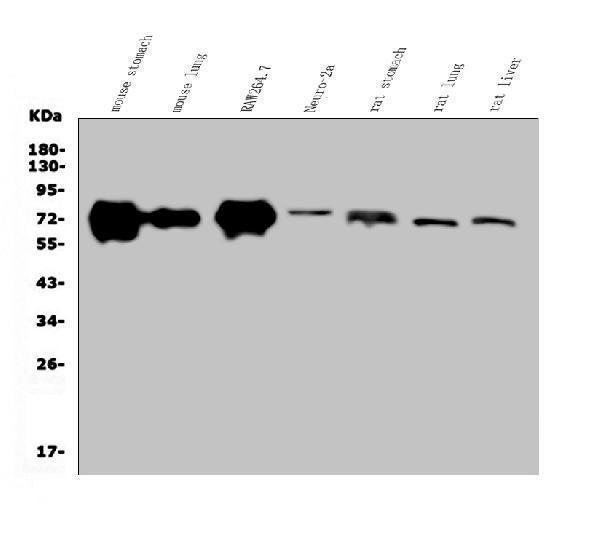
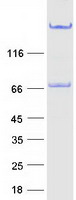
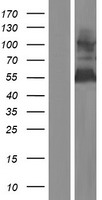
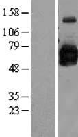
Mass (kDA):
45.333 kDA

| Human | |
|---|---|
| Location: | 9q21.33 |
| Sequence: | 9; NC_000009.12 (86026141..86100201, complement) |
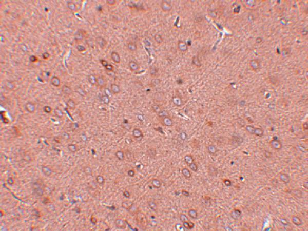
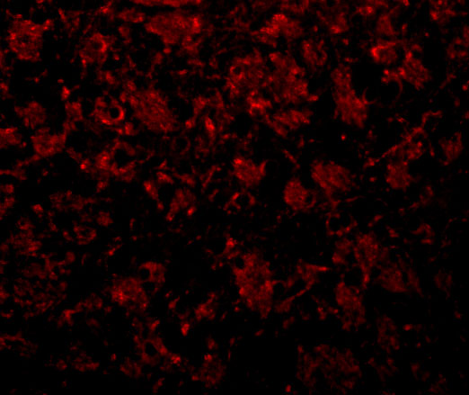
Widely expressed. Highly expressed in colon, prostate, trachea and stomach. Expressed at lower level in testis, muscle, lymphoid tissues, white blood cells and spleen. Predominantly expressed by cells of the epithelial lineage. Expressed at low level in normal liver. Expression significantly increases in virus (HBV, HCV) infected liver. Expression does not increase in liver disease due to non-viral causes (alcohol-induced liver disease, autoimmune hepatitis). Increased expression in hepatocytes appears to be a general feature of advanced liver disease. In liver tissue from patients with adult giant-cell hepatitis (GCH), it is strongly expressed in hepatocytes-derived syncytial giant cells. Constitutively expressed by biliary epithelial cells but not by hepatocytes.
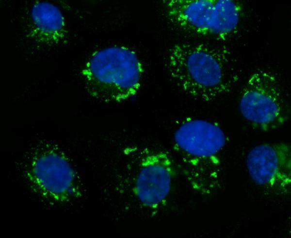
Golgi apparatus, cis-Golgi network membrane; Single-pass type II membrane protein. Early Golgi. Cycles via the cell surface and endosomes upon lumenal pH disruption.


PMID: 10831838 by Kladney R.D., et al. GP73, a novel Golgi-localized protein upregulated by viral infection.
PMID: 12191016 by Puri S., et al. Cycling of early Golgi proteins via the cell surface and endosomes upon lumenal pH disruption.