This website uses cookies to ensure you get the best experience on our website.
- Table of Contents
246 Citations 16 Q&As
161 Citations 4 Q&As
150 Citations 17 Q&As
153 Citations 16 Q&As
132 Citations 16 Q&As
127 Citations 16 Q&As
38 Citations 5 Q&As
50 Citations
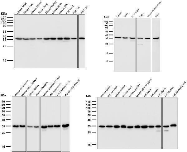
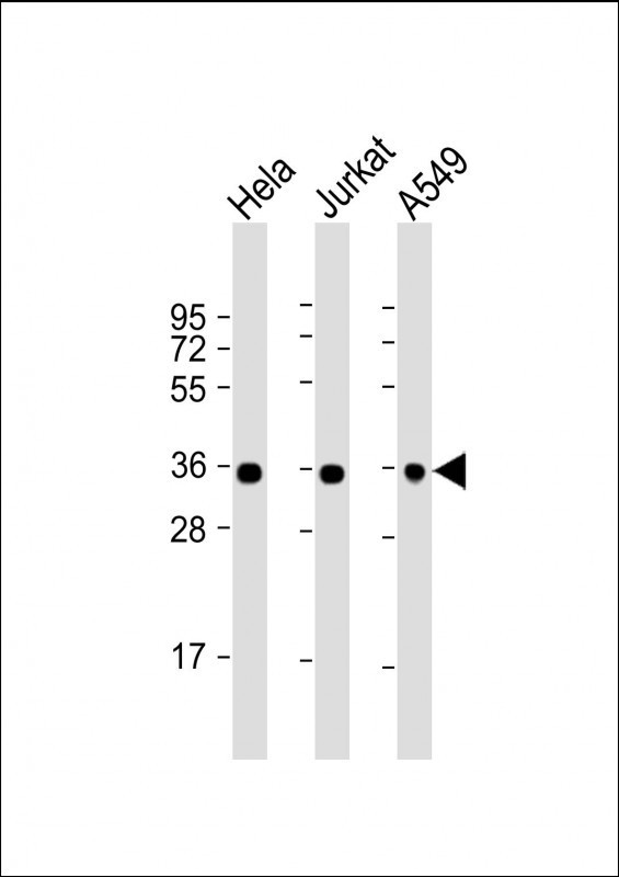
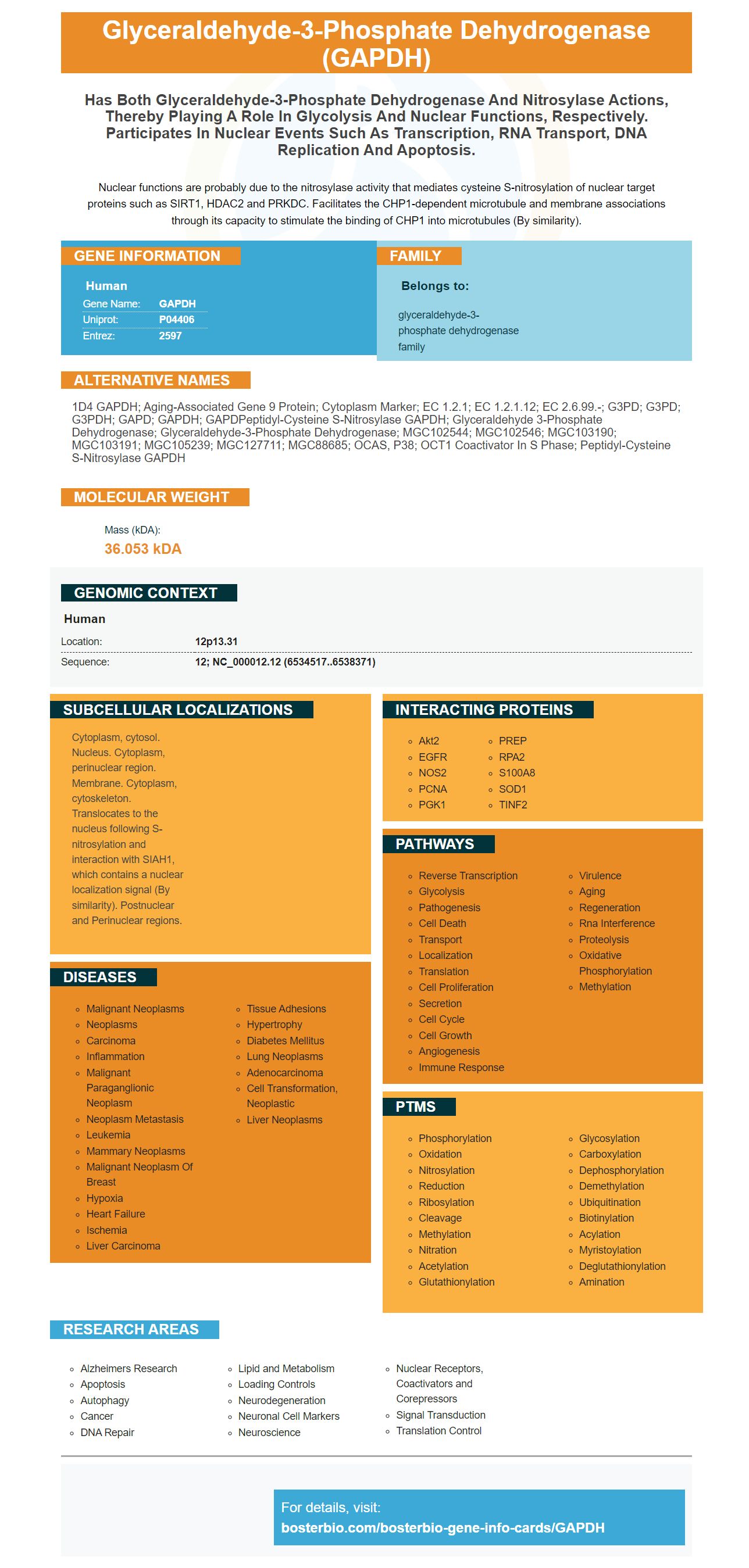
Facts about Glyceraldehyde-3-phosphate dehydrogenase.
Nuclear functions are probably due to the nitrosylase activity that mediates cysteine S-nitrosylation of nuclear target proteins such as SIRT1, HDAC2 and PRKDC. Facilitates the CHP1-dependent microtubule and membrane associations through its capacity to stimulate the binding of CHP1 into microtubules (By similarity).
Human | |
---|---|
Gene Name: | GAPDH |
Uniprot: | P04406 |
Entrez: | 2597 |
Belongs to: |
---|
glyceraldehyde-3-phosphate dehydrogenase family |
1D4 GAPDH; aging-associated gene 9 protein; Cytoplasm Marker; EC 1.2.1; EC 1.2.1.12; EC 2.6.99.-; G3PD; G3PD; G3PDH; GAPD; GAPDH; GAPDPeptidyl-cysteine S-nitrosylase GAPDH; glyceraldehyde 3-phosphate dehydrogenase; glyceraldehyde-3-phosphate dehydrogenase; MGC102544; MGC102546; MGC103190; MGC103191; MGC105239; MGC127711; MGC88685; OCAS, p38; OCT1 coactivator in S phase; Peptidyl-cysteine S-nitrosylase GAPDH
Mass (kDA):
36.053 kDA
Human | |
---|---|
Location: | 12p13.31 |
Sequence: | 12; NC_000012.12 (6534517..6538371) |
Cytoplasm, cytosol. Nucleus. Cytoplasm, perinuclear region. Membrane. Cytoplasm, cytoskeleton. Translocates to the nucleus following S-nitrosylation and interaction with SIAH1, which contains a nuclear localization signal (By similarity). Postnuclear and Perinuclear regions.
The GAPDH marker has many uses in research. The GAPDH protein is important in many different processes, including the study of oxygenative stress. This protein is essential for cell metabolism, and also plays a part in Alzheimer's disease and cancer. It is found in all tissues, and its concentrations can be determined by immunofluorescence, ELISA or Western Blot.
Boster Bio's own GAPDH marker has an ELISA that was developed by synthesizing. The full-length GAPDH genes are extremely conserved in various M. hyorhinis strains. The GAPDH gene was cloned to an expression vector called pET-28a, and then expressed in E. coli BL21 cells. SDS-PAGE, Western blotting using an anti-His antibody, as well as ELISA were used to determine the gene recombinant.
To determine if this ELISA can detect rGAPDH It first determines the binding between rGAPDH and a cell membrane protein. Cell membrane proteins were created using an commercial Membrane and Cytosol Protein Extraction Kit from Tiangen Biotech, China. Cell membrane proteins were added to a 96-well ELISA plate overnight at 4°C. The plate was then blocked using 5% BSA and incubated at 37°C for 2 hours.
Using an ELISA for Boster Bio's own GAPDH marker is an extremely sensitive and precise method for testing the presence of GAPDH. GAPDH is found on surface of pathogens, and has been identified in three Mycoplasma species: M. pneumoniae, M. hyorhinis and M. suis. GAPDH is specifically bound by the anti-rGAPDH antibody.
LmGAPDH has a nonglycolytic role in the HKO cell line. Scientists will be able to analyze the activity of this protein with other proteins based upon this knowledge. Glycosomal GAPDH plays a major component of trypanosomatids. This GAPDH is also able to be transported to organelles that are subcellular. The ELISA for Boster Bio's GAPDH marker can help identify the GAPDH marker in various species.
Different components of the ECM are bound to the rGAPDH protein. Matrigel, fibronectinand collagen, and laminin are used as substrates in the assay. Incubation was completed overnight with the rGAPDH antibody at a concentration of 12.5-100 mg/mL. The plate was then washed with PBST and proteins that were not bound were taken off.
Boster Bio's GHPD marker is detected using ELISA using the Luminol reagent, which is a fluorescent dye. The luminol reacts with the HRP to create an excited state. This product emits light once it has been returned to its normal state. Chemiluminescent reagents may have higher sensibility than colorimetric ones however, the intensity can be different.
The rGAPDH protein binds plasminogen. The rGAPDH is able to bind plasminogen in a non-specific manner by using an ELISA (or Western blot) to determine the GAPDH binding to the plasminogen. The rGAPDH bonds to plasminogen by binding to the GAPDH coated. The plasminogen is then activated. The signal is measured by OD405nm.
The results of the ELISA test for Boster Bio's EVs that are derived from GAPDH were in agreement with Western blot data. Results from mRNA and protein expression analysis indicate that GAPDH plays a crucial role in the development of Leishmania disease and infection in mice. Additionally, the EV-derived LmGAPDH plays a significant role in Leishmania-related the progression of disease in mice.
There are many uses for Boster Bio's GAPDH Marker. For instance when you're interested in determining the cause of disease of a particular bacterium you can look into the interaction of rGAPDH with plasminogen. This protein is found on the surface of three Mycoplasma species, including M. pneumoniae, M. hyopneumoniae, and M. suis. These bacteria also have cell-surface markers that suggest that GAPDH has a relationship with host cells.
Boster Bio: The Best uses of the Protein Detection Kit: The recombinant GAPDH gene was synthesized by using the Escherichiacoli's genome BL21(DE3). The bacterial cells were then maintained in a great broth (pH 28a) for 10 minutes. They were then removed by centrifugation at 6000g for 10 minutes. After centrifugation, the cell is free. rGAPDH positive recombinant plasmid could be detected using SDS-PAGE and Western Blotting using an anti-His antibody.
The results of this study also showed that the surface-exposed GAPDH plays an indispensable role in cytoadhesion M. hyorhinis. The anti-rGAPDH serum slowed down the adhesion of M. Hyorhinis to porcine PK-15 and human NCI H292 cells, however, other proteins were also identified as contributing to the adhesion process of M. hyorhinis.
The glyceraldehyde-3-phosphate dehydrogenase (GAPDH) is an essential housekeeping protein expressed in most cell types. GAPDH mRNA levels are frequently used as a reference in the study of mRNA expression. It can also be used as a loading control in Western Blot assays. Furthermore, GAPDH antibodies have multiple applications in immunoprecipitation, immunofluorescence, and immunohistochemistry.
As a surface-located molecule, GAPDH may have a greater range of applications than was previously believed. It could be an plasminogen receptor or as an adhesin, an adhesin produced by bacteria that assists in the breakdown of extracellular matrix. Its surface-localization makes it an valuable marker for assessing the degree of colonization of bacteria.
GAPDH antibody antibodies are used for many purposes, including cell-based analysis and genetic research. Certain research applications require a high level of specificity. In these situations monoclonal antibody is the best choice. Monoclonal antibodies are able to recognize a single epitope of antigen and have a lower background. Polyclonal antibodies recognize more that one epitope in the antigenic chain and typically have a greater affinity. However, they are diluted secondary antibodies that can be used together with monoclonal antibodies.
Subcutaneous injections were administered to boost monoclonal antibodies against GAPDH in rabbits. A female rabbit that was six months old received Freund's complete adjuvant. It was followed by three booster doses (15 mg) of recombinant LmGAPDH (15mg) at intervals of two weeks. The rabbits were bled two weeks after the last booster, and serum was collected to be used for Western blot analysis.
The EV fraction contains the LmGAPDH protein which is Leishmania major. It is believed that this protein could directly affect defenses of the host. The LmGAPDH gene is devoid of the N-terminal signal peptide. This suggests that LmGAPDH may be released via a non-conventional mechanism. Furthermore, the gene does not contain the peptide N-terminal signaling which is the hallmark of all temperature-induced secreted proteins.
The GAPDH gene has been found to be highly conserved between strains of M. hyorhinis. A synthetic GAPDH gene was constructed using the genome sequences of strain HUB-1, and then cloned into pET 28a (+). DNA sequencing was used to confirm the DNA sequence of the recombinant virus. In a subsequent study, E. coli cells were treated with IPTG at 1 mM concentration for 6 hours.
The rGAPDH protein binds plasminogen. The binding was determined through ELISA and Western Blots using an anti-plasminogen antibody. As an alternative to a negative control, BSA was used to detect nonspecific binding. After binding, plasminogen was activated to plasminogen with tPA. To determine the amount of plasminogen that was bound to the coating, OD405 num (or OD405) was used.
GAPDH is highly specific for the presence of plasminogen rGAPDH at the surface of pathogens. GAPDH is highly specific to host cells. This allows researchers to detect bacterial cell populations that have high levels of GAPDH. Furthermore the rGAPDH marker can detect GAPDH-related expression in a range of microorganisms, including M. pneumoniae, M. hyorhinis, and M. suis.
In a micro titer plate, rGAPDH is bound to the cell membrane protein. The commercial membrane protein extraction kit made by Tiangen Biotech, China was used to extract cytosol and membrane proteins. The membrane protein was coated onto the 96-well micro titer plate. The cells were then incubated for a period of 37°C using a blocking solution consisting of 5 % BSA. PBS was used as an additional positive control.
The GAPDH marker is useful in detecting the presence of mycoplasma through colony blot. It can be used for diagnostic tests, drug design, and in the laboratory diagnosis. Its low molecular weight enables it to recognize a wide range of pathogens, including mycoplasma. If you're looking for a top-quality and precise GAPDH marker, this may be the perfect choice.
The GAPDH marker has become an invaluable instrument for biomedical researchers and researchers alike. The protein was isolated from E. coli and purified using a PVDF membrane. Incubation with 10 mg/mL plasminogen in a PVDF membrane blocked the membrane prior to incubation with rGAPDH antibodies. Both secondary and primary antibodies were used to detect the protein. BSA was used in a third experiment as a negative control.
GAPDH is surface-localized. It is a plasminogen-dependent protein and has a higher possibility of being used as a subunit vaccine antigen. The recombinant version of the protein was shown to bind porcine PK-15 cells. It also binds to human NCI-H292 cell. Its binding ability is confirmed by studies that demonstrate GAPDH can bind to several ECM components.
PMID: 6096136 by Hanauer A., et al. The glyceraldehyde 3 phosphate dehydrogenase gene family: structure of a human cDNA and of an X chromosome linked pseudogene; amazing complexity of the gene family in mouse.
PMID: 6096821 by Arcari P., et al. The complete sequence of a full length cDNA for human liver glyceraldehyde-3-phosphate dehydrogenase: evidence for multiple mRNA species.
*Showing only the more recent 20. More publications can be found for each product on its corresponding product page