This website uses cookies to ensure you get the best experience on our website.
- Table of Contents
Facts about Myocilin.

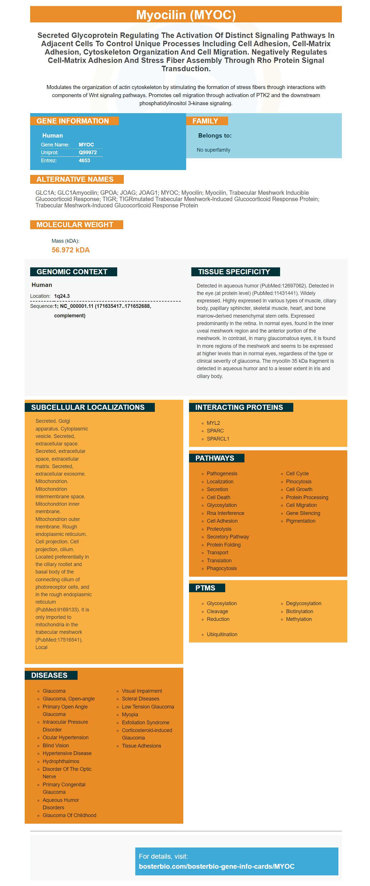
Modulates the organization of actin cytoskeleton by stimulating the formation of stress fibers through interactions with components of Wnt signaling pathways. Promotes cell migration through activation of PTK2 and the downstream phosphatidylinositol 3-kinase signaling.
| Human | |
|---|---|
| Gene Name: | MYOC |
| Uniprot: | Q99972 |
| Entrez: | 4653 |

| Belongs to: |
|---|
| No superfamily |

GLC1A; GLC1Amyocilin; GPOA; JOAG; JOAG1; MYOC; Myocilin; myocilin, trabecular meshwork inducible glucocorticoid response; TIGR; TIGRmutated trabecular meshwork-induced glucocorticoid response protein; Trabecular meshwork-induced glucocorticoid response protein
Mass (kDA):
56.972 kDA

| Human | |
|---|---|
| Location: | 1q24.3 |
| Sequence: | 1; NC_000001.11 (171635417..171652688, complement) |
Detected in aqueous humor (PubMed:12697062). Detected in the eye (at protein level) (PubMed:11431441). Widely expressed. Highly expressed in various types of muscle, ciliary body, papillary sphincter, skeletal muscle, heart, and bone marrow-derived mesenchymal stem cells. Expressed predominantly in the retina. In normal eyes, found in the inner uveal meshwork region and the anterior portion of the meshwork. In contrast, in many glaucomatous eyes, it is found in more regions of the meshwork and seems to be expressed at higher levels than in normal eyes, regardless of the type or clinical severity of glaucoma. The myocilin 35 kDa fragment is detected in aqueous humor and to a lesser extent in iris and ciliary body.
Secreted. Golgi apparatus. Cytoplasmic vesicle. Secreted, extracellular space. Secreted, extracellular space, extracellular matrix. Secreted, extracellular exosome. Mitochondrion. Mitochondrion intermembrane space. Mitochondrion inner membrane. Mitochondrion outer membrane. Rough endoplasmic reticulum. Cell projection. Cell projection, cilium. Located preferentially in the ciliary rootlet and basal body of the connecting cilium of photoreceptor cells, and in the rough endoplasmic reticulum (PubMed:9169133). It is only imported to mitochondria in the trabecular meshwork (PubMed:17516541). Local


PMID: 9280311 by Ortego J., et al. Cloning and characterization of subtracted cDNAs from a human ciliary body library encoding TIGR, a protein involved in juvenile open angle glaucoma with homology to myosin and olfactomedin.
PMID: 9169133 by Kubota R., et al. A novel myosin-like protein (myocilin) expressed in the connecting cilium of the photoreceptor: molecular cloning, tissue expression, and chromosomal mapping.