This website uses cookies to ensure you get the best experience on our website.
- Table of Contents
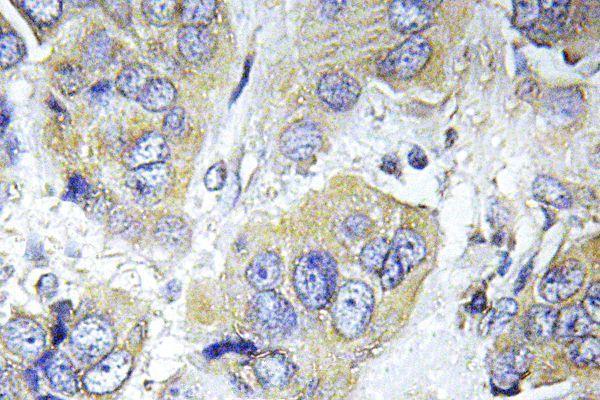
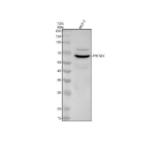
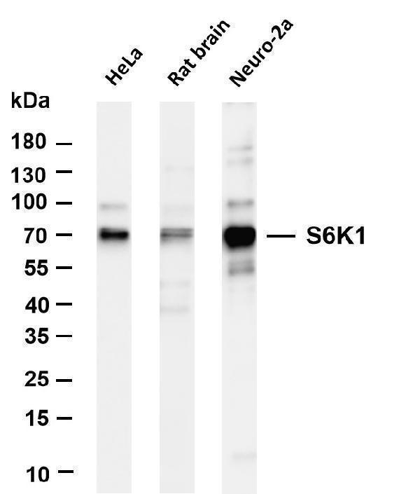
5 Citations 1 Q&As
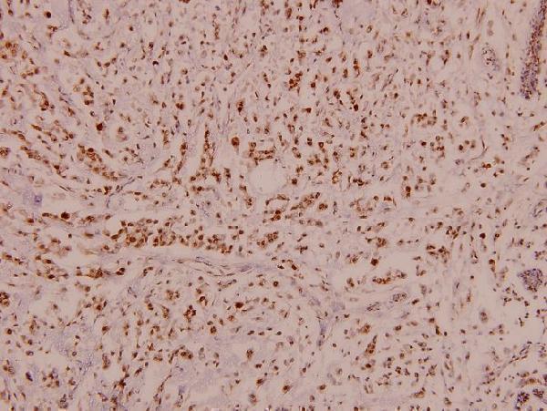
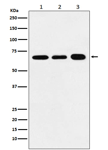
1 Citations 6 Q&As


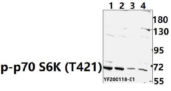
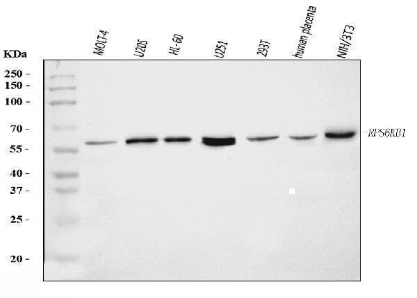
1 Citations
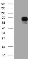
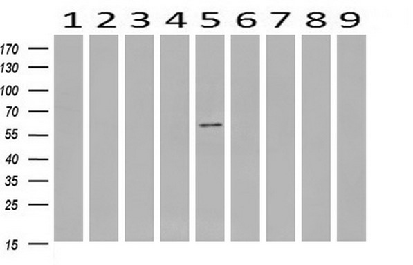
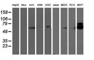
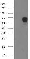
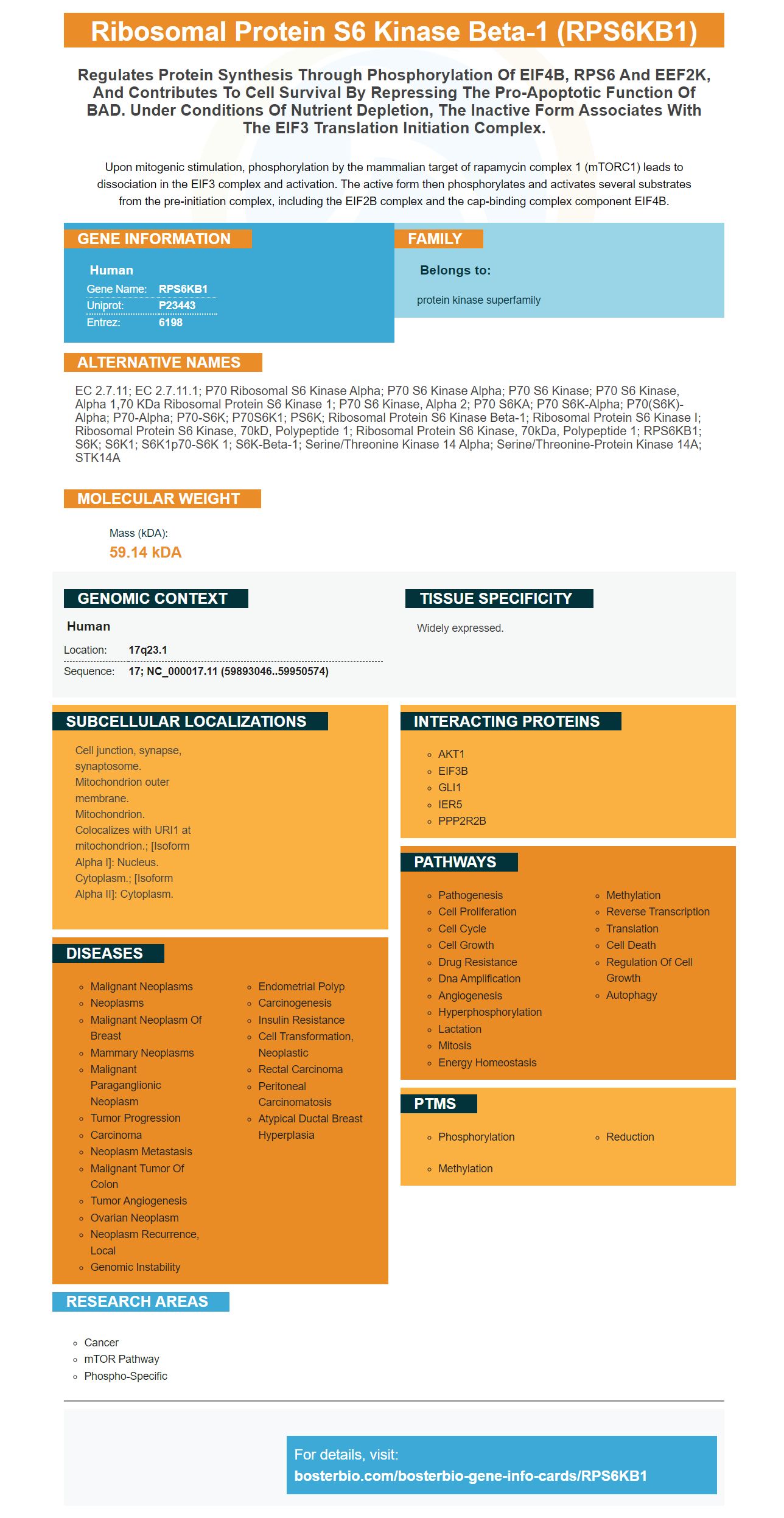
Facts about Ribosomal protein S6 kinase beta-1.

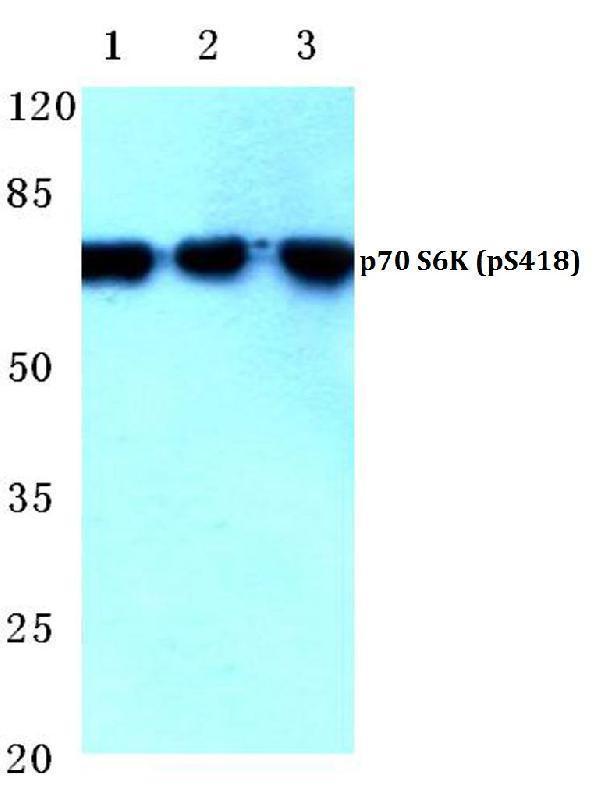
Upon mitogenic stimulation, phosphorylation by the mammalian target of rapamycin complex 1 (mTORC1) leads to dissociation in the EIF3 complex and activation. The active form then phosphorylates and activates several substrates from the pre-initiation complex, including the EIF2B complex and the cap-binding complex component EIF4B.
| Human | |
|---|---|
| Gene Name: | RPS6KB1 |
| Uniprot: | P23443 |
| Entrez: | 6198 |

| Belongs to: |
|---|
| protein kinase superfamily |

EC 2.7.11; EC 2.7.11.1; p70 ribosomal S6 kinase alpha; p70 S6 kinase alpha; p70 S6 Kinase; p70 S6 kinase, alpha 1,70 kDa ribosomal protein S6 kinase 1; p70 S6 kinase, alpha 2; p70 S6KA; p70 S6K-alpha; p70(S6K)-alpha; p70-alpha; p70-S6K; P70S6K1; PS6K; ribosomal protein S6 kinase beta-1; Ribosomal protein S6 kinase I; ribosomal protein S6 kinase, 70kD, polypeptide 1; ribosomal protein S6 kinase, 70kDa, polypeptide 1; RPS6KB1; S6K; S6K1; S6K1p70-S6K 1; S6K-beta-1; serine/threonine kinase 14 alpha; Serine/threonine-protein kinase 14A; STK14A
Mass (kDA):
59.14 kDA

| Human | |
|---|---|
| Location: | 17q23.1 |
| Sequence: | 17; NC_000017.11 (59893046..59950574) |
Widely expressed.
Cell junction, synapse, synaptosome. Mitochondrion outer membrane. Mitochondrion. Colocalizes with URI1 at mitochondrion.; [Isoform Alpha I]: Nucleus. Cytoplasm.; [Isoform Alpha II]: Cytoplasm.


PMID: 1922062 by Grove J., et al. Cloning and expression of two human p70 S6 kinase polypeptides differing only at their amino termini.
PMID: 9804755 by Gout I., et al. Molecular cloning and characterization of a novel p70 S6 kinase, p70 S6 kinase beta containing a proline-rich region.
*More publications can be found for each product on its corresponding product page