This website uses cookies to ensure you get the best experience on our website.
- Table of Contents
3 Citations 3 Q&As
1 Citations
1 Citations


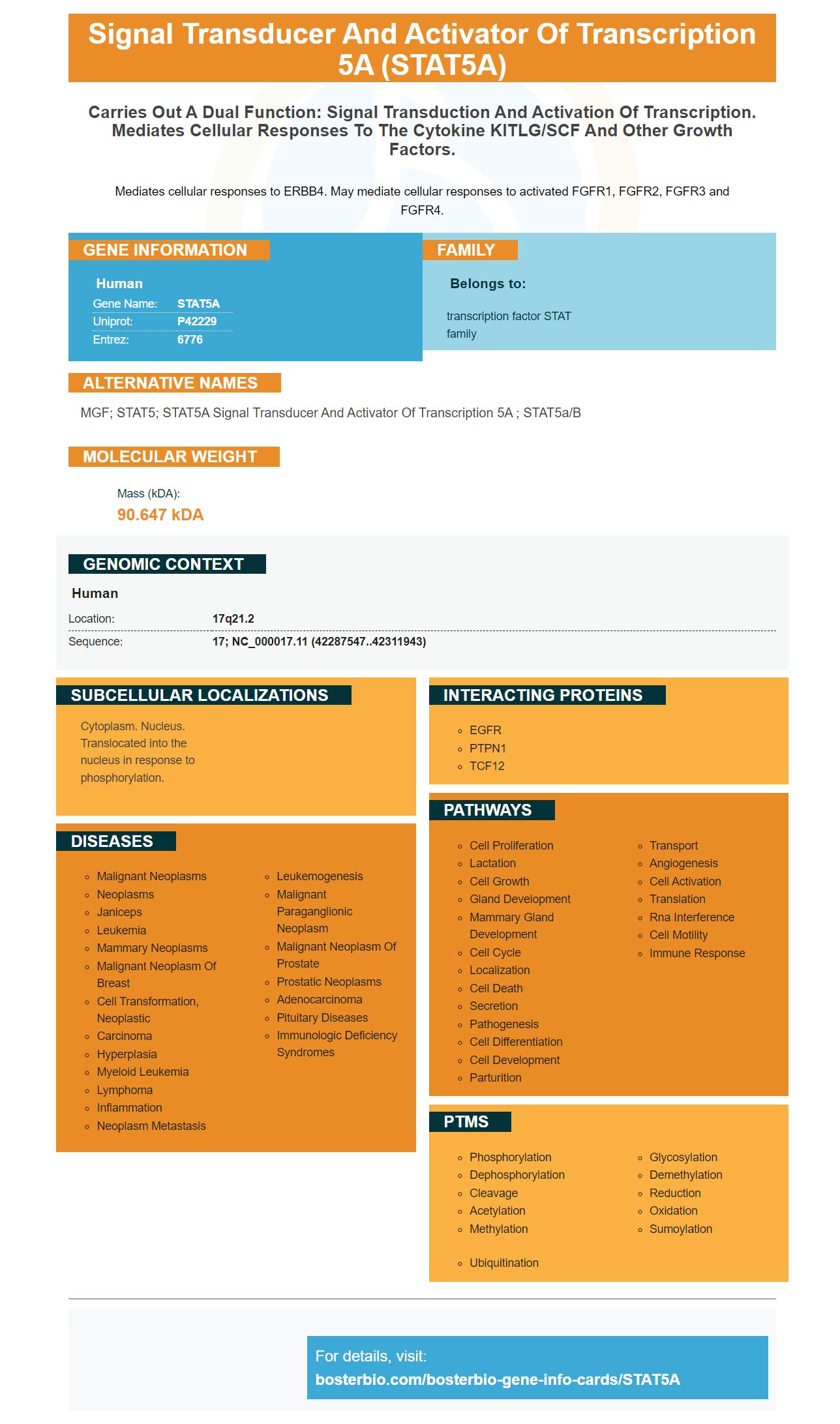
Facts about Signal transducer and activator of transcription 5A.

Mediates cellular responses to ERBB4. May mediate cellular responses to activated FGFR1, FGFR2, FGFR3 and FGFR4.
| Human | |
|---|---|
| Gene Name: | STAT5A |
| Uniprot: | P42229 |
| Entrez: | 6776 |

| Belongs to: |
|---|
| transcription factor STAT family |

MGF; STAT5; STAT5A signal transducer and activator of transcription 5A ; STAT5a/b
Mass (kDA):
90.647 kDA

| Human | |
|---|---|
| Location: | 17q21.2 |
| Sequence: | 17; NC_000017.11 (42287547..42311943) |
Cytoplasm. Nucleus. Translocated into the nucleus in response to phosphorylation.


The STAT5A Marker transcription factor is responsible for tumor growth and cell proliferation. Not many people realize that this gene is related to miR-339-5p or SNHG17. Here are three ways to use this gene. Find out how to use this gene in your classroom. Boster Bio's resources can be shared by educators.
The STAT5A marker is a transcription element that is expressed by oocytes in multiple stages of embryo development, including the two, four, and blastula. The protein is involved in two mechanisms that guarantee the survival and development of the embryo. Many sperm factors can cause low fertility. Incompatible sperm are also implicated in the low rate of fertilization, which results in the embryonic death before the blastocyst stage.
The well-known transcription element nuclear factor-kB is responsible for inflammation-induced tumorigenesis. The transcription factor regulates many immune reactions including those that promote apoptosis, inflammation, and other cell death. The STAT proteins are involved with a variety of immune reactions and they are secreted through crosstalks to NF-kb.
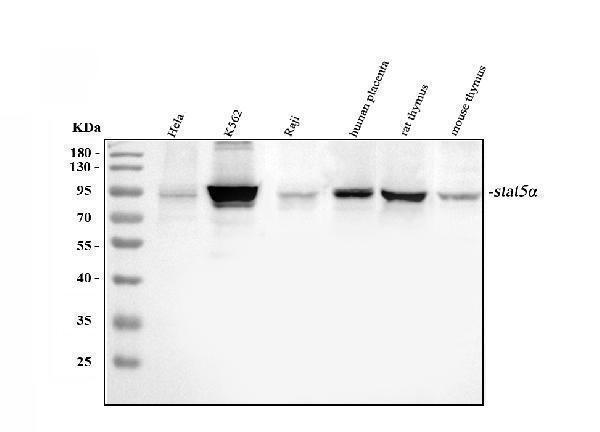
STAT5 plays a pivotal role during eosinophil differentiation. Western blotting was used in order to determine the function of STAT5 in eosinophil eosinophil division. Protein lysates from differentiating eosinophils have been prepared and subjected western blotting. Bcl-2 and p21 WAF/Cip1 were up-regulated early in the differentiation process and decreased during the final maturation phase.
Overexpression CA-STAT5A was associated with a significant increase at STAT5 in human bloodstream. However, STAT5a wasn't sufficient to promote proliferation of hepatocytes. CA-STAT5A significantly increased STAT5 bind to the PLF1 promoter area.
In vitro studies have also suggested that the STAT5-PLF1 cascade is essential for tube formation in EC. When activated, STAT5 induced EC formation. Forced expression of PLF1 alone prevented tube formation in DN-STAT5A-treated cells, whereas forced expression of PLF1 in the absence of serum induced tube formation resulted in a 10-fold increase in cell length.
Despite its role as a promoter of cancer growth, the STAT5A gene may also act as a tumor suppressor. It inhibits the growth of cancer cells by arresting the cell cycle and promoting apoptosis. In mouse models of colon cancer DLD-1, unphosphorylated human STAT5A had tumor suppressive properties. Unphosphorylated human STAT5A had similar effects on cancer cells to HP1a overexpression. Numerous cancers can be linked to unphosphorylated STAT5A and HP1a downregulations and somatic mutations.
The STAT5A marker is involved in T-cell Lymphoma. Its role may be responsible for heterochromatin formation, which could explain its involvement in the development of cancer. There are still many questions about its role in tumorigenesis. While STAT5A is a tumor suppressor, it is not a key factor in the development of tumors. Rb, p53, and STAT5A inhibit tumor development. STAT5A is a tumor suppressor in its own right.
The STAT5A marker regulates tumor growth and cell proliferation. It is involved in cell division, invasion, migration, and other functions in tumor cells. Mutation of STAT5a in mice delayed the progression of mammary cancer. Mutations in STAT5a caused tumors in mice by overexpression in mammary cells. In addition, hyperprolactinemia-induced antipsychotics induce STAT5A in the breast.
The cS5A mutations in tumor cells cause a phenotype called mature T-cell neoplasia. STAT5 activation promotes proliferation of CD8+T cells in mice. This leads to activation and activation the cytotoxic CD8-memory phenotype in mice. cS5A-mutated mice also have a higher number of gd T cells than cS5A+ mice.
Many cancers have been shown to be associated with dysregulated microRNAs. It has been associated with tumorigenesis and progression of AML. In AML, its expression is reduced. It has been associated with decreased cell growth, inhibition of cell-cycle progression, and induction cell apoptosis. It targets the sex-determining area Y-related high mobility box SOX4, a gene that is involved in cell proliferation.
The expression of SOX4 and miR-339-5p in AML cells was analyzed using reverse transcription-quantitative polymerase chain reaction (RT-qPCR) and western blotting. Spearman's correlation analysis showed that SOX4 protein and miR-339-5p were negatively related in AML. The findings suggest that miR-339-5p may directly target SOX4 in AML cells.
A miRNA mimic or suppressor was introduced into U87 cells by transfection. The miRNA mimics could be detected using qRTPCR. The relative expression level of miR-339-5p within the cancer cells was also compared to healthy controls. The miRNA mimics showed a significantly decreased level of expression in both glioma and healthy cells. Furthermore, qRT-PCR results supported the miRNA chip results and suggested that the miRNAs were correlated to one another.
Numerous studies have shown positive correlations between miR-339-5p levels and CDK2 gene expression. In addition, CDK2 is an important regulator of cell proliferation, and miR-339-5p suppressors decrease expression levels of these molecules. The cellular responses of these two genes are also positively correlated. MiR-339-5p and CDK2 expression are closely related to one another.
SNHG17 activates two distinct regulatory channels and promotes tumor cell growth and metastasis. SNHG17 promotes tumor growth by binding to the miR-328-3p/FOSL2-SNHG17 axis and by positively regulating the expression of SP1 and miR-328-3p. However, its role in tumorigenesis is not fully understood. Although the role SNHG17 may not be fully understood in tumorigenesis, its positive correlation to cancer progression (OS) and the upregulation other glial markers could explain the positive correlation.
RNA sequencing showed that SNHG17 was significantly increased in HCC cells. Knockdown experiments revealed that 1037 genes were differentially expressed following SNHG17 knockdown. These genes were found to overlap with SNHG17-related genes in the TCGA/LIHC dataset. SNHG17 expressed genes also influenced p53 and PI3K-Akt signaling pathways. These findings suggest that SNHG17 might be a potential therapeutic target for HCC.
The gene has substantial clinical value. SNHG17 expression levels are consistently associated with tumour size, TNM stage, and lymph node-metastasis. It is also linked to the development of chemical drug resistance. Consequently, SNHG17 may be an excellent biomarker for early diagnosis and a potential diagnostic test for GC. Its serum levels have a sensitivity level of 84.7%, and a specificity level of 78.2% making it a very useful biomarker for diagnosing GC.
It is interesting to note that this gene is also increased in PC cells. SNORA71B expression may also indicate poor prognosis. Kaplan Meier analysis was used to examine the association between SNORA71B levels and progression-free death. A Pearson's correlation curve was used to determine the correlation between SNORA71B expression and SNHG17. Moreover, SNHG17 was knocked down to inhibit PC cell growth and decrease SNORA71B levels.
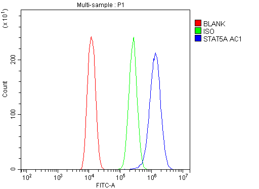
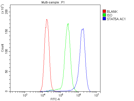
STAT5A is the primary antibody that can be used to treat B-cell lymphoma. It was recently validated in patients who were infected by the newly discovered HHV8. Flow Cytometric analysis has shown that the antibody induces homotypic adheresion, actin dependent cell death, and lymphosome-mediated cell deaths. STAT5A is also known to increase the immune response of patients with aggressive forms lymphoma.
During the second phase, patients receive a single antibody that targets the STAT5A protein. This antibody has proved to be highly effective in treating patients who have advanced forms of the disease. This antibody will be effective in treating patients with aggressive forms of the disease. Some patients will experience a high response while others will not. A course of chemotherapy is the first option for patients with this type lymphoma.
The STAT5A mark is a unique primary antibody for diagnosing B cell lymphoma. It is a monoclonal antibodies that detects IgA (and IgG) in the serum of patients suffering from B-cell cancer. It has also been tested in patients suffering from refractory B cell lymphoma.
The BosterBio STAT5A antibody marker can also be used in the detection of tumor cells. The antibody recognizes the STAT5A protein on a B-cell lymphoma. Besides the IgG and IgM antibodies, the STAT5A antibody also recognizes memory B cells. The antibody is highly specific for B-cell lymphoma. Patients should have an allergy test before starting therapy.
Boster Bio STAT5A's antigen-specific antibody markers are specifically designed to target human memory cells. Boster antibodies can be used to target human memory B cells. They are a great option for patients with B-cell lymphoma. It is important to mention that this antibody is only available on the US market.
PMID: 7719937 by Hou J., et al. Identification and purification of human Stat proteins activated in response to interleukin-2.
PMID: 11773439 by Aoki N., et al. A nuclear protein tyrosine phosphatase TC-PTP is a potential negative regulator of the PRL-mediated signaling pathway: dephosphorylation and deactivation of signal transducer and activator of transcription 5a and 5b by TC-PTP in nucleus.
*More publications can be found for each product on its corresponding product page