Click image to see more details
Product Info Summary
| SKU: | PA1840 |
|---|---|
| Size: | 100 μg/vial |
| Reactive Species: | Human, Mouse, Rat |
| Host: | Rabbit |
| Application: | Flow Cytometry, WB |
Customers Who Bought This Also Bought

Product info
Product Name
Anti-STAT5a Antibody Picoband®
SKU/Catalog Number
PA1840
BA3781-2 is an alternative SKU for this antibody, used in previous lots.
Size
100 μg/vial
Form
Lyophilized
Description
Boster Bio Anti-STAT5a Antibody catalog # PA1840. Tested in Flow Cytometry, WB applications. This antibody reacts with Human, Mouse, Rat. The brand Picoband indicates this is a premium antibody that guarantees superior quality, high affinity, and strong signals with minimal background in Western blot applications. Only our best-performing antibodies are designated as Picoband, ensuring unmatched performance.
Storage & Handling
Store at -20˚C for one year from date of receipt. After reconstitution, at 4˚C for one month. It can also be aliquotted and stored frozen at -20˚C for six months. Avoid repeated freeze-thaw cycles.
Cite This Product
Anti-STAT5a Antibody Picoband® (Boster Biological Technology, Pleasanton CA, USA, Catalog # PA1840)
Host
Rabbit
Contents
Each vial contains antibody formulated with stabilizing components, 0.9mg NaCl, 0.2mg Na2HPO4, 0.05mg Thimerosal, 0.05mg NaN3.
*This antibody is supplied in a stabilized formulation.
Compatibility with conjugation reactions depends on the chemistry of the conjugation method used.
For conjugation methods that are not compatible with the stabilizing components present in this formulation, a carrier-free antibody format is required.
Clonality
Polyclonal
Isotype
Rabbit IgG
Immunogen
A synthetic peptide corresponding to a sequence at the C-terminus of human STAT5a.
Cross-reactivity
No cross-reactivity with other proteins
Reactive Species
PA1840 is reactive to STAT5A in Human, Mouse, Rat
Observed Molecular Weight
95 kDa
Calculated molecular weight
90.6 kDa
Background of STAT5A
STAT5 (Signal transducer and activator of transcription 5) also known as STAT5A, MGF, is a protein that serve the dual function of signal transducers and activators of transcription in cells exposed to signaling polypeptides. Hou et al. (1995) cloned the human STAT5 cDNA from an umbilical vein endothelial cell library and found that it encodes a 794-amino acid polypeptide with a predicted mass of approximately 90.5 kD. To analyze the possible role of STAT5 in human GH-induced proliferation, Friedrichsen et al. (2001) expressed a dominant-negative STAT5 mutant, STAT5A-delta-749, in INS-1 cells under the control of a doxycycline-inducible promoter by stable transfection. Chromatin immunoprecipitation analysis confirmed direct binding of STAT5 to the HIF2A promoter 344 bp upstream of the HIF2A start site. STAT5 overexpression independently resulted in erythroid commitment in megakaryocytic-erythroid progenitors, which was abrogated by knockdown of GATA1. STAT5 is responsible for the immediate-early induction of the long isoform of the BCL2-related protein in erythroid cells through direct binding to the promoter of the BCLX gene.
Antibody Validation
Boster validates all antibodies on WB, IHC, ICC, Immunofluorescence, and ELISA with known positive control and negative samples to ensure specificity and high affinity, including thorough antibody incubations.
Application & Images
Applications
PA1840 is guaranteed for Flow Cytometry, WB Boster Guarantee
Recommend Dilution
| Application | Dilution | Species |
|---|---|---|
| Western blot | 0.1-0.5μg/ml | Human, Mouse, Rat |
| Flow Cytometry (Fixed) | 1-3μg/1x106 cells | Human |
Tested application
Suggested blocking solution with 5% non-fat milk or BSA; (*)Recommended protein loading: 20-40 µg per lane
Validation Images & Assay Conditions
Click image to see more details
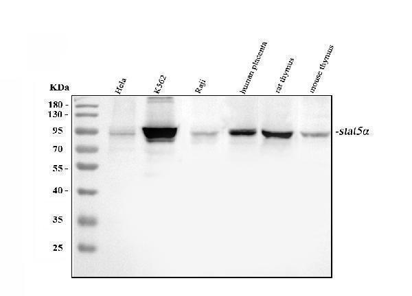
Western blot analysis of STAT5A using anti-STAT5A antibody (PA1840).
Electrophoresis was performed on a 5-20% SDS-PAGE gel at 70V (Stacking gel) / 90V (Resolving gel) for 2-3 hours. The sample well of each lane was loaded with 30 ug of sample under reducing conditions.
Lane 1: human Hela whole cell lysates,
Lane 2: human K562 whole cell lysates,
Lane 3: human Raji whole cell lysates,
Lane 4: human placenta tissue lysates,
Lane 5: rat thymus tissue lysates,
Lane 6: mouse thymus tissue lysates.
After electrophoresis, proteins were transferred to a nitrocellulose membrane at 150 mA for 50-90 minutes. Blocked the membrane with 5% non-fat milk/TBS for 1.5 hour at RT. The membrane was incubated with rabbit anti-STAT5A antigen affinity purified polyclonal antibody (Catalog # PA1840) at 0.5 μg/mL overnight at 4°C, then washed with TBS-0.1%Tween 3 times with 5 minutes each and probed with a goat anti-rabbit IgG-HRP secondary antibody at a dilution of 1:5000 for 1.5 hour at RT. The signal is developed using an Enhanced Chemiluminescent detection (ECL) kit (Catalog # EK1002) with Tanon 5200 system. A specific band was detected for STAT5A at approximately 95 kDa. The expected band size for STAT5A is at 91 kDa.

Click image to see more details
Flow Cytometry analysis of U20S cells using anti-STAT5A antibody (PA1840).
Overlay histogram showing U20S cells stained with PA1840 (Blue line). To facilitate intracellular staining, cells were fixed with 4% paraformaldehyde and permeabilized with permeabilization buffer. The cells were blocked with 10% normal goat serum. And then incubated with rabbit anti-STAT5A Antibody (PA1840,1μg/1x106 cells) for 30 min at 20°C. DyLight®488 conjugated goat anti-rabbit IgG (BA1127, 5-10μg/1x106 cells) was used as secondary antibody for 30 minutes at 20°C. Isotype control antibody (Green line) was rabbit IgG (1μg/1x106) used under the same conditions. Unlabelled sample without incubation with primary antibody and secondary antibody (Red line) was used as a blank control.

Click image to see more details
Flow Cytometry analysis of Jurkat cells using anti-STAT5A antibody (PA1840).
Overlay histogram showing Jurkat cells stained with PA1840 (Blue line). To facilitate intracellular staining, cells were fixed with 4% paraformaldehyde and permeabilized with permeabilization buffer. The cells were blocked with 10% normal goat serum. And then incubated with rabbit anti-STAT5A Antibody (PA1840,1μg/1x106 cells) for 30 min at 20°C. DyLight488 conjugated goat anti-rabbit IgG (BA1127, 5-10μg/1x106 cells) was used as secondary antibody for 30 minutes at 20°C. Isotype control antibody (Green line) was rabbit IgG (1μg/1x106) used under the same conditions. Unlabelled sample without incubation with primary antibody and secondary antibody (Red line) was used as a blank control.
Specific Publications For Anti-STAT5a Antibody Picoband® (PA1840)
Loading publications
Recommended Resources
Here are featured tools and databases that you might find useful.
- Boster's Pathways Library
- Protein Databases
- Bioscience Research Protocol Resources
- Data Processing & Analysis Software
- Photo Editing Software
- Scientific Literature Resources
- Research Paper Management Tools
- Molecular Biology Software
- Primer Design Tools
- Bioinformatics Tools
- Phylogenetic Tree Analysis
Customer Reviews
Have you used Anti-STAT5a Antibody Picoband®?
Share your experimental results or join a short interview to earn up to $1,000 in product credits or other rewards.
0 Reviews For Anti-STAT5a Antibody Picoband®
Customer Q&As
Have a question?
Find answers in Q&As, reviews.
Can't find your answer?
Submit your question
3 Customer Q&As for Anti-STAT5a Antibody Picoband®
Question
See below the WB image, lot number and protocol we used for subcutaneous adipose tissue using anti-STAT5a antibody PA1840. Please let me know if you require anything else.
Verified Customer
Verified customer
Asked: 2020-01-31
Answer
Thank you very much for the data. Our lab team are working to resolve this as quickly as possible, and we appreciate your patience and understanding! You have provided everything we needed. Please let me know if there is anything you need in the meantime.
Boster Scientific Support
Answered: 2020-01-31
Question
We are currently using anti-STAT5a antibody PA1840 for human tissue, and we are happy with the WB results. The species of reactivity given in the datasheet says human, mouse, rat. Is it true that the antibody can work on goat tissues as well?
Verified Customer
Verified customer
Asked: 2019-12-04
Answer
The anti-STAT5a antibody (PA1840) has not been tested for cross reactivity specifically with goat tissues, but there is a good chance of cross reactivity. We have an innovator award program that if you test this antibody and show it works in goat you can get your next antibody for free. Please contact me if I can help you with anything.
Boster Scientific Support
Answered: 2019-12-04
Question
I am interested in to test anti-STAT5a antibody PA1840 on rat subcutaneous adipose tissue for research purposes, then I may be interested in using anti-STAT5a antibody PA1840 for diagnostic purposes as well. Is the antibody suitable for diagnostic purposes?
Verified Customer
Verified customer
Asked: 2017-11-20
Answer
The products we sell, including anti-STAT5a antibody PA1840, are only intended for research use. They would not be suitable for use in diagnostic work. If you have the means to develop a product into diagnostic use, and are interested in collaborating with us and develop our product into an IVD product, please contact us for more discussions.
Boster Scientific Support
Answered: 2017-11-20
