This website uses cookies to ensure you get the best experience on our website.
- Table of Contents
and ELISA kits, proteins related to Cardiomyocytes.
Cardiomyocytes are specialized heart muscle cells responsible for the contractile function of the heart, enabling it to pump blood efficiently throughout the body. These highly dynamic cells exhibit unique properties, including the ability to generate and conduct electrical impulses, ensuring coordinated heartbeats. Understanding the biology and pathology of cardiomyocytes is crucial for advancing treatments for various heart diseases, such as cardiomyopathies, heart failure, and myocardial infarctions. Our research focuses on developing and utilizing specific antibodies that target key proteins and markers within cardiomyocytes. These antibodies facilitate detailed studies of cellular mechanisms, protein interactions, and disease pathways, paving the way for innovative therapeutic strategies. Whether you are a researcher, clinician, or biotech professional, our comprehensive range of cardiomyocyte-related antibodies is designed to support and accelerate your heart research endeavors.

Anti-Connexin 43/GJA1 Antibody Picoband®, Figure 5. IF analysis of GJA1 using anti-GJA1 antibody (A00599).
GJA1 was detected in immunocytochemical section of U20...
Anti-Troponin I Rabbit Monoclonal AntibodyImmunohistochemical analysis of paraffin-embedded human heart, using Troponin I Antibody.

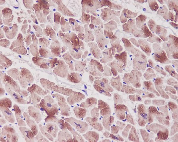
Anti-Cardiac Troponin T (TNNT2) Mouse Monoclonal Antibody, Immunohistochemical staining of paraffin-embedded Carcinoma of Human lung tissue using anti-TNNT2 mouse monoclonal antibody.
| Protein Name | Gene Name | Function |
|---|---|---|
| Troponin I | TNNI3 | Regulates cardiac muscle contraction by inhibiting actin-myosin interaction. |
| Troponin T | TNNT2 | Anchors the troponin complex to the muscle thin filament, playing a key role in muscle contraction. |
| Myosin Heavy Chain 7 | MYH7 | Essential for sarcomere structure and cardiac muscle contraction. |
| Connexin 43 | GJA1 | Forms gap junctions facilitating electrical coupling between cardiomyocytes. |
| SERCA2a | ATP2A2 | Pumps calcium into the sarcoplasmic reticulum, regulating cardiac muscle relaxation. |
| Phospholamban | PLN | Regulates SERCA2a activity, modulating calcium uptake in cardiomyocytes. |
| Natriuretic Peptide A | NPPA | Involved in blood pressure regulation and fluid balance. |
| Natriuretic Peptide B | NPPB | Plays a role in cardiovascular homeostasis and blood pressure regulation. |
| Alpha-Actinin-2 | ACTN2 | Crosslinks actin filaments in the sarcomere, maintaining muscle structure. |
| Titin | TTN | Contributes to the elasticity and structural integrity of the sarcomere. |
| Ryanodine Receptor 2 | RYR2 | Regulates calcium release from the sarcoplasmic reticulum during muscle contraction. |
| Calsequestrin 2 | CASQ2 | Binds calcium in the sarcoplasmic reticulum, aiding in calcium storage and release. |
| Desmin | DES | Maintains the structural integrity of cardiomyocytes by linking the sarcomere to the cell membrane. |
| Beta-Adrenergic Receptor 1 | ADRB1 | Mediates heart rate and contractility responses to sympathetic stimulation. |
| Lamin A/C | LMNA | Provides structural support to the nucleus and is involved in gene regulation. |
| Myosin Light Chain 2v | MYL2 | Plays a role in the regulation of cardiac muscle contraction. |
| Sodium-Calcium Exchanger 1 | SLC8A1 | Regulates intracellular calcium levels by exchanging sodium and calcium ions. |
| Cardiac Troponin C | TNNC1 | Binds calcium ions, initiating muscle contraction in cardiomyocytes. |
| Beta-Myosin Heavy Chain | MYH7 | Critical for force generation and contraction velocity in cardiac muscle. |
| Calcium/Calmodulin-Dependent Protein Kinase II Delta | CAMK2D | Regulates various aspects of cardiac function, including excitation-contraction coupling. |
Calcium handling and excitation-contraction coupling are fundamental processes within cardiomyocytes that underlie the heart’s ability to contract and pump blood effectively. This sub-research area focuses on the precise regulation and dynamics of calcium ions (Ca²⁺) within the cardiac muscle cells. During each heartbeat, an electrical signal, or action potential, propagates through the cardiomyocyte, triggering the release of Ca²⁺ from the sarcoplasmic reticulum into the cytoplasm. This sudden increase in intracellular calcium concentration binds to troponin, initiating the interaction between actin and myosin filaments, leading to muscle contraction. The subsequent reuptake of calcium into the sarcoplasmic reticulum by the SERCA pump and extrusion through the Na⁺/Ca²⁺ exchanger is critical for muscle relaxation. Dysregulation of calcium handling can lead to impaired cardiac function and is implicated in various cardiac pathologies, including heart failure and arrhythmias. Understanding the molecular mechanisms governing calcium dynamics offers potential therapeutic targets for enhancing cardiac performance and treating heart disease.
Mitochondrial function and energy metabolism are vital aspects of cardiomyocyte physiology, as the heart requires a constant and substantial supply of energy to sustain its continuous contractions. This research sub-area delves into the intricate processes by which mitochondria produce adenosine triphosphate (ATP) through oxidative phosphorylation, driven by the electron transport chain and the Krebs cycle. In cardiomyocytes, mitochondria are densely packed and strategically positioned to efficiently meet the high energy demands of the heart. Additionally, this area examines the role of mitochondrial dynamics, including fission and fusion, and mitophagy in maintaining mitochondrial quality and function. Impairments in mitochondrial energy metabolism are linked to various cardiac diseases, such as ischemia-reperfusion injury, cardiomyopathies, and heart failure. Moreover, metabolic flexibility, the ability of cardiomyocytes to switch between substrates like fatty acids and glucose, is critical for optimal heart function. Research in this domain seeks to elucidate the molecular pathways regulating mitochondrial efficiency and resilience, offering insights into novel interventions for enhancing cardiac energy metabolism and preventing metabolic dysfunction in heart disease.