Annexin V/Propidium Iodide (PI) staining is one of the most widely used methods for detecting and quantifying apoptosis. This guide explains the principles behind the technique, step-by-step protocols, common pitfalls, and best practices to ensure accurate and reproducible results.
Introduction to Apoptosis and Its Importance in Research
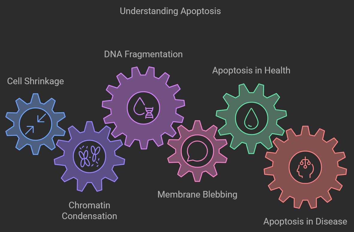
Understanding Apoptosis: Key Roles in Health and Disease
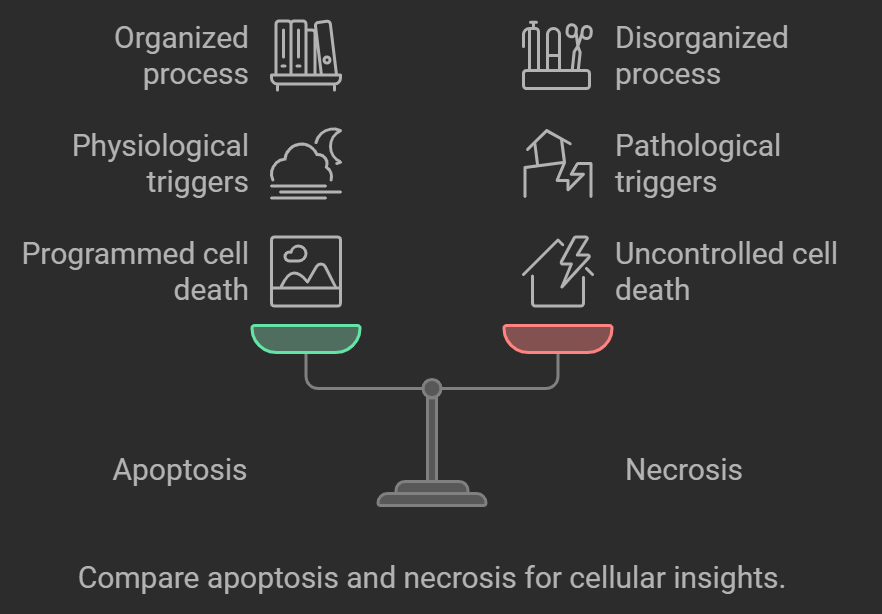
Apoptosis is a tightly regulated process of programmed cell death that is crucial for development, immune system function, and the elimination of damaged or diseased cells. It involves a cascade of biochemical events leading to characteristic cellular changes, such as cell shrinkage, chromatin condensation, DNA fragmentation, and membrane blebbing. Unlike necrosis, which is a form of traumatic cell death resulting from acute cellular injury, apoptosis is a controlled and energy-dependent process that does not elicit an inflammatory response.
In health, apoptosis ensures the removal of unwanted or potentially harmful cells, maintaining tissue homeostasis. In the immune system, for instance, apoptosis eliminates autoreactive lymphocytes, preventing autoimmune diseases. During development, it shapes organs and tissues by removing excess cells.
In disease contexts, dysregulation of apoptosis is implicated in a variety of conditions:
Cancer: Defective apoptosis allows cancer cells to survive beyond their normal lifespan, contributing to tumor growth and resistance to chemotherapy.
Neurodegenerative Diseases: Excessive apoptosis of neurons is a hallmark of diseases like Alzheimer's, Parkinson's, and Huntington's disease.
Autoimmune Disorders: Insufficient apoptosis of immune cells can lead to the survival of autoreactive cells, causing autoimmune reactions.
Infectious Diseases: Pathogens can manipulate apoptosis to evade the immune system or induce cell death.
Understanding apoptosis is thus critical for unraveling disease mechanisms and developing therapeutic interventions.
Why Accurate Detection of Apoptosis Matters in Cellular Studies
Accurate apoptosis detection is essential for disease mechanism studies, drug screening, and cell viability assessment. It allows researchers to distinguish between apoptotic, necrotic, and healthy cells, leading to more reliable interpretations in biomedical research.
Accurate detection of apoptosis is vital for several reasons:
Disease Pathway Elucidation: Identifying apoptotic cells helps in understanding disease progression and the underlying mechanisms of cell death.
Drug Development: Assessing apoptosis is essential for evaluating the efficacy and cytotoxicity of new drugs, particularly in cancer therapy.
Cell Viability Assessment: In cell culture and tissue engineering, monitoring apoptosis ensures the health and viability of cell populations.
Research Applications: Apoptosis studies contribute to fields like immunology, developmental biology, and neuroscience.
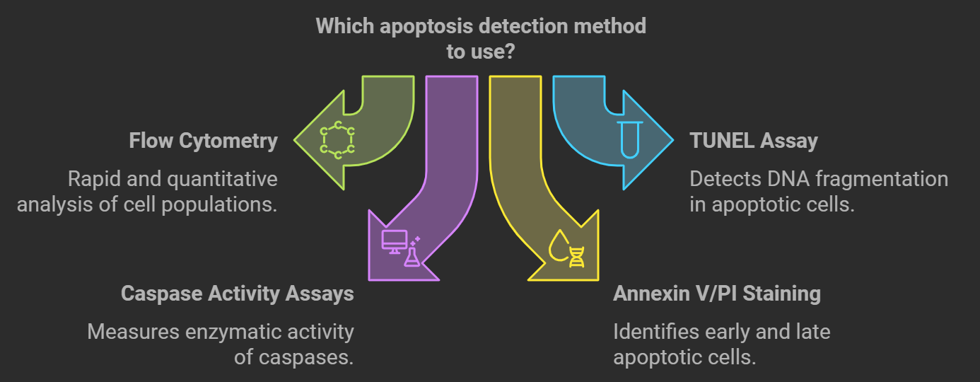
Overview of Apoptosis Detection Methods
Reliable apoptosis detection methods enable researchers to distinguish between apoptotic, necrotic, and healthy cells, providing insights into cellular responses under various experimental conditions.
Flow Cytometry for Apoptosis: A Reliable Detection Method
Flow cytometry is a powerful analytical technique that allows for the rapid quantification of physical and chemical characteristics of individual cells within a heterogeneous population. It offers several advantages for apoptosis detection:
Quantitative Analysis: Provides precise measurements of apoptotic cell percentages within a population.
High Throughput: Analyzes thousands of cells per second, enabling statistically robust data.
Multiparametric Capability: Simultaneously measures multiple markers, allowing for detailed characterization of cell states.
Sensitivity: Detects subtle changes in cell properties, aiding in the early detection of apoptosis.
By combining flow cytometry with specific apoptosis markers like Annexin V and PI, researchers can effectively distinguish between different stages of cell death.
Common Markers for Apoptosis Detection: Annexin V and PI Explained
Annexin V: Detecting Early Apoptosis
Annexin V is a calcium-dependent phospholipid-binding protein with a high affinity for phosphatidylserine (PS), a phospholipid normally localized to the inner leaflet of the plasma membrane. During early apoptosis, PS translocates to the outer leaflet, exposing it to the extracellular environment. Annexin V labeled with a fluorochrome can bind to externalized PS, serving as a marker for early apoptotic cells.
Propidium Iodide (PI): A Marker for Late Apoptosis and Necrosis
Propidium Iodide is a membrane-impermeable DNA-binding dye that intercalates into nucleic acids. In viable cells with intact membranes, PI cannot penetrate and, therefore, does not stain these cells. However, in late apoptotic or necrotic cells with compromised membrane integrity, PI enters the cell and binds to DNA, emitting fluorescence upon excitation.
By using Annexin V and PI simultaneously, researchers can categorize cells into:
Viable Cells: Annexin V negative / PI negative
Early Apoptotic Cells: Annexin V positive / PI negative
Late Apoptotic or Necrotic Cells: Annexin V positive / PI positive
Necrotic Cells: Annexin V negative / PI positive (though this is less common)
This dual-staining approach provides a comprehensive view of cell health within a sample.
How Annexin V/PI Staining Works in Apoptosis Detection
The Science Behind Annexin V and Phosphatidylserine Binding
In healthy cells, phosphatidylserine is confined to the cytoplasmic side of the plasma membrane due to the activity of translocases and flippases. Early in apoptosis, these enzymes become inactive while scramblases activate, leading to the externalization of PS. This externalization serves as an "eat-me" signal to phagocytes for the removal of dying cells.
Annexin V binds specifically to PS in the presence of calcium ions. By conjugating Annexin V to a fluorescent dye (e.g., FITC, PE), it becomes possible to detect PS exposure using flow cytometry. The intensity of fluorescence correlates with the amount of PS on the cell surface, indicating the progression of apoptosis.
Propidium Iodide (PI) as a Marker for Cell Viability and Necrosis
PI is excluded by live cells with intact plasma membranes. When the membrane integrity is lost, as in late apoptosis or necrosis, PI can penetrate the cell and bind to DNA. Upon excitation, PI-DNA complexes emit red fluorescence. By analyzing PI staining, researchers can identify cells that have lost membrane integrity, distinguishing between early apoptotic (PI negative) and late apoptotic or necrotic cells (PI positive).
Interpreting Results: Early Apoptosis vs. Late Apoptosis vs. Necrosis
When analyzing Annexin V/PI staining results in flow cytometry, cells are typically displayed in a two-dimensional dot plot with Annexin V fluorescence on one axis and PI fluorescence on the other:
Quadrant Q1 (Annexin V negative / PI positive): Necrotic cells that have lost membrane integrity but do not show PS externalization.
Quadrant Q2 (Annexin V positive / PI positive): Late apoptotic or secondary necrotic cells with PS externalization and compromised membranes.
Quadrant Q3 (Annexin V negative / PI negative): Viable, healthy cells with intact membranes and no PS externalization.
Quadrant Q4 (Annexin V positive / PI negative): Early apoptotic cells with PS externalization but intact membranes.
By interpreting the distribution of cells across these quadrants, researchers can quantify the proportions of cells in different stages of apoptosis and necrosis, providing valuable insights into cellular responses.
Step-by-Step Protocol: for Annexin V/PI Staining
Materials Needed for Annexin V/PI Staining
List all necessary reagents, buffers, controls, and equipment, including calcium-containing binding buffer, fluorochrome-conjugated Annexin V, PI stock solution, and control cell samples.
Cells: Cultured cells or cell suspension from tissues.
Annexin V Conjugate: Fluorescently labeled Annexin V (e.g., Annexin V-FITC, Annexin V-PE).
Propidium Iodide (PI): Stock solution, typically at 50 µg/mL.
Binding Buffer:Binding Buffer: Calcium-containing buffer (e.g., 10 mM HEPES, 140 mM NaCl, 2.5 mM CaCl₂, pH 7.4).
Flow Cytometer: Equipped with appropriate lasers and filters for the chosen fluorochromes.
Controls:
Unstained Cells: For setting flow cytometer baseline.
Single-Stained Controls: Cells stained with only Annexin V or PI for compensation.
Positive Control: Cells treated to induce apoptosis (e.g., with staurosporine).
Negative Control: Untreated healthy cells.
Optional:
Apoptosis Inducer: To create a positive control.
Centrifuge Tubes: 5 mL polystyrene round-bottom tubes.
Ice Bath: To keep cells cold during staining.
Step-by-Step Guide to Annexin V/PI Staining
Step 1: Cell Preparation
Induce Apoptosis (Optional Positive Control):
Treat cells with an apoptosis-inducing agent (e.g., staurosporine) and incubate for the desired time.
Harvest Cells:
Adherent Cells: Detach gently using non-enzymatic methods (e.g., EDTA) to preserve membrane integrity.
Suspension Cells: Collect directly.
Wash Cells:
Centrifuge at 300 x g for 5 minutes at room temperature.
Discard supernatant and resuspend cells in cold PBS.
Repeat wash to remove residual media and serum proteins.
Adjust Cell Concentration:
Resuspend cells in binding buffer at a concentration of 1 x 10⁶ cells/mL.
Step 2: Staining with Annexin V and PI
Aliquot Cells:
Transfer 100 µL of cell suspension (1 x 10⁵ cells) into flow cytometry tubes.
Add Annexin V Conjugate:
Add 5 µL of Annexin V-FITC (or appropriate volume per manufacturer's instructions).
Add Propidium Iodide:
Add 5 µL of PI solution (50 µg/mL stock).
Gently Mix:
Gently vortex or tap the tubes to mix the staining solution with the cells.
Step 3: Incubation and Sample Handling
Incubate:
Incubate the cells at room temperature for 15 minutes in the dark.
Step 4: Final Steps Before Flow Cytometry
Add Binding Buffer:
Add 400 µL of binding buffer to each tube.
Keep Samples on Ice:
To prevent further progression of apoptosis, keep samples on ice if there is a delay before analysis.
Proceed to Flow Cytometry:
Analyze samples promptly using a flow cytometer.
Tips to Avoid Common Errors
Use Fresh Reagents: Ensure that Annexin V and PI solutions are fresh and properly stored.
Avoid Cell Aggregation: Gently pipette to create a single-cell suspension.
Maintain Calcium Levels: Annexin V binding requires calcium; ensure that binding buffer contains calcium ions.
Minimize Time After Staining: Analyze samples within one hour to prevent changes in staining patterns.
Protect from Light: Fluorochromes are light-sensitive; keep samples protected to prevent photobleaching.
Setting Up Controls and Compensation for Flow Cytometry
Essential Controls for Accurate Apoptosis Detection
Single-Stained Controls: Required for setting compensation to correct spectral overlap between fluorochromes.
Fluorescence Minus One (FMO) Controls: Helps in setting gates by staining with all fluorochromes except one.
Positive Control: Cells induced to undergo apoptosis to validate the staining protocol.
Negative Control: Healthy cells to establish baseline staining.
Compensation Techniques to Minimize Fluorescence Spillover
Adjust the flow cytometer settings using single-stained controls to correct for fluorescence spillover between detection channels. Proper compensation ensures accurate separation between the different cell populations.
Troubleshooting Annexin V/PI Staining in Flow Cytometry
Common Issues and Solutions in Apoptosis Detection
Address problems like high background, weak signal, non-specific binding, and cell aggregation with targeted fixes such as reagent titration, calcium verification, and gentle handling.
High Background Fluorescence:
Possible Causes: Non-specific binding, over-concentration of fluorochromes.
Solutions: Titrate Annexin V and PI concentrations; include blocking steps if necessary.
Weak Staining Signal:
Possible Causes: Low expression of PS externalization, expired reagents.
FAQs on Annexin V/PI Staining for Apoptosis Detection
Yes, Annexin V/PI staining can help differentiate between early apoptosis, late apoptosis, and necrosis based on the staining patterns:
Early Apoptosis: Annexin V positive / PI negative.
Late Apoptosis/Necrosis: Annexin V positive / PI positive.
Necrosis: Typically Annexin V negative / PI positive, but interpretation requires caution as late apoptotic cells may also show this pattern.
Fluorochrome Compatibility: Choose an Annexin V conjugate with a fluorochrome compatible with your flow cytometer.
Signal Intensity: Select fluorochromes with high brightness for better sensitivity.
Supplier Reputation: Use validated products from reputable suppliers.
Application Data: Review product datasheets and user reviews for performance in similar applications.
Early Membrane Compromise: Cells in very early apoptosis may not have externalized PS yet.
Non-Apoptotic PS Exposure: Some non-apoptotic cells might expose PS under certain conditions.
Necrotic Cells: Late apoptotic and necrotic cells can both be Annexin V positive / PI positive, requiring additional markers to distinguish.
Temperature Sensitivity: PS externalization can be induced by cold temperatures; avoid processing cells on ice unless specified.
Conclusion: Enhancing Apoptosis Research with Accurate Flow Cytometry Analysis
Annexin V/PI staining combined with flow cytometry is a robust and reliable method for detecting and quantifying apoptosis in various cell types. By understanding the principles behind Annexin V and PI staining, carefully optimizing protocols, and adhering to best practices, researchers can obtain accurate and reproducible results. This, in turn, enhances the quality of apoptosis research, contributing to our understanding of cellular processes in health and disease. Accurate detection of apoptosis is indispensable for advancing therapeutic strategies, studying disease mechanisms, and exploring the intricacies of cell biology.
By mastering the techniques outlined in this guide, scientists and researchers can confidently employ Annexin V/PI staining in their work, leading to meaningful discoveries and innovations in biomedical research.