This website uses cookies to ensure you get the best experience on our website.


- Table of Contents
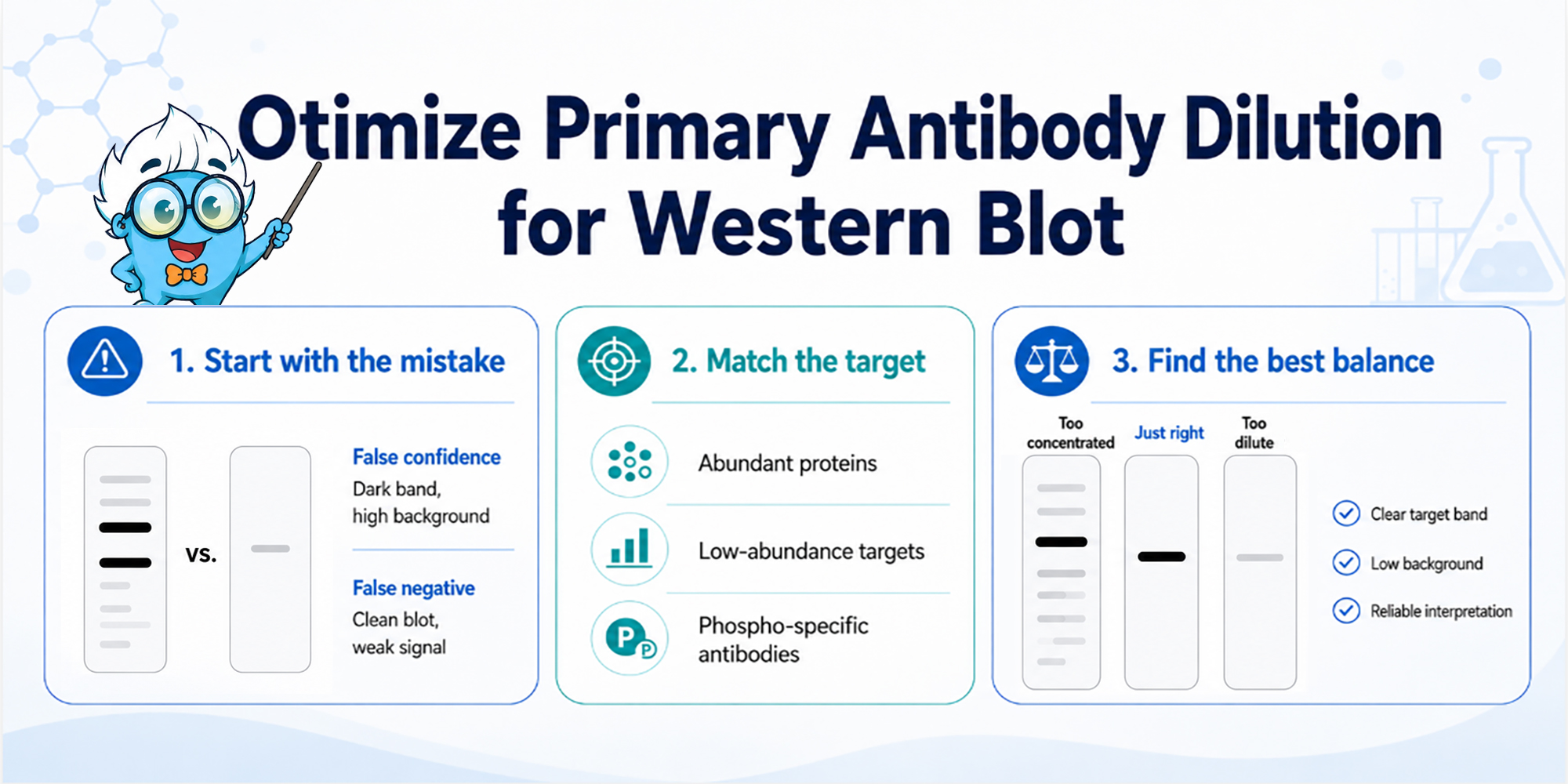
More primary antibody is not a shortcut to a better Western blot. A stronger band only helps if the rest of the membrane stays clean enough to trust.
To optimize primary antibody dilution for Western blot, start from the datasheet range and test a small dilution series. Choose the condition with the best signal-to-background balance, not simply the darkest band. Too much primary antibody can raise haze, extra bands, and saturation; too little can make low-abundance targets look weak or absent.
Primary antibody dilution controls more than target intensity. It changes the balance between specific binding and weak off-target binding that should ideally wash away. At high concentration, low-affinity interactions are more likely to survive washing and then become visible after secondary antibody amplification. At very low concentration, true target binding can fall close to the detection limit, where small differences in transfer, exposure, or handling start to look like biology.
That is why primary dilution should be optimized as an assay window, not as a single "strong enough" condition. For broader context, Boster's Western Blotting Antibody Concentration Optimization and Western Blotting Troubleshooting Guide are useful next references.
To find that usable assay window, it helps to first define which type of error the experiment is most likely to create.
Primary antibody dilution creates two different risks. One is a false negative: the antibody is too dilute, the target is low abundance, and a real band looks weak or absent. This is easy to miss with phospho-specific targets, cleaved proteins, transcription factors, membrane proteins, or samples where expression is condition-dependent.
The other risk is false confidence. The antibody is too concentrated, the expected band becomes darker, but the blot also gains haze, extra bands, or fast saturation. The image looks stronger, but the interpretation becomes weaker.
A useful first question is simple: what failure would be worse for this experiment—missing a real target, or trusting a dirty signal?
A concentrated primary antibody often improves the target band first.
The effect becomes even more pronounced after incubation with the secondary antibody, which amplifies both specific and nonspecific binding events. In a Western blot, apparent sensitivity should therefore be evaluated together with overall membrane cleanliness rather than band intensity alone.
This can make a concentrated primary antibody look helpful at first. The problem is that Western blot detection does not amplify only the binding you want.
If the primary antibody binds weakly to off-target proteins, those interactions may be hard to see at a moderate dilution. At a higher concentration, more of that weak binding can remain on the membrane after washing. The secondary antibody then amplifies it. By the time ECL is added, the blot may show more signal everywhere, not just at the expected molecular weight.
This is where background can be misread as "better sensitivity." If both the target band and surrounding noise rise together, the assay is losing separation between true signal and background. For detection-side issues, Boster's ECL Western blot substrate guide may help separate antibody dilution from detection chemistry.
The opposite mistake is choosing the cleanest condition without proving that the target can still be detected. A very dilute primary antibody can make the membrane look beautiful. It can also push the target below the detection threshold.
A clean negative is only meaningful when the positive control works. If the positive control is faint, if the target only appears after a long exposure, or if the same sample gives unstable results across repeats, the dilution may be too low.
Do not interpret absence before checking the basics: protein loading, transfer, antibody validation for the sample species, and whether the model actually expresses the target. If transfer is uncertain, check Boster's guide on transfer quality in Western blot and right antibody for Western blot guide are useful next references.
This issue is particularly relevant when studying low-level protein expression. A dilution that appears acceptable for abundant proteins may fail to detect subtle changes in protein targets expressed near the detection limit.
Once these two risks are clear, the next step is to design a dilution series that separates the antibody variable from the rest of the workflow.
A useful titration should change one main variable: the primary antibody dilution. For reliable Western blot analysis, keep the rest of the workflow as consistent as possible, including sample preparation, electrophoresis, transfer, blocking, secondary antibody incubation, detection chemistry, and exposure settings. Ideally, compare dilutions using samples prepared from the same batch of cell lysates, with the same protein load and the same electrophoresis buffer system, such as SDS Sample Buffer and Tris-Glycine SDS Running Buffer.
Start with the datasheet recommendation when available.
If a tested condition such as 1:1000 does not give a clean, interpretable result, use the blot pattern to guide the next step: test more dilute conditions when background, extra bands, or saturation are the main problem, and test stronger conditions when the target is weak but the membrane is clean. If the pattern is unclear, test a small range on both sides of the starting point to define the usable signal-to-background window.
The goal is not to find the darkest possible band in one attempt. The goal is to create a small comparison range that shows how the target signal and background change as the primary antibody becomes more or less concentrated.
Use smaller dilution steps when the result is close but not ideal. For example, if 1:1000 gives a clear target band but slightly elevated background, testing nearby conditions such as 1:1500 or 1:2000 may help refine the balance.
Use wider steps when you have almost no signal or overwhelming background. For abundant targets, include more dilute conditions early. For low-abundance targets, include one stronger condition, but do not assume concentration alone will solve the problem.
Keep the rest of the system fixed: same lysate, same protein load, same gel and transfer condition, same blocker, same incubation time, same secondary antibody, same ECL substrate, and the same exposure series. If those variables move together, the titration stops telling you what the primary antibody is doing.
Consistency is particularly important in Western blotting because changes in reagents can affect apparent antibody performance. When comparing dilutions, use the same blocking buffer, identical incubation conditions, and the same primary antibody dilution buffer throughout the experiment. Researchers commonly prepare antibody solutions in Tris Buffered Saline or Phosphate Buffered Saline supplemented with protein stabilizers such as Bovine serum albumin. Maintaining consistent reagent composition ensures that observed differences arise from the antibody dilution rather than changes in assay conditions.
The membrane type should also remain unchanged during optimization. Both nitrocellulose membrane and PVDF membrane formats are widely used in Western blot workflows, but they differ in protein-binding characteristics and background behavior. Switching between a nitrocellulose membrane and a PVDF membrane during optimization can complicate interpretation because signal intensity may change independently of antibody concentration.
The best primary antibody dilution gives you room to interpret the blot. A good condition should show the expected band at a short or moderate exposure, keep background low, limit unexplained bands, and avoid early saturation. The target should be comfortably above the detection limit but still within the linear response range.
Both ends of the window are risky. Near the detection limit, small handling differences can look like biological change. Near saturation, real differences are compressed or lost. For comparison work, Boster's Western blot quantification guide and total protein normalization guide are useful next references.
This principle is especially important for quantitative western blots, where accurate comparison depends on maintaining signal intensity within the linear range of detection. A suitable loading control should be included to verify that observed differences reflect biology rather than variation in sample handling.
Different targets need different dilution strategies. In Western blotting experiments that compare subtle changes in protein expression, repeated runs and a stable loading control may be needed to confirm that band intensity differences reflect biology rather than unequal sample loading. The same primary antibody dilution that works well for an abundant total protein may be too weak for a low-abundance target, or too nonspecific for a phospho-specific, cleavage-specific, or isoform-specific antibody.
Abundant total proteins usually tolerate a more dilute primary antibody. If several dilutions show the expected band, choose the cleaner condition.
Low-abundance proteins may need a stronger primary condition, but only if signal improves faster than background. If both rise together, concentration is not giving you a better assay window. Better extraction, improved transfer, increased sample load, or more sensitive detection may be the real fix.
Phospho-specific, cleavage-specific, and isoform-specific targets need a different level of caution. For these targets, band position and biological controls matter as much as intensity. Treatment controls, inhibitor controls, or positive and negative lysates can be more useful than chasing a darker band.
Primary dilution is worth optimizing when the blot changes meaningfully across the series. If one dilution gives a cleaner membrane while preserving the expected band, you have useful information. If one stronger condition rescues a weak target without lifting background too much, that also helps.
If the same problem appears across every dilution, the dilution itself is probably not the limiting variable.
At this stage, it is often useful to review the entire Western blot workflow, including membrane transfer efficiency, wash conditions, and secondary antibody concentration. Problems introduced during transfer may persist regardless of the primary antibody dilution selected.
If all dilutions are dirty, look at blocking, washing, secondary antibody concentration, ECL exposure, membrane drying, or sample overload. If all dilutions are weak, check sample integrity, target expression, protein loading, positive control selection, and membrane transfer conditions, including transfer duration, current settings, and preparation of Tris-Glycine Transfer Buffer. Inadequate transfer can reduce detectable protein expression even when the primary antibody dilution is appropriate.
A practical rule: adjust primary dilution when it changes the signal-to-background relationship. Move elsewhere in the workflow when it only changes how bad the same problem looks. For blocking-related background, Boster's Western blot blocking optimization guide may help separate antibody dilution issues from blocking, washing, and membrane-handling conditions.
| What you see | What it suggests | What to test next |
|---|---|---|
| Weak target, clean membrane | Primary may be too dilute, or target is low abundance | Test a stronger primary condition |
| Strong target, haze also increases | Primary may be outside the useful range | Test a more dilute primary |
| Extra bands increase with concentration | Low-affinity off-target binding is being amplified | Dilute primary; check specificity |
| Target only appears after long exposure | Signal is near the detection limit | Titrate primary and exposure range |
| Target saturates quickly | Signal is outside the linear range | Dilute primary or shorten exposure |
| All dilutions are dirty | Blocking, washing, secondary, ECL, or sample overload issue | Check non-primary variables |
| All dilutions are weak | Transfer, sample, positive control, or detection issue | Check upstream workflow |
Although this article focuses on antibody titration, successful Western blotting depends on multiple interconnected variables. Membrane selection, blocking buffer composition, sample preparation, transfer efficiency, and secondary antibody performance all contribute to final signal quality. Researchers working with challenging protein targets may benefit from evaluating each variable independently before concluding that antibody dilution is the primary cause of poor results.
What primary antibody dilution should I use for Western blot?
Start with the datasheet range, then test a small dilution series around it. The best dilution is the one with the strongest interpretable signal, not simply the darkest band.
How do I know if my primary antibody is too concentrated?
The target may become darker, but haze, extra bands, or saturation also increase. If signal and background rise together, the antibody is probably too concentrated for that workflow.
Can too much primary antibody cause high background?
Yes. High primary antibody concentration can allow weak off-target binding to survive washing. The secondary antibody and ECL detection then amplify that background.
Should I adjust primary antibody dilution for quantification?
Yes. For densitometry, the target signal should stay within the linear response range. A saturated band is not reliable for comparing expression between samples.
Does membrane type affect primary antibody optimization?
Yes. Nitrocellulose membranes and PVDF-based membranes can produce different signal and background characteristics. For consistent Western blot analysis, membrane type should remain fixed throughout optimization experiments.
Can reagent selection influence antibody dilution results?
Yes. The composition of the primary antibody dilution buffer, Tris Buffered Saline, Tris Buffered Saline with Tween 20, and the chosen blocking buffer can affect antibody binding, background levels, and overall assay performance in a Western blot.
Primary antibody dilution should be optimized for interpretation, not intensity. A useful dilution gives a clear expected band, controlled background, limited extra bands, and enough exposure range to compare samples honestly.
Start from the datasheet range, test a small series, and keep the rest of the workflow stable. If the dilution series changes the signal-to-background balance, use that information to choose the working condition. If every dilution fails in the same way, the real problem is probably not the primary antibody concentration.
Optimizing dilution is one component of a broader Western blotting strategy. Consistent handling of cell lysates, appropriate loading control selection, effective blocking buffer conditions, reliable secondary antibody performance, and suitable detection chemistry are equally important for generating reproducible data. Careful optimization is especially valuable when measuring subtle differences in protein expression across experimental groups.
Unlike techniques such as immunohistochemical analysis, which often require antigen retrieval before target detection, Western blot workflows rely on protein extraction and electrophoretic separation to evaluate target abundance and specificity.
For related workflow support, explore Boster's Western blot reagents.







