This website uses cookies to ensure you get the best experience on our website.


- Table of Contents
Plan chromogenic AGPAT4 IHC in paraffin sections around the cytoplasmic tissue pattern and ER membrane annotation (HPA tissue IHC; UniProt). Use high-staining bronchial respiratory epithelium and unstained adipocytes as tissue controls, and assess skeletal muscle cautiously because the two sources disagree (HPA tissue IHC; UniProt).

Expected localisation, validated protocols, controls and antibodies — the at-a-glance facts below, then the full design guide.
| Expected localisation | Cytoplasmic IHC; ER membrane protein (HPA tissue IHC; UniProt) | |
| Staining pattern | Cytoplasmic staining in most tissues (HPA tissue IHC) | |
| Antigen retrieval | Tris-EDTA pH 9.0 HIER, heat-mediated (datasheet A11729) | |
| Positive control | Bronchus+4 more · see all | |
| Negative control | Adipose tissue+4 more · see all |
| Fixation | Keep fixation consistent across paraffin sections (standard IHC practice; not target-specific); Matched tissue-IHC evidence does not establish the fixation claim. Validate the specimen-specific method before use. (selected-SKU IHC image A11729) | |
| Caveat | Skeletal myocytes lack IHC staining despite reported high expression (HPA tissue IHC; UniProt) | |
| Regulation | Staining-related regulation is unreported (UniProt) | |
| Isoform / epitope | Two isoforms; map the antibody epitope across both (UniProt) |
The catalog antibody’s IHC-P protocol (datasheet A11729) is accompanied by two published AGPAT4 IHC protocols (PMC11485110; PMC7099097).
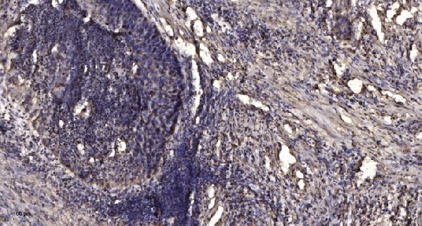
| Sample | Paraffin-embedded human Squamous cell carcinoma of lung tissue; fixative not specified (datasheet A11729) |
| Fixation | Image fixative and duration unreported (datasheet A11729); verify before use. |
| Sectioning | 4–5 µm sections on charged slides (standard) |
| Deparaffinisation | Xylene, graded ethanol series to water (standard) |
| Antigen retrieval | Heat retrieval: Tris-EDTA pH 9.0 (datasheet A11729); 20 min, 95–100 °C (standard) |
| Peroxidase block | 3% H2O2, 10 min, room temperature (standard) |
| Blocking | 10% normal serum of the secondary host, 30 min, room temperature (standard) |
| Primary antibody | Rabbit anti-AGPAT4, 1:50 recommended; image 1:200 (datasheet A11729) |
| Primary incubation | Overnight at 4 °C (standard) |
| Detection | HRP-polymer secondary, DAB chromogen 5–10 min (standard) |
| Counterstain | Hematoxylin, blue, dehydrate and mount (standard) |
| Expected result | AGPAT4-positive staining in respiratory epithelial cells of bronchus (HPA tissue IHC: High). HPA tissue profile: Cytoplasmic expression in most tissues. No signal in the no-primary control. |
AGPAT4 is an endoplasmic reticulum membrane protein with 4 transmembrane segments (UniProt Q9NRZ5 topology). In paraffin IHC, expect predominantly cytoplasmic staining in respiratory epithelium, neurons, glandular cells, cardiomyocytes, and kidney tubules (HPA: tissue IHC). HPA rates the tissue pattern Approved, with medium consistency against RNA data and external verification pending (HPA: tissue IHC reliability).
| Cytoplasmic staining in the expected cells, with a clear difference from adjacent unstained areas. | This fits HPA’s broad cytoplasmic IHC profile. Respiratory epithelial cells, neurons, colon and fallopian tube glandular cells, cardiomyocytes, and kidney tubular cells are listed as High; compare the relevant cell population within each section (HPA: tissue IHC). |
| Predominantly nuclear or extracellular staining, with little cytoplasmic signal. | Reassess staining specificity and section quality: that distribution does not match the reported IHC profile or membrane assignment (HPA: cytoplasmic expression in most tissues; UniProt Q9NRZ5: endoplasmic reticulum membrane). Nucleolar localisation has been reported by ICC-IF, so compartment judgments should remain application specific (HPA: subcellular ICC-IF). |
| Strong staining in cells listed as Not detected, such as adipocytes or skeletal myocytes. | Treat this as an unexpected IHC result and check controls for cross-reactivity or endogenous detection activity (HPA: adipocytes and skeletal myocytes Not detected; general IHC practice). UniProt reports high expression in skeletal muscle, so the myocyte discrepancy warrants investigation rather than an automatic artefact call (UniProt Q9NRZ5: tissue specificity). |
| Color extends diffusely across tissue and cell-free areas, obscuring cell boundaries. | The slide cannot establish a cell-specific AGPAT4 pattern when background obscures localisation (general IHC practice). Reassess blocking, washes, detection controls, and counterstain before scoring; HPA’s expected readout is cytoplasmic expression in most tissues (HPA: tissue IHC profile; general IHC practice). |
| No staining in an expected positive cell population. | A blank result in bronchial respiratory epithelium, heart cardiomyocytes, or kidney tubules conflicts with their High HPA IHC entries (HPA: tissue IHC). Check that the expected cells are present and review antibody, retrieval, and detection controls before interpreting the specimen as negative (general IHC practice). |
| Membrane topology and compartment | AGPAT4 has 4 transmembrane segments and an endoplasmic reticulum membrane assignment (UniProt Q9NRZ5 topology and subcellular location). HPA describes the paraffin IHC pattern as cytoplasmic; these sources do not define a paraffin IHC epitope or predict antigen-retrieval conditions (HPA: tissue IHC profile; UniProt Q9NRZ5). |
| Tissue evidence and its limit | HPA lists High staining in several epithelial, neuronal, cardiac, and renal cell populations, but rates antibody–RNA consistency as medium and notes external verification is pending (HPA: tissue IHC and reliability). Use its cell-level observations as reference patterns, not a guarantee for every specimen. |
| Skeletal muscle discrepancy | Skeletal myocytes are Not detected by HPA tissue IHC, while UniProt describes AGPAT4 expression as highest in skeletal muscle (HPA: skeletal muscle IHC; UniProt Q9NRZ5: tissue specificity). Keep assay type and sampled cell population explicit when evaluating this disagreement. |
| Antibody validation | HPA053287 is Approved for IHC and ICC; HPA060525 is Approved for ICC, with no IHC status supplied (HPA: antibody validation). The tissue IHC profile is Approved, with medium antibody–RNA consistency and external verification pending (HPA: tissue IHC reliability). |
| IF/ICC Q: Should punctate Golgi staining match paraffin IHC? | A: HPA reports mainly Golgi localisation, with additional nucleolar and vesicular localisation, in ICC-IF; its tissue IHC profile is broadly cytoplasmic (HPA: subcellular ICC-IF; HPA: tissue IHC). Assess each application against its own reported pattern. |
| Situation | Likely cause | Next action |
|---|---|---|
| Expected positive cells remain blank. | The assay may have failed, or the expected cell population may be absent from the section (general IHC practice). HPA lists bronchial respiratory epithelial cells, cardiomyocytes, and kidney tubular cells as High (HPA: tissue IHC). | Confirm the relevant cells on the section, then review a positive control and the staining run’s retrieval, primary-antibody, and detection steps (general IHC practice). Do not score an unverified blank section as an AGPAT4 negative. |
| Signal appears mainly nuclear or outside cells. | That distribution conflicts with broad cytoplasmic tissue IHC staining and the endoplasmic reticulum membrane assignment (HPA: tissue IHC profile; UniProt Q9NRZ5: subcellular location). | Inspect morphology and a detection control, and reassess the primary-antibody staining pattern (general IHC practice). Keep HPA’s additional nucleolar ICC-IF localisation specific to that application (HPA: subcellular ICC-IF). |
| Unexpected cells stain strongly. | Cross-reactivity or endogenous detection activity is possible (general IHC practice). This matters especially when adipocytes or skeletal myocytes stain despite HPA listing them as Not detected (HPA: tissue IHC). | Compare with appropriate control sections and the expected positive cell population (general IHC practice). For skeletal muscle, record the conflict with UniProt’s high tissue-expression statement before drawing a conclusion (UniProt Q9NRZ5: tissue specificity). |
| Diffuse background masks cell boundaries. | Nonspecific binding or detection background can prevent reliable localisation assessment (general IHC practice); HPA’s reported tissue pattern is cytoplasmic (HPA: tissue IHC profile). | Review blocking, wash conditions, detection controls, and counterstain so that cellular signal can be distinguished from background (general IHC practice). Reassess localisation only after cell boundaries are readable. |
| Signal is weak in a cell population expected to be High. | The observed result differs from that HPA tissue IHC entry, but HPA reports only medium antibody–RNA consistency and pending external verification (HPA: tissue IHC and reliability). | Check cell identity and compare an independently processed positive section; then review retrieval and detection performance (general IHC practice). Report the observed intensity without assuming the HPA level applies to every specimen. |
| ICC-IF shows Golgi puncta while paraffin IHC looks broadly cytoplasmic. | The two HPA datasets describe different application-level appearances: mainly Golgi in ICC-IF and cytoplasmic expression in most tissue IHC sections (HPA: subcellular ICC-IF; HPA: tissue IHC). | Interpret each image against its matching HPA application record. Do not require paraffin IHC to resolve the Golgi, nucleolar, or vesicular detail reported for ICC-IF (HPA: subcellular ICC-IF; HPA: tissue IHC). |


Comprehensive Human Protein Atlas IHC scoring per tissue (reliability: Approved — Medium consistency between antibody staining and RNA expression data. Pending external verification.). Rows are taken directly from the HPA tissue chart — click any row's HPA link to view the source.
| Tissue | Cell type | Level | Evidence | Source |
|---|---|---|---|---|
| Bronchus | Respiratory epithelial cells | High | Protein (IHC) | HPA → |
| Caudate | Neuronal cells | High | Protein (IHC) | HPA → |
| Cerebral cortex | Neuronal cells | High | Protein (IHC) | HPA → |
| Colon | Glandular cells | High | Protein (IHC) | HPA → |
| Fallopian tube | Glandular cells | High | Protein (IHC) | HPA → |
| Tissue | Cell type | Level | Evidence | Source |
|---|---|---|---|---|
| Adipose tissue | Adipocytes | Not detected | Protein (IHC) | HPA → |
| Skeletal muscle | Myocytes | Not detected | Protein (IHC) | HPA → |
| Smooth muscle | Smooth muscle cells | Not detected | Protein (IHC) | HPA → |
| Soft tissue | Fibroblasts | Not detected | Protein (IHC) | HPA → |
| Spleen | Cells in red pulp | Not detected | Protein (IHC) | HPA → |
Troubleshoot AGPAT4 staining by checking retrieval, cell type, compartment and controls before comparing staining intensity across paraffin sections.
A11729 has a human paraffin-section IHC image (IHC caption). Its listed reactivity includes human, mouse and rat (catalog); no IF image is supplied (catalog).
A11729 will render with IHC data from a paraffin-embedded human lung squamous cell carcinoma section (IHC caption). Its listed applications are IHC and WB, and its listed reactivity is human, mouse and rat (catalog).
Which to pick: Choose A11729 for paraffin-section tissue IHC; its image caption reports Tris-EDTA retrieval at pH 9.0 and an overnight primary incubation at 1:200 (IHC caption). The caption does not report the fixative, and the catalog does not report a clone (IHC caption; catalog). For IF/ICC, A11729 has no listed application or image; for mouse or rat IHC, its reactivity is listed, but the supplied IHC image shows human tissue only (catalog; IHC caption).