This website uses cookies to ensure you get the best experience on our website.


- Table of Contents
IN VITRO FUNCTIONAL TESTING
Custom cellular testing for potency, viability and compound profiling.
Explore Our ServicesBoster runs cell-based assays as part of its assay services, through Boster and its partner labs. The core menu uses standard cell lines and primary cells.
| Study Type | What It Measures | What It Helps Decide |
|---|---|---|
| Bioactivity / potency | Functional dose-response of a biologic or small molecule | Activity relative to a reference standard |
| Proliferation & anti-proliferation | Growth promotion or inhibition in responsive cell lines | Cytokine or growth-factor activity |
| Cytotoxicity & viability | Loss of membrane integrity or metabolic activity | Early safety flags, hit triage |
| Apoptosis | Markers of programmed cell death | Whether apoptosis-associated changes contribute to the observed cell-death response |
| Signaling / reporter | Pathway activation via luciferase, GFP, or NFAT/CRE reporters | Pathway modulation and support for mechanism-of-action studies |
| Cytokine secretion & immune function | Cytokine release, immune cell activation or suppression | Immunomodulatory activity |
Antiviral cytopathic-effect assays, stem-cell-derived models and organoids are available on request, with feasibility confirmed at quote. Projects outside these formats go through custom development.
Cell-based work varies by nature, so each project starts with a written plan that fixes the design before any plates are run.
Deliverables are agreed before testing and may include:
EC50/IC50 estimates include confidence intervals and fit statistics where estimable. Results that do not support a reliable estimate are identified in the report.
A cell-based assay measures a biological response in intact, living cells rather than in an isolated protein or receptor.
Biochemical assays measure target binding or biochemical activity, such as enzyme inhibition, in a defined system. Cell-based assays complement these measurements by assessing responses within a cellular context, where membrane permeability, signaling, metabolism and gene expression can influence the result. A compound can bind a receptor tightly and still fail to trigger the downstream biology a program needs.
Each assay reads one or more functional endpoints: cell number or viability, reporter or pathway activation, cytokine secretion, migration or differentiation. Results are usually reported as a dose-response relationship rather than a single yes/no answer.
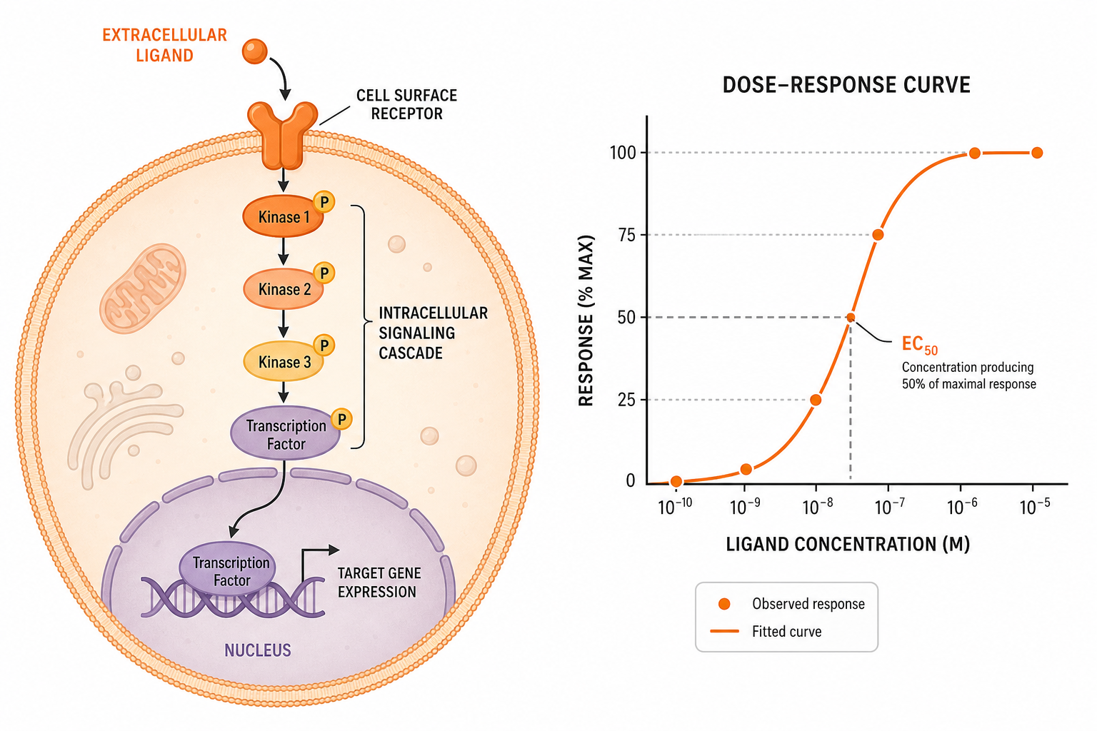
Why it matters: a functional cell-based result adds functional context that binding data lack, which helps decide which compounds and targets are worth taking further.
The cell model sets how closely a result reflects tissue biology, and how much the study costs.
| Model | Biological Relevance | Throughput | Typical Variability |
|---|---|---|---|
| Immortalized cell lines | Moderate; genetically stable but drifted from native tissue | High | Low |
| Donor-derived primary cells | High; keeps native receptor and signaling profile | Moderate | Moderate to high, donor dependent |
| Stem-cell-derived models (on request) | High; reaches cell types that are hard to isolate | Low to moderate | Moderate, batch dependent |
| Organoids (on request) | Highest; multicellular, tissue-like structure | Low | High |
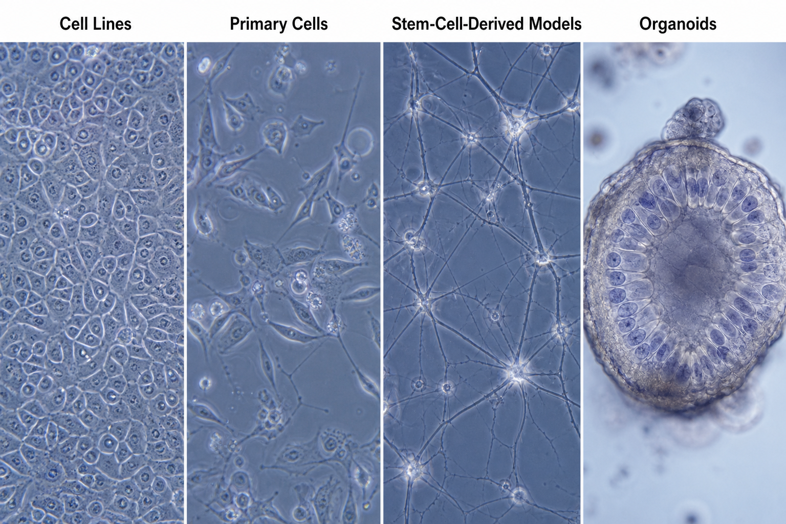
Immortalized cell lines are cheap, consistent and easy to scale, but genetic drift over many passages can shift receptor density and signaling. Donor-derived primary cells keep a more native profile, at the cost of donor-to-donor variability, which donor selection and pooling help control.
Stem-cell-derived models and organoids give access to cell types such as cardiomyocytes, neurons and hepatocytes, or to multicellular tissue structure. They bring lower throughput, higher cost and longer development, so the right model is the one that fits the biological question, not the most complex one.
When a target or pathway has no established assay, Boster builds one in five stages.
In a cell-based assay, functional potency describes the amount or concentration of a test article needed to produce a defined cellular response under specified assay conditions. It provides information distinct from binding affinity.
A potency assay measures a functional endpoint, such as proliferation, reporter activation or cytokine release, across a concentration range. Depending on the assay and study objective, curve fitting may provide an EC50, an IC50, or a relative potency estimate against a suitable reference standard. EC50 and IC50 values depend on the assay conditions and the response definition used.
Relative potency compares the amount or concentration of a test article needed to produce a defined response with that of a reference standard. Reporting requires suitable assay performance and assessment of dose–response comparability, including parallelism where appropriate.
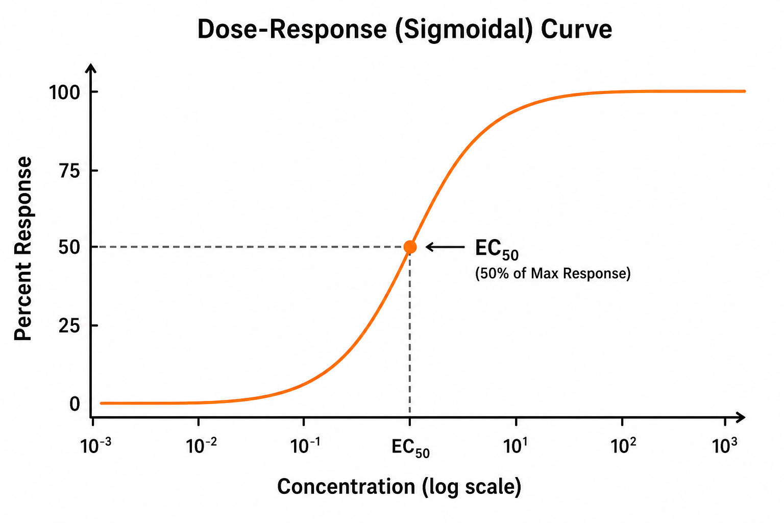
Binding assays characterize molecular interactions. Cell-based potency assays measure the resulting functional response in the selected cellular system.
Cytotoxicity testing shows early whether a compound harms cells at concentrations close to those needed for activity.
Cell viability describes the proportion or relative abundance of living cells, assessed through indicators such as membrane integrity or metabolic activity. Viability is a prerequisite for cell division, while proliferation assays measure whether and how much cells divide. Depending on the method, results may be reported as the percentage of viable cells or as a signal relative to an untreated or vehicle control:
A parallel viability readout helps determine whether a drop in a functional signal reflects biological modulation, reduced viable cell numbers, or both. Without this assessment, proliferation and signaling results can be misinterpreted.
Reduced cell growth over time relative to untreated controls.
Apoptosis-associated changes assessed alongside membrane integrity and other relevant endpoints to help characterize cell death.
Activity against unrelated cell types or pathways outside the intended mechanism.
Finding a cytotoxicity problem at the hit stage, in cells, costs far less than finding it later in an animal study.
Immune-cell assays expose a responsive cell population to a test article and measure a defined functional outcome.

The endpoint is usually cytokine secretion, proliferation or activation-marker expression in T cells, NK cells or monocytes. Secreted cytokines can be measured as a panel through Boster's multiplex assay services. These assays help assess immunosuppressive or immunostimulatory activity.
Antiviral cytopathic effect (CPE) inhibition assays are available on request. Cells are challenged with a virus that damages them, and the readout is whether the test article, such as an interferon or antiviral candidate, protects them, typically reported as the concentration that protects 50% of cells.
A note on scope: these assays measure defined endpoints in specific cell/stimulus or cell/virus pairings. Read results within the context of the model used, not as evidence of broader antiviral or immunomodulatory effect.
The assay format and cell model change with the question at each stage of a discovery program.
| Pipeline Stage | Typical Assay Use |
|---|---|
| Hit confirmation | Confirmatory and orthogonal assays to remove false positives and rank hits |
| Lead optimization | Dose-response potency across chemical analogs |
| Mechanism of action | Pathway reporter or signaling assays |
| Target validation | Assess whether target modulation produces the expected cellular response |
| Safety profiling | Cytotoxicity, viability and off-target screening |
Primary screening of compound libraries runs through Boster's compound screening services; cell-based assays then confirm and rank the hits.
The readout sets an assay's sensitivity, throughput and how much detail one run captures.
| Platform | Best Suited For | Trade-off |
|---|---|---|
| Plate reader (absorbance/colorimetric) | Simple viability or proliferation readouts at scale | Lower sensitivity, population average only |
| Luminescence | ATP-based viability, reporter-gene assays | High sensitivity; usually one endpoint per well |
| Fluorescence | Live/dead staining, pathway reporters, kinetic assays | Susceptible to compound autofluorescence |
| Flow cytometry | Multi-parameter, single-cell immune or viability profiling | Lower throughput per sample; more complex data |
| High-content imaging | Morphological and spatial endpoints, multiplexed phenotypes | Larger data volumes; longer analysis |
To quantify protein levels or phosphorylation directly in fixed cells on the plate, see Boster's In-Cell Western service. Multiplexing, such as reading viability alongside a functional signal from the same well, saves cells and catches cytotoxicity that could mask a real signaling result.
Automated liquid handling and 384-well formats help when a study needs more compounds, conditions or replicates than manual pipetting handles reliably. Smaller volumes are more sensitive to evaporation, edge effects and pipetting error, so an assay moved to a smaller format needs extra optimization first.
Automated and miniaturized formats are available on request. For larger screening campaigns, explore our high-throughput screening (HTS) services. Small or exploratory panels are often faster to set up and troubleshoot by hand.

A cell-based result is only as credible as the controls and analysis behind it. Boster builds these into each study plan.
Positive, negative and vehicle controls alongside replicates per condition, to separate real signal from assay noise.
Assay-specific criteria agreed before testing, such as control performance, replicate variability, signal-to-background ratio, or Z′-factor for suitable screening formats.
Consistency checked across independent runs and cell lots, not only within one experiment.
Nonlinear regression for suitable concentration–response datasets, with confidence intervals and fit statistics where estimable. Responses that do not support a reliable EC50 or IC50 estimate are identified in the report.
Dose-response curves and summary tables you can read without reprocessing raw data.
Methods, raw data and any deviations documented, so results can be reviewed or reproduced.
What good reporting looks like: your scientists can trace every EC50 or IC50 value back to the raw dose-response data, with the acceptance criteria the run was judged against stated plainly.
Yes. We can assess projects using customer-provided cell lines, assay protocols or SOPs. Depending on the method and receiving laboratory, the project may involve direct testing, assay transfer or additional optimization.
Routine testing uses an established assay with defined conditions and acceptance criteria. Custom development may include model selection, feasibility testing, optimization and assay qualification; your quote distinguishes development work from sample testing.
Tell us about your cells, compounds and the endpoint you need, and we will reply with a study plan and quote.
Email services@bosterbio.com or call (888) 466-3604.
GLP and other quality requirements are confirmed for each project before quotation.