ALMS1 / Centrosome-associated protein ALMS1 · IHC design guide

Design Immunohistochemistry for ALMS1

In paraffin-section IHC, ALMS1 shows cytoplasmic staining in a subset of testis cells (HPA tissue IHC). This guide gives the catalog antibody’s 1:100–1:300 dilution range (datasheet A03225) and highlights presumed off-target staining (HPA tissue IHC).

Evidence assembled Oct 2026 · For research use; verify linked source records and product datasheet before use
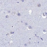
Immunohistochemistry protocol sheet for ALMS1 (IHC for ALMS1): expected localisation Cytoplasmic staining in a subset of testis cells (HPA tissue IHC), antibody A03225, validated IHC image, and IHC protocol steps
Printable ALMS1 IHC protocol sheet — expected localisation Cytoplasmic staining in a subset of testis cells (HPA tissue IHC), antibody A03225, controls and protocol steps. Open the full ALMS1 IHC guide →

ALMS1 Immunohistochemistry Experimental Design Guide

Expected localisation, validated protocols, controls and antibodies — the at-a-glance facts below, then the full design guide.

Must know before staining
Expected localisation Cytoplasmic staining in a subset of testis cells (HPA tissue IHC)
Staining pattern Subset of testis cells shows cytoplasmic staining (HPA tissue IHC)
Antigen retrieval Tris-EDTA pH 9.0 HIER, heat-mediated (datasheet A03225)
Positive control ⓘ Appendix+4 more · see all
Negative control ⓘ Adipose tissue+4 more · see all
Important caveats
Reasons your staining may differ from the expected pattern.
Fixation Keep fixation consistent across sections (standard IHC practice; not target-specific)
Caveat Presumed off-target staining may complicate scoring (HPA tissue IHC)
Regulation No staining regulator established (UniProt)
Isoform / epitope 3 isoforms; epitope coverage is unspecified (UniProt)
Section 1

Recommended ALMS1 IHC & IF Protocols

Compare the catalog antibody’s IHC-P protocol with the published mouse paraffin-section protocol (datasheet: A03225; PMC1761047: methods).

Recommended immunohistochemistry (IHC-P) protocol parameters
SampleParaffin-embedded human Gastric adenocarcinoma tissue; fixative not specified (datasheet A03225)
FixationImage fixative and duration unreported (datasheet A03225); verify before use.
Sectioning4–5 µm sections on charged slides (standard)
DeparaffinisationXylene, graded ethanol series to water (standard)
Antigen retrievalHeat retrieval: Tris-EDTA pH 9.0 (datasheet A03225); 20 min, 95–100 °C (standard)
Peroxidase block3% H2O2, 10 min, room temperature (standard)
Blocking10% normal serum of the secondary host, 30 min, room temperature (standard)
Primary antibodyRabbit anti-ALMS1, 1:100-1:300 (datasheet A03225)
Primary incubationOvernight at 4 °C (standard)
DetectionHRP-polymer secondary, DAB chromogen 5–10 min (standard)
CounterstainHematoxylin, blue, dehydrate and mount (standard)
Expected resultALMS1-positive staining in endocrine cells of appendix (HPA tissue IHC: High). HPA tissue profile: Cytoplasmic expression in a subset of cells in testis. No signal in the no-primary control.
💡Decision noteStart with heat-mediated Tris-EDTA retrieval at pH 9.0 (datasheet: A03225); the published protocol does not specify retrieval (PMC1761047: methods).
Section 2

What Is the Expected ALMS1 Staining Pattern?

ALMS1 is an intracellular protein associated with centrosomes and basal bodies, with no transmembrane segment (UniProt Q8TCU4). In paraffin-section IHC, HPA describes cytoplasmic staining in a subset of testis cells and high staining in elongated or late spermatids (HPA tissue IHC). HPA rates the tissue profile Enhanced, while noting medium agreement with RNA data and presumed off-target staining that was disregarded (HPA tissue IHC).

What am I looking at on my slide?
A subset of testis cells shows strong cytoplasmic staining, including elongated or late spermatids (HPA tissue IHC).This matches the reported testis pattern (HPA tissue IHC). ALMS1 associates with centrosomes and basal bodies (UniProt Q8TCU4); routine chromogenic IHC need not resolve those structures as separate puncta (standard IHC interpretation). Judge the stained cell population as well as its compartment.
Staining is confined to nuclei or outlines the cell surface, without a credible cytoplasmic component.That distribution conflicts with the reported cytoplasmic testis pattern (HPA tissue IHC) and ALMS1's intracellular, non-transmembrane annotation (UniProt Q8TCU4). Treat it as suspect and compare with a known-positive section and detection controls (standard IHC practice).
Strong staining appears mainly in cells reported as not detected, such as adipocytes (HPA tissue IHC).Investigate cross-reactivity or endogenous detection activity (standard IHC practice). HPA's 'not detected' refers to staining in those cells; it does not prove absolute protein absence, particularly because UniProt reports expression in adipose tissue (HPA tissue IHC; UniProt Q8TCU4).
Color is spread uniformly across cells and surrounding tissue, obscuring cell boundaries.A broad deposit that does not follow the reported cell-selective pattern is difficult to score as ALMS1 (HPA tissue IHC; standard IHC interpretation). Check background with a no-primary control and review blocking, washes and chromogen development (standard IHC practice).
There is no staining in the expected subset of testis cells (HPA tissue IHC).The run has not reproduced the reported high staining in elongated or late spermatids (HPA tissue IHC). Check that those cells are present, then review antibody use and detection controls (standard IHC practice). Absence on one section alone does not establish absent ALMS1.
💡Expected ALMS1 appearanceCall a positive result when a subset of testis cells, including elongated or late spermatids, shows high cytoplasmic staining (HPA tissue IHC); isolated nuclear, surface-only or indiscriminate staining warrants review (HPA tissue IHC; UniProt Q8TCU4; standard IHC interpretation).
How each factor affects the staining
IHC evidence and antibody choiceThe tissue profile is Enhanced but has medium agreement with RNA data and notes disregarded presumed off-target binding (HPA tissue IHC). HPA043200 is IHC Enhanced; HPA035276 has no supplied IHC status (HPA antibodies). Use the validated profile as a guide, not proof that every colored cell is specific.
Cell selection across tissuesHPA reports high staining in lung macrophages, placental cytotrophoblasts and intestinal endocrine cells, as well as testis spermatids (HPA tissue IHC). Other cells in those sections need separate interpretation; HPA's high level is cell-specific, not a claim of uniform tissue staining.
Intracellular target and isoformsALMS1 has no transmembrane segment or annotated signal peptide and has three annotated isoforms (UniProt Q8TCU4). The supplied evidence gives no antibody epitope or isoform coverage, so a membrane-only pattern or isoform-specific explanation cannot be established from this record.
IF/ICC Q: Should its finer localization appear as puncta in IHC?A: HPA supports centrosome, basal-body, cytosol and flagellar-centriole localization by ICC-IF (HPA subcellular). UniProt also places ALMS1 at proximal centriole and basal-body ends (UniProt Q8TCU4). These findings help assess compartment, but chromogenic section IHC may not resolve individual structures (standard IHC interpretation).
Why is my staining missing, weak or wrong?
SituationLikely causeNext action
No color in the expected testis cell subset (HPA tissue IHC).The relevant cells may be absent from the section, or the IHC run may have failed (standard IHC practice).Confirm cell identity on the counterstain; check an HPA-reported positive cell population and the run's detection control before interpreting absence (HPA tissue IHC; standard IHC practice).
Only nuclear or membrane-rim staining is visible.The distribution conflicts with the reported cytoplasmic pattern and non-transmembrane topology (HPA tissue IHC; UniProt Q8TCU4).Review the no-primary control and compare a known-positive section; score the unusual compartment cautiously (standard IHC practice).
Strong signal appears in adipocytes reported as not detected (HPA tissue IHC).Cross-reactivity or endogenous detection activity is possible (standard IHC practice); HPA's designation is assay-specific (HPA tissue IHC).Use a no-primary control and inspect whether signal tracks tissue features or the expected cell population (standard IHC practice; HPA tissue IHC).
Diffuse brown color masks the reported cell-selective pattern (HPA tissue IHC).Background from detection chemistry or excessive chromogen development is possible (standard IHC practice).Inspect a no-primary control; review blocking, washes and development time before scoring the section (standard IHC practice).
Testis staining is weaker than the reported high level (HPA tissue IHC).Differences in antibody use or detection conditions may reduce visible signal (standard IHC practice).Verify that elongated or late spermatids are present, then review the validated antibody's IHC instructions and the run control (HPA tissue IHC; HPA antibodies; standard IHC practice).
A lung section stains cells beyond macrophages (HPA tissue IHC).HPA's high lung finding is assigned to macrophages, so other colored cells need independent assessment (HPA tissue IHC).Identify stained cells using morphology and counterstain, and compare detection controls before extending the positive call (standard IHC practice).

Sample controls for ALMS1 IHC & IF

🧪Run colon first and look for staining in its endocrine cells (HPA: High in colon endocrine cells); use adrenal glandular cells as the negative tissue comparator (HPA: Not detected in adrenal glandular cells). Surrounding non-endocrine cells on the colon slide should lack a comparable enriched signal, but should not be treated as ALMS1-null (HPA: High in colon endocrine cells; UniProt Q8TCU4 tissue specificity).
Positive control tissue: Appendix (Endocrine cells, HPA High)
Negative control tissue: Adipose tissue (HPA Not detected)
ICC-IF cell lines (HPA subcellular resource): HPA ICC-IF images show ALMS1 in HAP1, Rh30, U2OS, U-251MG, hTERT-RPE1, ASC52telo, RPTEC/TERT1, hTERT-RPE1 (serum starved), Sperm, with annotated localisation: Centrosome (supported), Basal body (supported), Cytosol (supported), Flagellar centriole (supported) (HPA subcellular).
Technical controls: Include no-primary (secondary-only) and host-species- and clonality-matched isotype controls, plus ALMS1 knockout tissue or a validated peptide-block control where available (standard IHC practice). For chromogenic colon IHC, quench endogenous peroxidase and check endogenous biotin if using avidin–biotin detection (standard IHC practice).
⚠️Feasibility: The A03225 paraffin-section caption reports Tris–EDTA retrieval at pH 9.0 and a 1:200 primary dilution overnight at 4°C, but gives no fixative; a target-specific fixation window, fixation effect, and retrieval dependency are unreported (A03225 tissue-IHC caption). IF may make small centrosomal and basal-body puncta easier to resolve, but the supplied evidence does not establish that IF or frozen sections are easier overall (UniProt Q8TCU4 subcellular; HPA subcellular; standard microscopy practice). In colon, endogenous peroxidase in infiltrating leukocytes can create misleading chromogenic signal (standard IHC practice).

HPA tissue IHC evidence for ALMS1

Comprehensive Human Protein Atlas IHC scoring per tissue (reliability: Enhanced — Medium consistency between antibody staining and RNA expression data. Presumed off target binding observed and disregarded.). Rows are taken directly from the HPA tissue chart — click any row's HPA link to view the source.

Positive expression · recommended positive controls

TissueCell typeLevelEvidenceSource
Appendix Endocrine cells High Protein (IHC) HPA →
Colon Endocrine cells High Protein (IHC) HPA →
Duodenum Endocrine cells High Protein (IHC) HPA →
Lung Macrophages High Protein (IHC) HPA →
Placenta Cytotrophoblasts High Protein (IHC) HPA →

Undetected expression · recommended negative controls

TissueCell typeLevelEvidenceSource
Adipose tissue Adipocytes Not detected Protein (IHC) HPA →
Adrenal gland Glandular cells Not detected Protein (IHC) HPA →
Bone marrow Hematopoietic cells Not detected Protein (IHC) HPA →
Bronchus Respiratory epithelial cells Not detected Protein (IHC) HPA →
Caudate Glial cells Not detected Protein (IHC) HPA →
Section 3

Advanced ALMS1 IHC Tips

Troubleshoot ALMS1 staining in paraffin sections by checking retrieval, cell type, subcellular pattern and assay controls before scoring.

What retrieval should I try first when ALMS1 staining is weak?
Start with heat-mediated Tris-EDTA retrieval at pH 9.0 for paraffin sections (datasheet A03225). The selected tissue image used antibody at 1:200, incubated at 4°C overnight, so reproduce those conditions before changing several variables at once (datasheet A03225; standard IHC practice). If staining remains weak, compare a shorter and longer heating interval on adjacent sections while keeping cooling and detection consistent (standard IHC practice). Score cellular staining against a matched control because ALMS1 can localize to cytosol, centrosomes and basal bodies, and these patterns may be difficult to distinguish in chromogenic sections (HPA subcellular; standard IHC practice).
Could fixation explain weak or uneven ALMS1 staining?
Target-specific fixation sensitivity is unknown: the selected paraffin-section caption does not state a fixative or fixation time (datasheet A03225). Record the fixative, processing history and section age for each case, then compare similarly handled sections before attributing a difference to ALMS1 abundance (standard IHC practice). If archival material is variable, use adjacent sections and a shared retrieval and detection run to separate specimen variation from staining variation (standard IHC practice). Do not infer a preferred fixative from ALMS1 phosphorylation, lack of a transmembrane segment or reported tissue staining; none supplies a fixation comparison (UniProt Q8TCU4; HPA tissue IHC).
Where should convincing ALMS1 chromogenic staining appear?
Expect a mainly cytoplasmic pattern, with ALMS1 associated with centrosomes and basal bodies at the base of primary cilia (UniProt Q8TCU4; HPA subcellular). These small structures may be unresolved in routine chromogenic sections, so assess the staining compartment and cell identity together rather than demanding visible puncta (standard IHC practice). HPA reports cytoplasmic staining in a subset of testis cells and high staining in elongated or late spermatids, while also reporting high staining in several endocrine cell populations (HPA tissue IHC). Predominantly nuclear staining deserves scrutiny because the HPA nucleoplasmic assignment is uncertain and its tissue profile notes presumed off-target binding (HPA subcellular; HPA tissue IHC).
How could isoforms or epitope choice affect ALMS1 staining?
ALMS1 has 3 annotated isoforms, but the supplied caption does not identify this antibody’s epitope or establish which isoforms it recognizes (UniProt Q8TCU4; datasheet A03225). Treat a negative chromogenic result as antibody-dependent until epitope coverage and tissue controls have been checked (standard IHC practice). The record lists several phosphoserines, including positions 464 and 1189, yet it provides no evidence that these modifications alter this antibody’s binding after retrieval (UniProt Q8TCU4). If an independently validated antibody to a distinct epitope is available, compare cellular patterns on serial sections and document any disagreement before assigning isoform-specific biology (standard IHC practice).
How should I check ALMS1 localisation with multiplex immunofluorescence?
Use IF as a separate assay: HPA has ICC/IF images, while the selected A03225 caption documents paraffin-section chromogenic staining only (HPA subcellular; datasheet A03225). Multiplex ALMS1 with a marker identifying the expected cell population, such as endocrine cells in colon, and compare within marked cells (HPA tissue IHC; standard IF practice). Choose a far-red fluorophore and include an unstained section to assess tissue autofluorescence before interpreting small puncta (standard IF practice). Because ALMS1 has no transmembrane segment and is reported at cytosolic centrosomes and basal bodies, optimize mild permeabilisation for intracellular access without assuming the IHC conditions transfer directly (UniProt Q8TCU4; standard IF practice).
How can I reduce diffuse or misleading ALMS1 staining?
Check a no-primary control and inspect unstained tissue before assigning diffuse brown signal to ALMS1 (standard IHC practice). In a peroxidase and DAB workflow, block endogenous peroxidase and keep chromogen development comparable across sections; these are general detection controls, not ALMS1-specific validation (standard IHC practice). The selected image used primary and secondary antibodies at 1:200, with secondary incubation for 45 minutes at room temperature; treat those as starting conditions for that image (datasheet A03225). Because HPA reports presumed off-target binding, require the expected cellular distribution and clean controls before accepting widespread staining as specific (HPA tissue IHC; standard IHC practice).
How should I score ALMS1 across heterogeneous paraffin sections? ⚠ ANSWER MARKED FOR VERIFICATION
Define the eligible cell population before scoring, since HPA reports high staining in specific endocrine cells, lung macrophages and late spermatids rather than uniform staining across every tissue compartment (HPA tissue IHC). For a defined population, report the percentage of positive cells and an H-score from 0–300 using intensity categories, with the scoring threshold fixed across the comparison (standard IHC practice). For sparse positive populations, report positive-cell density per mm² alongside the number of eligible cells examined (standard IHC practice). Normalize comparisons to the same cell population, sampled tissue area and staining run, and review control sections for background (standard IHC practice).
How do I distinguish true ALMS1 signal from artefact?
Favor reproducible staining in a plausible cell population and compartment: ALMS1 is associated with cytosol, centrosomes and basal bodies, and HPA reports high staining in selected endocrine cells and late spermatids (UniProt Q8TCU4; HPA subcellular; HPA tissue IHC). Treat isolated nuclear dominance cautiously because HPA labels nucleoplasmic localisation uncertain and reports presumed off-target tissue binding (HPA subcellular; HPA tissue IHC). Exclude section-edge staining, necrotic regions and signal present in a no-primary control before counting positive cells (standard IHC practice). Check endogenous peroxidase contribution in chromogenic detection, then confirm that the cellular pattern persists in comparable sections before making a biological interpretation (standard IHC practice).
Boster reagents

Best ALMS1 / Centrosome-associated protein ALMS1 IHC Antibodies

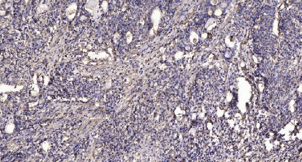
A03225 has an IHC figure from paraffin-embedded human gastric adenocarcinoma (catalog IHC image caption). IF and mouse reactivity are listed, but no IF figure is supplied (catalog: applications, reactivity, IF image alts).

Real IHC data Immunohistochemical analysis of paraffin-embedded human Gastric adenocarcinoma. 1, Antibody was diluted at 1:200 (4° overnight). 2, Tris-EDTA, pH9.0 was used for antigen retrieval. 3, Secondary antibody was diluted at 1:200 (room temperature, 45min).
Anti-Alstrom syndrome protein 1 ALMS1 Antibody
Cat # A03225

A03225 is listed for IHC and IF in human and mouse (catalog: applications, reactivity). Its IHC figure shows paraffin-embedded human gastric adenocarcinoma; no IF image is supplied (catalog: IHC and IF image alts).

Which to pick: Choose A03225 for tissue IHC: its own caption documents paraffin sections, pH 9.0 Tris-EDTA retrieval and a 1:200 overnight primary incubation; the fixative is unreported (catalog IHC image caption). For IF, A03225 lists a 1:50 dilution, but ICC-specific validation and an IF figure are unreported (catalog: applications, IF dilution, IF image alts). For work across species, A03225 lists human and mouse reactivity, while its displayed IHC evidence is human only and clonality is unreported.

Each figure is that product's own IHC / IF validation image from its datasheet.

References

  1. UniProt Consortium. UniProt entry Q8TCU4 (ALMS1_HUMAN, Centrosome-associated protein ALMS1).
  2. Human Protein Atlas. ALMS1 tissue IHC expression (reliability: Enhanced).
  3. Human Protein Atlas. ALMS1 subcellular location (ICC-IF): Mainly localized to the cytosol, centrosome, basal body and flagellar centriole. In addition localized to the nucleoplasm, microtubules, mid piece and end piece..
  4. Human Protein Atlas. ALMS1 antibody validation summary (2 antibodies).
  5. Mutations in Alström protein impair terminal differentiation of cardiomyocytes. Nature communications 2014 — PMC3992616.
  6. A role for Alström syndrome protein, alms1, in kidney ciliogenesis and cellular quiescence. PLoS genetics 2007 — PMC1761047.
  7. GLUT4 defects in adipose tissue are early signs of metabolic alterations in Alms1GT/GT, a mouse model for obesity and insulin resistance. PloS one 2014 — PMC4192353.
  8. Triple Vectors Expand AAV Transfer Capacity in the Retina. Molecular therapy : the journal of the American Society of Gene Therapy 2018 — PMC5835116.
  9. PubMed PMID:11941370 — UniProt-cited evidence.
  10. PubMed PMID:15815621 — UniProt-cited evidence.
  11. PubMed PMID:9205841 — UniProt-cited evidence.