ASAH1 / Acid ceramidase · IHC design guide

Design Immunohistochemistry for ASAH1

Plan paraffin section IHC for ASAH1 using the catalog antibody A02055-1 and its documented 1 μg/mL starting concentration (datasheet A02055-1). Use the high lysosomal staining reported in lung macrophages to guide interpretation, while comparing cell types within each section (HPA tissue IHC).

Evidence assembled Oct 2026 · For research use; verify linked source records and product datasheet before use
Immunohistochemistry protocol sheet for ASAH1 (IHC for ASAH1): expected localisation Lysosomal staining is expected in tissue (HPA tissue IHC), antibody A02055-1, validated IHC image, and IHC protocol steps
Printable ASAH1 IHC protocol sheet — expected localisation Lysosomal staining is expected in tissue (HPA tissue IHC), antibody A02055-1, controls and protocol steps. Open the full ASAH1 IHC guide →

ASAH1 Immunohistochemistry Experimental Design Guide

Expected localisation, validated protocols, controls and antibodies — the at-a-glance facts below, then the full design guide.

Must know before staining
Expected localisation Lysosomal staining is expected in tissue (HPA tissue IHC)
Staining pattern High lysosomal staining in lung macrophages (HPA tissue IHC)
Antigen retrieval EDTA pH 8.0 HIER, heat-mediated (datasheet A02055-1)
Positive control ⓘ Adrenal gland+4 more · see all
Negative control ⓘ Soft tissue
Important caveats
Reasons your staining may differ from the expected pattern.
Fixation Keep fixation conditions consistent across sections. (standard IHC practice; not target-specific)
Caveat Staining varies with the cells present in each tissue (HPA tissue IHC)
Regulation Expression is heart enriched; regulation is unreported (UniProt)
Isoform / epitope 3 isoforms; α/β processing may affect epitope mapping (UniProt)
Section 1

Recommended ASAH1 IHC & IF Protocols

The catalog antibody uses EDTA pH 8.0 HIER (datasheet A02055-1). Published ASAH1 IHC protocols below cover kidney biopsy, mouse xenograft, and skin/melanoma sections (PMC8583658; PMC11518951; PMC8081993).

Recommended immunohistochemistry (IHC-P) protocol parameters
SampleParaffin-embedded human melanoma tissue; fixative not specified (datasheet A02055-1)
FixationImage fixative and duration unreported (datasheet A02055-1); verify before use.
Sectioning4–5 µm sections on charged slides (standard)
DeparaffinisationXylene, graded ethanol series to water (standard)
Antigen retrievalHeat retrieval: EDTA pH 8.0 (datasheet A02055-1); 20 min, 95–100 °C (standard)
Peroxidase block3% H2O2, 10 min, room temperature (standard)
Blocking10% goat serum (datasheet A02055-1)
Primary antibodyRabbit anti-ASAH1, 0.5-1μg/ml (datasheet A02055-1)
Primary incubationOvernight at 4 °C (datasheet A02055-1)
DetectionStreptavidin-biotin complex (SABC), DAB chromogen (datasheet A02055-1)
CounterstainHematoxylin, blue, dehydrate and mount (standard)
Expected resultASAH1-positive staining in glandular cells of adrenal gland (HPA tissue IHC: High). HPA tissue profile: High expression in lysosomes. No signal in the no-primary control.
💡Decision noteStart with EDTA pH 8.0 HIER for the catalog antibody (datasheet A02055-1). Use citrate pH 6.0 when reproducing the corresponding published protocols (PMC11518951; PMC8081993).
Section 2

What Is the Expected ASAH1 Staining Pattern?

ASAH1 should appear mainly as cytoplasmic lysosomal staining in paraffin-section IHC (HPA: high expression in lysosomes; UniProt Q13510: lysosome). High staining is reported in cardiomyocytes, kidney tubular cells, and lung macrophages, among other cells (HPA: tissue IHC). Interpret the pattern with appropriate controls: HPA rates its tissue IHC evidence Supported, with medium consistency between staining and RNA data (HPA: reliability). ASAH1 has no transmembrane segment (UniProt Q13510: topology).

What am I looking at on my slide?
Distinct cytoplasmic granules in cardiomyocytes, kidney tubular cells, or lung macrophages.This fits lysosomal enrichment and the reported High staining in these cells (HPA: tissue IHC and lysosomal profile). Compare signal with adjacent cells on the same section; the reported level describes those cell populations, not uniform staining of every cell (HPA: cell-specific tissue IHC).
Predominantly crisp cell-surface outlines or isolated nuclear staining, without a lysosomal pattern.Question a cell-surface-only result: ASAH1 has no transmembrane segment, while its principal reported location is lysosomal (UniProt Q13510: topology and localization; HPA: lysosomal profile). Nuclear localization is also annotated, so nuclear signal alone is inconclusive; assess its cell type and controls (UniProt Q13510: nucleus; general IHC practice).
Strong staining in peripheral nerve or uniformly strong staining across low-staining cell types.Consider cross-reactivity or detection background before assigning ASAH1 expression (general IHC practice). HPA reports peripheral nerve in soft tissue as Not detected and squamous epithelial cells in oral mucosa, esophagus, and vagina as Low; these are comparisons, not absolute exclusion criteria (HPA: tissue IHC).
Diffuse color across extracellular space, empty areas, or many unrelated cell types.Treat this as possible nonspecific or endogenous detection signal when it obscures cellular localization (general chromogenic IHC practice). Very low secretion is annotated for ASAH1, but that annotation alone does not establish a diffuse extracellular staining pattern (UniProt Q13510: secretion). Check the detection control before scoring the diffuse signal (general IHC practice).
No detectable staining in an expected high-staining cell population.First confirm that the relevant cells are present: HPA reports High staining in heart cardiomyocytes, kidney tubular cells, and lung macrophages (HPA: tissue IHC). A blank result in one specimen does not by itself establish absent ASAH1; assess a known-positive section and the staining run (general IHC practice).
💡Expected ASAH1 appearanceCall a positive IHC result when the expected cells show discernible cytoplasmic, lysosome-associated staining, potentially High in HPA-listed cell populations; diffuse field-wide color or exclusive sharp cell-surface outlining warrants control-based review (HPA: tissue IHC and lysosomal profile; UniProt Q13510: lysosome and topology; general IHC practice).
How each factor affects the staining
Cell type and tissue selectionASAH1 is broadly expressed, with higher expression in heart (UniProt Q13510: tissue specificity). HPA reports High staining in eight listed tissue and cell-type pairs, including adrenal glandular cells and caudate glial cells; choose a control by the specified cell population rather than by tissue name alone (HPA: tissue IHC).
Processing and antibody epitopeASAH1 is processed from a precursor into disulfide-linked alpha and beta chains (UniProt Q13510: processing and subunit). The payload gives no antibody epitope, so it cannot establish which form the catalog antibody recognizes or predict how processing affects its IHC signal; check epitope documentation before making that inference.
Localization evidence and validationHPA tissue IHC describes high expression in lysosomes and rates the tissue staining Supported (HPA: tissue IHC). Its subcellular ICC-IF entry says Membrane but gives no main location or cell-line images; it cannot resolve a membrane-only pattern here (HPA: subcellular ICC-IF). Antibody HPA005468 is Supported for IHC, with no ICC status listed (HPA: antibody validation).
Antigen retrievalRetrieval conditions can affect chromogenic IHC staining in general (general IHC practice). Neither supplied source establishes ASAH1-specific retrieval requirements or fixation sensitivity; select and assess retrieval using the antibody's IHC-P instructions and a known-positive section, without treating a change in signal as proof of an ASAH1-specific fixation effect.
Why is my staining missing, weak or wrong?
SituationLikely causeNext action
Known-positive cells remain blank.The run may have a detection, antibody, or retrieval problem; the blank section alone does not identify which (general IHC practice).Check a known-positive section and detection controls, then review the catalog antibody's IHC-P conditions before adjusting the run (general IHC practice). Heart cardiomyocytes or kidney tubular cells are reported High by HPA (HPA: tissue IHC).
Color covers the slide or masks cell boundaries.Nonspecific staining or endogenous detection activity is possible in chromogenic IHC (general IHC practice).Inspect the no-primary control and the pattern within tissue-free areas; review blocking, washing, and detection steps if background persists (general IHC practice). Do not score diffuse color as lysosomal staining without discernible cellular signal (HPA: lysosomal profile; general IHC practice).
Only a sharp cell-surface rim appears.That pattern needs scrutiny because ASAH1 has no annotated transmembrane segment (UniProt Q13510: topology); the HPA ICC-IF Membrane summary lacks images and a main-location assignment (HPA: subcellular ICC-IF).Compare with the expected cytoplasmic lysosomal pattern and controls, and check whether the IHC-validated antibody reproduces the result (HPA: tissue IHC and antibody validation; general IHC practice).
Peripheral nerve stains as strongly as the expected positive cells.This conflicts with HPA's Not detected peripheral-nerve observation; cross-reactivity or local detection background is possible (HPA: soft-tissue IHC; general IHC practice).Identify the stained cell type, compare a positive section, and inspect the no-primary control before assigning the signal to ASAH1 (general IHC practice).
Signal appears chiefly nuclear.UniProt includes nucleus among ASAH1 locations, but HPA describes the tissue IHC profile as lysosomal; a nuclear-only slide cannot be judged from either annotation alone (UniProt Q13510: localization; HPA: tissue IHC).Assess the stained cell population, cytoplasmic signal, and controls together; avoid declaring either specificity or artifact from nuclear color alone (general IHC practice).
Q: Can this IHC pattern serve as an IF/ICC protocol?HPA lists no ICC validation for HPA005468 and no cell-line ICC-IF images in the supplied record (HPA: antibody validation and subcellular ICC-IF).A: Use an IF/ICC-specific guide and validation before interpreting fluorescent localization; this IHC tissue pattern supplies an expectation, not IF/ICC conditions (HPA: tissue IHC and ICC-IF record; general IF practice).

Sample controls for ASAH1 IHC & IF

🧪Run adrenal gland first: glandular cells should stain (HPA: High in adrenal gland glandular cells). Use soft tissue peripheral nerve as the negative tissue (HPA: Not detected); on the adrenal slide, assess nonglandular cells for absent or lower staining as an internal background reference (standard IHC practice).
Positive control tissue: Adrenal gland (Glandular cells, HPA High)
Negative control tissue: Soft tissue (HPA Not detected)
ICC-IF cell lines (HPA subcellular resource): HPA carries no ICC-IF cell line for ASAH1; derive a cell-line control from the positive tissue's cell type (Glandular cells) and confirm it by RNA or western blot first.
Technical controls: Include no-primary (secondary-only), matched rabbit IgG isotype, and ASAH1-knockout tissue controls (caption: rabbit primary antibody; standard IHC practice). With the caption’s biotin-based DAB detection, check endogenous peroxidase and biotin background in the adrenal section (caption: SABC with DAB; standard IHC practice).
⚠️Feasibility: A target-specific fixation window or fixation effect is unreported, and the selected A02055-1 paraffin-section caption does not state a fixative (caption: fixative not stated). Heat retrieval in EDTA at pH 8.0 was used in that IHC example; whether retrieval is required has not been established by the supplied evidence (caption: EDTA retrieval). The supplied evidence does not establish that frozen sections or IF are easier; endogenous biotin could complicate adrenal staining with the caption’s SABC method, so inspect the negative controls for that artefact (caption: SABC; standard IHC practice).

HPA tissue IHC evidence for ASAH1

Comprehensive Human Protein Atlas IHC scoring per tissue (reliability: Supported — Medium consistency between antibody staining and RNA expression data.). Rows are taken directly from the HPA tissue chart — click any row's HPA link to view the source.

Positive expression · recommended positive controls

TissueCell typeLevelEvidenceSource
Adrenal gland Glandular cells High Protein (IHC) HPA →
Caudate Glial cells High Protein (IHC) HPA →
Cerebral cortex Neuronal cells High Protein (IHC) HPA →
Epididymis Glandular cells High Protein (IHC) HPA →
Gallbladder Glandular cells High Protein (IHC) HPA →

Undetected expression · recommended negative controls

TissueCell typeLevelEvidenceSource
Soft tissue Peripheral nerve Not detected Protein (IHC) HPA →
Section 3

Advanced ASAH1 IHC Tips

Troubleshoot ASAH1 staining in paraffin sections by checking retrieval, cellular pattern and assay controls before comparing signal between samples.

What retrieval should I try first when ASAH1 staining is weak in paraffin sections?
Start with heat-mediated retrieval in EDTA, pH 8.0 (datasheet A02055-1). The selected paraffin-section example used this retrieval before overnight incubation with 1 µg/ml primary antibody at 4°C, so check that both steps match before changing conditions (datasheet A02055-1). If signal remains weak, compare a second retrieval condition on adjacent sections while keeping detection and exposure comparable (standard IHC practice). Record tissue integrity and background alongside signal, because excessive heating can damage morphology and make staining harder to interpret (standard IHC practice). Judge any gain by whether the expected lysosomal pattern becomes clearer, rather than by overall darkness alone (HPA: high expression in lysosomes; standard IHC practice).
Could fixation explain weak or uneven ASAH1 staining?
The selected ASAH1 paraffin-section caption does not state a fixative, so target-specific fixation sensitivity is unknown (datasheet A02055-1). Document each specimen’s fixative and processing history, then compare sections with similar handling before attributing different signal to biology (standard IHC practice). For uneven staining, inspect section adherence, morphology, folds and edges, and repeat staining on an intact section when those features coincide with the signal change (standard IHC practice). Keep retrieval at EDTA, pH 8.0 during this comparison so fixation history is the main variable under review (datasheet A02055-1; standard IHC practice). Do not infer a fixation effect from ASAH1’s lysosomal distribution or glycosylation sites (UniProt Q13510; HPA: lysosomal expression).
Where should credible ASAH1 chromogenic staining appear?
Look for intracellular, granular staining compatible with lysosomes, while assessing which cell type carries the signal (HPA: high expression in lysosomes; UniProt Q13510: lysosome). ASAH1 has a signal peptide and no transmembrane segment, and its lysosomal delivery depends on the mannose-6-phosphate receptor (UniProt Q13510 topology and localisation). UniProt also records nuclear and cytoplasmic localisation and extremely low secretion, so an unusual pattern merits validation rather than automatic dismissal (UniProt Q13510 localisation). Compare suspected positive cells with a matched negative control and tissue morphology, using identical chromogen development for that comparison (standard IHC practice). Diffuse staining across unrelated structures without a plausible cellular pattern warrants investigation of background or detection chemistry (standard IHC practice).
How could epitope placement affect interpretation of ASAH1 IHC?
Check the antibody’s documented immunogen or mapped epitope before claiming that staining represents every ASAH1 form (standard IHC practice). ASAH1 has 3 listed isoforms, and its precursor yields an alpha chain at residues 22–142 and a beta chain at 143–395 (UniProt Q13510 processing and isoforms). An epitope in one processed chain could report that chain’s accessibility without proving equal detection of the other chain or every isoform (UniProt Q13510 processing; standard IHC interpretation). Six annotated glycosylation sites lie in the beta-chain region, making epitope accessibility a question to test empirically (UniProt Q13510 glycosylation and processing). Compare independently validated reagents or orthogonal evidence before assigning isoform-specific staining (standard IHC practice).
How should I assess ASAH1 localisation by IF alongside this IHC result?
Use the separate IF/ICC workflow to test whether ASAH1 puncta overlap a marker identifying the expected cell type, such as a macrophage identity marker when examining lung macrophages (HPA: high in lung macrophages; standard IF practice). Choose fluorophores and imaging channels after checking tissue autofluorescence in unstained controls, then compare single-label controls before interpreting multiplex overlap (standard IF practice). Because ASAH1 has no transmembrane segment and is delivered to lysosomes, test permeabilisation sufficient to expose a lysosomal luminal epitope if the antibody binds there (UniProt Q13510 topology and localisation; standard IF practice). Confirm the antibody’s epitope before assigning which membrane must be traversed, and compare punctate signal with the IHC lysosomal expectation (HPA: lysosomal expression; standard IF practice).
How can I distinguish ASAH1 signal from chromogenic background?
Run a section omitting primary antibody and inspect it beside the test section for staining from the detection system (standard IHC practice). The selected example used biotinylated goat anti-rabbit secondary antibody, a streptavidin-biotin complex and DAB; background in that workflow should be checked at each detection stage (datasheet A02055-1; standard IHC practice). Include a peroxidase block as a general chromogenic IHC step and assess whether endogenous enzyme activity persists in the control (standard IHC practice). The example used 10% goat serum block and 1 µg/ml primary antibody; use those documented settings as comparison points when adjusting blocking or primary concentration (datasheet A02055-1).
How should I score ASAH1 staining across paraffin sections? ⚠ ANSWER MARKED FOR VERIFICATION
Define the cell population and anatomical region before scoring, then apply the same inclusion rules to every section (standard IHC practice). An H-score can combine staining intensity with the percentage of positive cells, while percentage positive alone is useful when intensity is unreliable (standard IHC practice). For discrete positive cells, report counts per mm² of evaluable tissue and retain the cell-type annotation, because ASAH1 signal can differ between tissue compartments (standard IHC practice; HPA: high in lung macrophages and kidney tubular cells). Normalise to viable tissue area or the number of relevant cells, and exclude folds, damaged edges and necrosis using preset criteria (standard IHC practice). Keep retrieval, chromogen development and scoring thresholds consistent across compared sections (standard IHC practice).
What would make an apparent ASAH1-positive area unconvincing?
A convincing result has cellular staining compatible with ASAH1’s predominantly lysosomal profile and remains distinguishable from the no-primary control (HPA: lysosomal expression; standard IHC practice). High signal in lung macrophages or kidney tubular cells has supporting tissue-level context, whereas HPA reports no detection in peripheral nerve within soft tissue (HPA: lung, kidney and soft tissue IHC). Examine unexpected nuclear, extracellular or uniformly diffuse staining carefully: UniProt lists nuclear localisation and extremely low secretion, so compartment alone cannot establish either specificity or artefact (UniProt Q13510 localisation). Edge-concentrated staining, necrotic areas and signal reproduced without primary antibody favour processing or endogenous-enzyme artefacts (standard IHC practice). Confirm disputed patterns with appropriate controls and independent evidence before making a biological claim (standard IHC practice).
Boster reagents

Best ASAH1 / Acid ceramidase IHC Antibodies

Two anti-ASAH1 antibodies have IHC images from human, mouse, or rat tissue and IF images from human cells or rat tissue (catalog image captions).

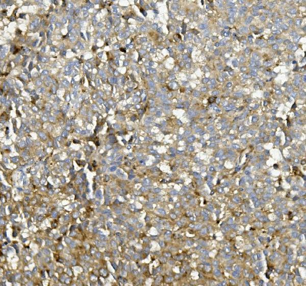
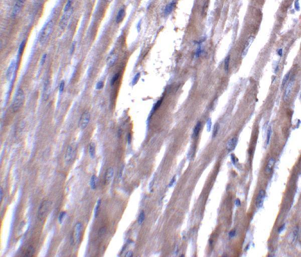
Real IHC data IHC analysis of ASAH1 using anti-ASAH1 antibody (A02055-1). ASAH1 was detected in paraffin-embedded section of human melanoma tissue. Heat mediated antigen retrieval was performed in EDTA buffer (pH8.0, epitope retrieval solution). The tissue section was blocked with 10% goat serum. The tissue section was then incubated with 1μg/ml rabbit anti-ASAH1 Antibody (A02055-1) overnight at 4°C. Biotinylated goat anti-rabbit IgG was used as secondary antibody and incubated for 30 minutes at 37°C. The tissue section was developed using Strepavidin-Biotin-Complex (SABC) (Catalog # SA1022) with DAB as the chromogen.
Anti-ASAH1 ® Antibody
Cat # A02055-1
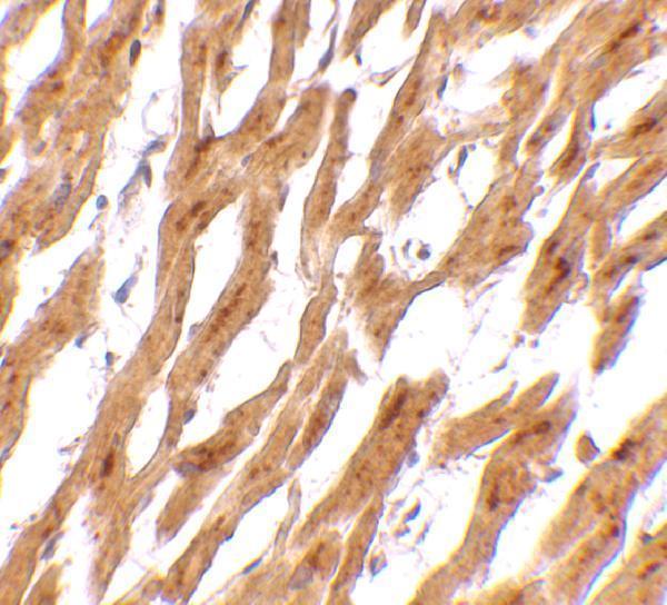
Real IHC data Immunohistochemistry of ASAH1 in human heart tissue with ASAH1 antibody at 2.5 μg/mL.
Anti-Acid ceramidase ASAH1 Antibody
Cat # A02055

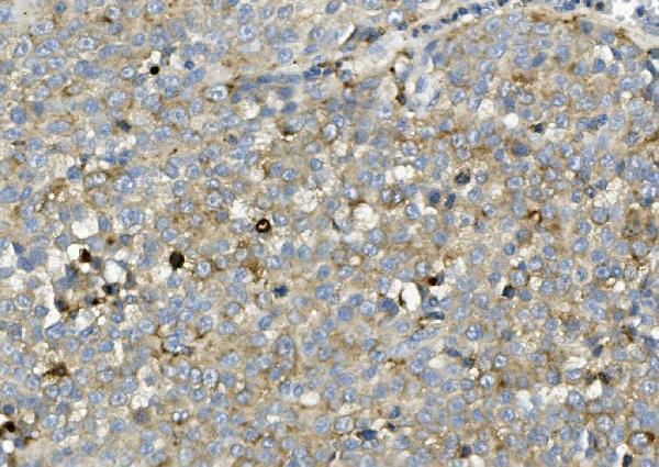
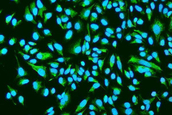
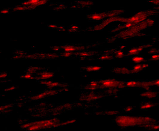
A02055-1 has paraffin-section IHC images from human melanoma, human thyroid cancer, and mouse kidney, plus IF in HeLa cells (catalog image captions). A02055 has IHC images from human and rat heart and IF images from human heart cells and rat heart tissue (catalog image captions).

Which to pick: For paraffin-section IHC, choose A02055-1 when its documented EDTA pH 8.0 retrieval and 1 μg/mL staining conditions suit your starting protocol (A02055-1 IHC captions); its fixative is unreported (A02055-1 IHC captions). For IF/ICC, A02055-1 has an ICC/IF application and a HeLa-cell image, while A02055 has IF images from human heart cells and rat heart tissue (catalog applications and IF captions). Both list human, mouse, and rat reactivity, but the pictured IHC evidence covers human and mouse for A02055-1 and human and rat for A02055; A02055’s IHC caption does not report the fixative (catalog reactivity and IHC captions).

Each figure is that product's own IHC / IF validation image from its datasheet.

References

  1. UniProt Consortium. UniProt entry Q13510 (ASAH1_HUMAN, Acid ceramidase).
  2. Human Protein Atlas. ASAH1 tissue IHC expression (reliability: Supported).
  3. Human Protein Atlas. ASAH1 subcellular location (ICC-IF): Membrane.
  4. Human Protein Atlas. ASAH1 antibody validation summary (1 antibodies).
  5. α-Galactosidase a Deficiency in Fabry Disease Leads to Extensive Dysregulated Cellular Signaling Pathways in Human Podocytes. International journal of molecular sciences 2021 — PMC8583658.
  6. Acid ceramidase and its inhibitors: a de novo drug target and a new class of drugs for killing glioblastoma cancer stem cells with high efficiency. Oncotarget 2017 — PMC5762539.
  7. Targeting acid ceramidase enhances antitumor immune response in colorectal cancer. Journal of advanced research 2024 — PMC11518951.
  8. N-acylsphingosine amidohydrolase 1 promotes melanoma growth and metastasis by suppressing peroxisome biogenesis-induced ROS production. Molecular metabolism 2021 — PMC8081993.
  9. PubMed PMID:8955159 — UniProt-cited evidence.
  10. PubMed PMID:10993717 — UniProt-cited evidence.
  11. PubMed PMID:10610716 — UniProt-cited evidence.