This website uses cookies to ensure you get the best experience on our website.


- Table of Contents
Plan chromogenic IHC on paraffin sections using nuclear staining as the expected ATF3 pattern in most tissues (HPA tissue IHC). Colon glandular cells show medium staining, while esophageal squamous epithelial cells have no detected staining; interpret controls in light of low consistency between antibody staining and RNA expression (HPA tissue IHC).

Expected localisation, validated protocols, controls and antibodies — the at-a-glance facts below, then the full design guide.
| Expected localisation | Nuclear in most tissues (HPA tissue IHC) | |
| Staining pattern | Nuclear staining in most tissues (HPA tissue IHC) | |
| Antigen retrieval | Tris-EDTA pH 9.0 HIER, heat-mediated (datasheet A00904-2) | |
| Positive control | Fallopian tube+4 more · see all | |
| Negative control | Esophagus+4 more · see all |
| Fixation | Keep fixation consistent across sections (standard IHC practice; not target-specific) | |
| Caveat | Antibody staining and RNA expression show low consistency (HPA tissue IHC) | |
| Regulation | Expression regulation is not annotated (UniProt) | |
| Isoform / epitope | Five isoforms; epitope coverage is unspecified (UniProt) |
Compare the catalog antibody’s IHC protocol (datasheet: A00904-2) with four published ATF3 chromogenic IHC protocols (PMC2924561; PMC3772181; PMC12837017; PMC5564541).
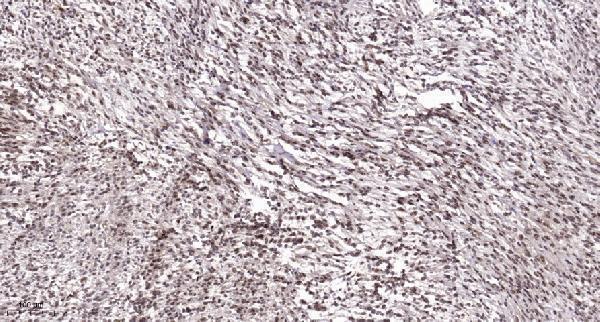
| Sample | Paraffin-embedded human Colon cancer tissue; fixative not specified (datasheet A00904-2) |
| Fixation | Image fixative and duration unreported (datasheet A00904-2); verify before use. |
| Sectioning | 4–5 µm sections on charged slides (standard) |
| Deparaffinisation | Xylene, graded ethanol series to water (standard) |
| Antigen retrieval | Heat retrieval: Tris-EDTA pH 9.0 (datasheet A00904-2); 20 min, 95–100 °C (standard) |
| Peroxidase block | 3% H2O2, 10 min, room temperature (standard) |
| Blocking | 10% normal serum of the secondary host, 30 min, room temperature (standard) |
| Primary antibody | Rabbit anti-ATF3, 1:100 recommended; image 1:200 (datasheet A00904-2) |
| Primary incubation | Overnight at 4 °C (standard) |
| Detection | HRP-polymer secondary, DAB chromogen 5–10 min (standard) |
| Counterstain | Hematoxylin, blue, dehydrate and mount (standard) |
| Expected result | ATF3-positive staining in ciliated cells (tip of cilia) of fallopian tube (HPA tissue IHC: High). HPA tissue profile: Nuclear expression in most tissues. No signal in the no-primary control. |
ATF3 is a nuclear, DNA-binding transcription factor with no transmembrane segment (UniProt P18847). In paraffin-section IHC, expect nuclear staining across many tissues, including appendix glandular cells and cerebral cortex neurons (HPA tissue IHC). The tissue IHC profile is Approved, but antibody staining has low consistency with RNA expression; use the listed cell types as reference patterns, not guarantees for every specimen (HPA tissue IHC).
| Distinct nuclear signal in appendix or colon glandular cells, or in cerebral cortex neurons, with relatively clear surrounding tissue. | This fits the reported Medium staining in those cell types (HPA tissue IHC) and ATF3's nuclear location (UniProt P18847). Judge intensity against a matched control slide; the HPA categories describe observed staining, not a required intensity for every preparation (HPA tissue IHC). |
| Predominantly diffuse cytoplasmic or membranous staining, without convincing nuclear signal. | That distribution conflicts with the principal nuclear location (UniProt P18847; HPA subcellular). Check the control and detection background before calling it ATF3. HPA also reports High staining at fallopian-tube ciliary tips, so an isolated nonnuclear feature needs context rather than automatic dismissal (HPA tissue IHC). |
| Strong signal in esophageal squamous cells, cardiomyocytes, or skeletal-muscle myocytes. | These cell types are listed as Not detected (HPA tissue IHC). Recheck tissue identification and whether staining follows cell boundaries, pigment, or reagent deposition; cross-reactivity or endogenous detection activity is possible (standard IHC practice). A single discrepancy does not establish a new ATF3 tissue pattern. |
| Broad, hazy chromogen over cells and extracellular areas, with poor nuclear contrast. | This is an uninterpretable localization pattern, since the expected principal compartment is nuclear (UniProt P18847; HPA tissue IHC). Uneven blocking, washing, or chromogen development can contribute to nonspecific background (standard IHC practice). Compare a no-primary control before scoring any nuclei. |
| No nuclear signal in an appendix, colon, or cerebral cortex section selected as a positive control. | HPA reports Medium staining in the specified glandular or neuronal cells (HPA tissue IHC), so verify that those cells are present and that the control run worked. Absence in one section is not definitive: the HPA tissue profile is Approved with low staining-to-RNA consistency (HPA tissue IHC). |
| Tissue and cell selection | HPA reports nuclear expression in most tissues, with Medium staining in appendix and colon glandular cells and Not detected staining in esophageal squamous cells (HPA tissue IHC). Select controls by the named cell population, then verify it is present on the section. |
| Antibody evidence | The HPA tissue profile is Approved, while its staining shows low consistency with RNA expression; HPA001562 is IHC Approved and ICC Supported (HPA tissue IHC; HPA antibodies). These labels support use as a reference but do not resolve every unexpected signal. |
| Isoforms and epitope | UniProt lists 5 ATF3 isoforms and a bZIP domain at residues 86–149 (UniProt P18847). The supplied evidence does not locate the catalog antibody's epitope, so it cannot establish which isoforms that antibody detects. |
| Fixation and antigen retrieval | Target-specific fixation effects are not established by the supplied assay evidence. Verify with a matched IHC source before attributing a result to fixation. |
| IF/ICC Q: What localization should I expect? | A: HPA supports nucleoplasm and nucleoli; it marks basal body, vesicles, and centrosome as uncertain (HPA subcellular). Those ICC-IF observations can guide interpretation, but this IHC section does not specify an IF/ICC protocol. |
| Situation | Likely cause | Next action |
|---|---|---|
| A candidate positive tissue has little or no nuclear chromogen. | The sampled section may lack the HPA-listed cell type, or the IHC run may have failed (HPA tissue IHC; standard IHC practice). | Locate the relevant glandular or neuronal cells, examine the matched positive control, and check the catalog antibody's IHC-P staining conditions before interpreting the specimen (HPA tissue IHC; standard IHC practice). |
| An expected negative cell type stains strongly. | HPA lists esophageal squamous cells, cardiomyocytes, and skeletal-muscle myocytes as Not detected (HPA tissue IHC); nonspecific binding or endogenous detection activity may mimic signal (standard IHC practice). | Confirm cell identity, compare a no-primary control, and review blocking and detection steps (standard IHC practice). Treat persistent staining as unresolved until its nuclear localization and reproducibility are checked (UniProt P18847; standard IHC practice). |
| Staining is mainly cytoplasmic or outlines cell membranes. | That pattern does not match ATF3's principal nuclear localization (UniProt P18847; HPA subcellular), though HPA records a ciliary-tip signal in fallopian tube (HPA tissue IHC). | Check the tissue and cell type, examine nuclear counterstain alignment, and compare controls (standard IHC practice). Document any fallopian-tube ciliary-tip staining separately from nuclear scoring (HPA tissue IHC). |
| Hazy staining obscures nuclear boundaries. | Background from blocking, washing, or detection can make compartment scoring unreliable (standard IHC practice). | Compare a no-primary control and review wash, blocking, and chromogen-development steps (standard IHC practice). Score ATF3 only where individual nuclei can be distinguished, consistent with its nuclear localization (UniProt P18847). |
| Only a few cells stain in a tissue expected to show broader expression. | HPA describes nuclear expression in most tissues but also reports low consistency between antibody staining and RNA expression (HPA tissue IHC). Differences in sampled cell populations can change the apparent distribution (standard IHC practice). | Identify and record the stained cell types and their nuclear intensity, then compare the same populations across sections and controls (standard IHC practice). Avoid converting the broad HPA profile into a universal positive-cell threshold (HPA tissue IHC). |
| An ICC-IF image shows a nucleolar punctum or a basal-body-like focus. | Nucleolar localization is supported, whereas basal-body localization is uncertain in HPA's ICC-IF summary (HPA subcellular). | Interpret the finding in the dedicated IF/ICC guide and compare it with a nuclear marker and appropriate controls (standard IF practice). Do not use that image alone to define the expected chromogenic IHC pattern (HPA subcellular; HPA tissue IHC). |


Comprehensive Human Protein Atlas IHC scoring per tissue (reliability: Approved — Low consistency between antibody staining and RNA expression data.). Rows are taken directly from the HPA tissue chart — click any row's HPA link to view the source.
| Tissue | Cell type | Level | Evidence | Source |
|---|---|---|---|---|
| Fallopian tube | Ciliated cells (tip of cilia) | High | Protein (IHC) | HPA → |
| Appendix | Glandular cells | Medium | Protein (IHC) | HPA → |
| Bone marrow | Hematopoietic cells | Medium | Protein (IHC) | HPA → |
| Bronchus | Ciliated cells (cell body) | Medium | Protein (IHC) | HPA → |
| Caudate | Neuronal cells | Medium | Protein (IHC) | HPA → |
| Tissue | Cell type | Level | Evidence | Source |
|---|---|---|---|---|
| Esophagus | Squamous epithelial cells | Not detected | Protein (IHC) | HPA → |
| Heart muscle | Cardiomyocytes | Not detected | Protein (IHC) | HPA → |
| Lymph node | Non-germinal center cells | Not detected | Protein (IHC) | HPA → |
| Ovary | Ovarian stroma cells | Not detected | Protein (IHC) | HPA → |
| Skeletal muscle | Myocytes | Not detected | Protein (IHC) | HPA → |
Troubleshoot ATF3 staining in paraffin sections by checking retrieval, nuclear localisation, controls and scoring before interpreting biological differences.
Two anti-ATF3 antibodies have paraffin-section IHC images from human cancers (catalog image captions); one also lists IF and mouse reactivity (catalog: A00904-2 applications and reactivity).
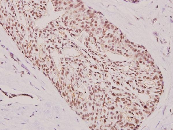
A00904-1 lists IHC for human and rat and shows paraffin-embedded human breast carcinoma at 1:100 (catalog applications, reactivity and image caption). A00904-2 lists IHC and IF for human, mouse and rat and shows paraffin-embedded human colon cancer at 1:200 (catalog applications, reactivity and image caption).
Which to pick: For tissue IHC, choose A00904-1 for its breast carcinoma example or A00904-2 for its colon cancer example; both captions specify paraffin-embedded tissue, but neither reports the fixative (catalog image captions). For IF/ICC or mouse samples, choose A00904-2 because it lists IF and mouse reactivity; no IF image is supplied (catalog applications, reactivity and image captions). A00904-1 is described as pAb in its IHC caption; both antibodies have rabbit hosts, and neither catalog entry specifies a clone (catalog image caption and host/clone fields).