ATG3 / Ubiquitin-like-conjugating enzyme ATG3 · IHC design guide

Design Immunohistochemistry for ATG3

Plan chromogenic IHC on paraffin sections using the IHC-validated antibody at a starting concentration of 5 μg/mL (datasheet: A01768). Compare staining with the often granular cytoplasmic pattern reported across most tissues (HPA tissue IHC).

Evidence assembled Oct 2026 · For research use; verify linked source records and product datasheet before use
Immunohistochemistry protocol sheet for ATG3 (IHC for ATG3): expected localisation Cytoplasmic staining, often granular (HPA tissue IHC), antibody A01768, validated IHC image, and IHC protocol steps
Printable ATG3 IHC protocol sheet — expected localisation Cytoplasmic staining, often granular (HPA tissue IHC), antibody A01768, controls and protocol steps. Open the full ATG3 IHC guide →

ATG3 Immunohistochemistry Experimental Design Guide

Expected localisation, validated protocols, controls and antibodies — the at-a-glance facts below, then the full design guide.

Must know before staining
Expected localisation Cytoplasmic staining, often granular (HPA tissue IHC)
Staining pattern Often granular cytoplasm across most tissues (HPA tissue IHC)
Antigen retrieval Citrate pH 6.0 HIER, 95–98 °C, 20 min (rule: cytoplasmic / membrane antigen)
Positive control ⓘ Adrenal gland+4 more · see all
Negative control ⓘ None in HPA (detected in all 45 tissues); use no-primary + isotype controls
Important caveats
Reasons your staining may differ from the expected pattern.
Fixation Keep fixation consistent across sections (standard IHC practice; not target-specific). Selected-image fixative and duration unreported (datasheet A01768); verify before use.
Caveat Staining and RNA show medium consistency (HPA tissue IHC)
Regulation No specific induction reported (UniProt)
Isoform / epitope 2 isoforms; epitope effects are unresolved (UniProt)
Section 1

Recommended ATG3 IHC & IF Protocols

Compare the catalog antibody’s IHC-P protocol (datasheet) with a published ATG3 paraffin-section IHC protocol (PMC6911302).

Recommended immunohistochemistry (IHC-P) protocol parameters
SampleTissue sections; selected-image fixative not specified (standard IHC workflow)
FixationImage fixative and duration unreported (datasheet A01768); verify before use.
Sectioning4–5 µm sections on charged slides (standard)
DeparaffinisationXylene, graded ethanol series to water (standard)
Antigen retrievalHeat-induced epitope retrieval in citrate buffer, pH 6.0, 20 min at 95–98 °C (standard rule: cytoplasmic / membrane antigen)
Peroxidase block3% H2O2, 10 min, room temperature (standard)
Blocking10% normal serum of the secondary host, 30 min, room temperature (standard)
Primary antibodyRabbit anti-ATG3, 5 μg/mL (datasheet A01768)
Primary incubationOvernight at 4 °C (standard)
DetectionHRP-polymer secondary, DAB chromogen 5–10 min (standard)
CounterstainHematoxylin, blue, dehydrate and mount (standard)
Expected resultATG3-positive staining in glandular cells of adrenal gland (HPA tissue IHC: High). HPA tissue profile: Cytoplasmic expression often with a granular pattern in most tissues. No signal in the no-primary control.
💡Decision noteStart with citrate pH 6.0 HIER at 95–98 °C for 20 min (page antigen retrieval); the paper does not specify retrieval conditions (PMC6911302).
Section 2

What Is the Expected ATG3 Staining Pattern?

ATG3 is expected mainly in the cytoplasm, often with a granular pattern across many tissues (HPA tissue IHC; UniProt Q9NT62: cytoplasm). Look for staining in glandular, hematopoietic, neuronal and other documented cell populations (HPA tissue IHC). ATG3 has no transmembrane segment (UniProt Q9NT62: topology). HPA rates tissue IHC reliability as Enhanced, while reporting medium consistency between antibody staining and RNA expression (HPA tissue IHC).

What am I looking at on my slide?
Cytoplasmic, sometimes granular, staining appears in adrenal or appendix glandular cells, or bone marrow hematopoietic cells (HPA tissue IHC: High).This matches the reported compartment and cell populations (HPA tissue IHC). Judge staining within the identified cells; nearby unstained structures are not, by themselves, evidence of assay failure (general IHC practice).
Signal is confined to nuclei, with no convincing cytoplasmic staining.A nuclear-only pattern conflicts with the reported cytoplasmic IHC profile and UniProt localization (HPA tissue IHC; UniProt Q9NT62). Treat it as suspect and check controls before interpreting it as ATG3 (general IHC practice).
Strong color appears chiefly in a cell population documented as low, such as lung alveolar type II cells or soft-tissue fibroblasts (HPA tissue IHC: Low).The distribution is unexpected relative to HPA, but Low does not mean absent (HPA tissue IHC). Cross-reactivity or endogenous chromogen-generating activity is possible; assess morphology and detection controls (general IHC practice).
Color spreads diffusely across cells and surrounding tissue, obscuring cell boundaries.The slide cannot be scored confidently for the reported cytoplasmic pattern (HPA tissue IHC; general IHC practice). Excess detection background or inadequate blocking or washing may contribute (general IHC practice).
A documented high-staining population, such as duodenal glandular cells or cerebral-cortex neurons, shows no detectable signal (HPA tissue IHC: High).First suspect an assay or specimen problem rather than concluding ATG3 is absent (general IHC practice). HPA reports High staining in those populations, but its tissue IHC has medium staining-to-RNA consistency (HPA tissue IHC).
💡Expected ATG3 appearanceCall a result positive when identifiable cells show cytoplasmic, often granular staining, especially in documented High populations (HPA tissue IHC); isolated nuclear-only color or diffuse acellular color is suspect (UniProt Q9NT62: cytoplasm; general IHC practice).
How each factor affects the staining
Compartment and topologyUniProt places ATG3 in the cytoplasm and reports no transmembrane segment (UniProt Q9NT62). HPA tissue IHC describes often-granular cytoplasmic staining; membrane color alone should not define an IHC positive (HPA tissue IHC; general IHC practice).
Choice of comparison tissueHPA reports High staining in appendix and gallbladder glandular cells, skin melanocytes and lymph-node non-germinal-center cells (HPA tissue IHC). It reports Low staining in cerebellar granular-layer cells and ovarian stroma cells; use those as contrasts, not absolute negatives (HPA tissue IHC).
Strength of tissue evidenceHPA labels tissue IHC reliability Enhanced and describes medium consistency with RNA (HPA tissue IHC). Both listed antibodies have Enhanced IHC validation (HPA antibodies: HPA040471, CAB037260); this supports pattern checking, not certainty for every specimen (general IHC practice).
Isoforms and processingUniProt lists two ATG3 isoforms, one chain spanning residues 1–314, and no signal peptide or propeptide (UniProt Q9NT62). Without an antibody epitope map, these facts cannot establish isoform-specific staining or a cleavage-dependent IHC pattern (general IHC practice).
IF/ICC Q: should the same compartment appear?A: HPA ICC-IF supports cytosol and plasma-membrane localization, with images in A-431, U-251MG and U2OS (HPA subcellular). This informs IF interpretation but does not establish a membrane-only pattern in paraffin IHC (HPA tissue IHC; general IHC practice).
Why is my staining missing, weak or wrong?
SituationLikely causeNext action
No stain in a documented High population (HPA tissue IHC).The specimen or IHC detection workflow may have failed; a blank slide alone cannot assign a target-specific cause (general IHC practice).Run a documented High tissue alongside a detection control; verify retrieval, antibody application and chromogen development against the chosen assay instructions (HPA tissue IHC; general IHC practice).
Only nuclear staining is visible.The pattern conflicts with cytoplasmic localization (UniProt Q9NT62; HPA tissue IHC); nonspecific binding or detection background may be involved (general IHC practice).Review cell morphology and a primary-antibody omission control, then reassess whether any convincing cytoplasmic signal remains (general IHC practice).
A documented Low population appears as strong as the chosen High comparator (HPA tissue IHC).This may reflect specimen variation, cross-reactivity or endogenous detection activity; HPA Low is not a negative designation (HPA tissue IHC; general IHC practice).Compare the two populations under the same staining run and inspect an appropriate detection control before assigning specificity (general IHC practice).
Brown chromogen covers the section with little cellular definition.Excess background may arise from detection reagents, blocking or washing (general IHC practice).Check the primary-antibody omission control and review blocking, washes and chromogen development using the assay instructions (general IHC practice).
Granular staining is mistaken for puncta marking autophagosomes.HPA describes often-granular ATG3 cytoplasmic IHC, while UniProt describes ATG3's role in autophagosome formation (HPA tissue IHC; UniProt Q9NT62); morphology alone cannot identify an organelle (general IHC practice).Score ATG3 distribution as cytoplasmic or granular; require an independently validated organelle marker before assigning puncta to autophagosomes (general IHC practice).
Membrane-edge color dominates, with little cytoplasmic IHC signal.HPA supports plasma-membrane localization in ICC-IF, but describes cytoplasmic expression in tissue IHC (HPA subcellular; HPA tissue IHC). The assays provide different pattern evidence.Interpret the paraffin section against the tissue IHC pattern and controls; do not use ICC-IF membrane localization alone to validate an IHC result (HPA tissue IHC; HPA subcellular; general IHC practice).

Sample controls for ATG3 IHC & IF

🧪Run bone marrow first: hematopoietic cells should stain (HPA: High in hematopoietic cells). HPA detects ATG3 in all 45 scored tissues, so there is no supported negative tissue; use no-primary and isotype controls, and expect cells without specific staining on the positive slide to show counterstain only, without assuming any cell population is ATG3-negative (HPA: no negative tissues).
Positive control tissue: Adrenal gland (Glandular cells, HPA High)
Negative control tissue: None in HPA: ATG3 is detected in all 45 scored tissues. Use a no-primary (secondary-only) and an isotype control instead.
ICC-IF cell lines (HPA subcellular resource): HPA ICC-IF images show ATG3 in A-431, U-251MG, U2OS, with annotated localisation: Plasma membrane (supported), Cytosol (supported) (HPA subcellular).
Technical controls: Include a no-primary, secondary-only section and an isotype control matched to the primary antibody’s host species and immunoglobulin class or subclass; use matched ATG3-knockout material as a biological negative, if available (standard IHC practice). In bone marrow, quench endogenous peroxidase and check for residual chromogen-like signal in the negative controls (HPA: bone marrow positive; standard IHC practice).
⚠️Feasibility: No target-specific fixation window or fixation effect is reported in the supplied evidence, and the fixative for the A01768 kidney IHC caption is unreported (selected A01768 caption). Antigen-retrieval dependence is also unreported, so retrieval conditions require empirical optimization for paraffin IHC (standard IHC practice). HPA provides ICC-IF images in A-431, U-251MG and U2OS, but the evidence does not establish that IF or frozen sections are easier; endogenous peroxidase is a practical concern in bone marrow IHC (HPA: ICC-IF cell lines; HPA: bone marrow positive; standard IHC practice).

HPA tissue IHC evidence for ATG3

Comprehensive Human Protein Atlas IHC scoring per tissue (reliability: Enhanced — Medium consistency between antibody staining and RNA expression data.). Rows are taken directly from the HPA tissue chart — click any row's HPA link to view the source.

Positive expression · recommended positive controls

TissueCell typeLevelEvidenceSource
Adrenal gland Glandular cells High Protein (IHC) HPA →
Appendix Glandular cells High Protein (IHC) HPA →
Bone marrow Hematopoietic cells High Protein (IHC) HPA →
Cerebral cortex Neuronal cells High Protein (IHC) HPA →
Duodenum Glandular cells High Protein (IHC) HPA →

Undetected expression · recommended negative controls

TissueCell typeLevelEvidenceSource
None in HPA: ATG3 is detected in all 45 scored tissues. Use a no-primary (secondary-only) and an isotype control instead.
Section 3

Advanced ATG3 IHC Tips

Troubleshoot ATG3 chromogenic IHC in paraffin sections by checking retrieval, staining pattern, controls and scoring before interpreting biological differences.

What retrieval conditions should I try first when ATG3 staining is weak?
Start with citrate buffer at pH 6.0, heated to 95–98 °C for 20 min (page retrieval specification). Run a known positive section beside the test section and keep cooling and subsequent wash times consistent across both slides (standard IHC practice; HPA: ATG3 is detected in most tissues). If staining remains weak, compare a separately validated retrieval condition on adjacent sections while holding antibody concentration and detection chemistry constant (standard IHC practice). Excessive retrieval can damage tissue morphology or increase background, so judge any gain in cytoplasmic signal alongside section integrity and a no-primary control (standard IHC practice; UniProt Q9NT62: cytoplasmic localisation).
How should I troubleshoot possible fixation effects on ATG3 staining?
Target-specific ATG3 sensitivity to fixation is unknown from the supplied evidence, and the A01768 kidney caption does not state a fixative (A01768 tissue-IHC caption). Record fixation conditions for each specimen, then compare sections processed with matched retrieval and detection settings before attributing staining differences to biology (standard IHC practice). Include a positive control section in each run and inspect whether weak signal coincides with poor morphology or uneven staining across the section (standard IHC practice; HPA: cytoplasmic staining occurs in most tissues). Do not infer ATG3 fixation tolerance from its cytoplasmic localisation, lack of a transmembrane segment or HPA staining profile (UniProt Q9NT62 topology; HPA tissue IHC).
Where should convincing ATG3 chromogenic staining appear in tissue?
Expect predominantly cytoplasmic staining, often granular, when assessing tissue sections (UniProt Q9NT62: cytoplasm; HPA tissue IHC: cytoplasmic expression often with a granular pattern). ATG3 lacks an annotated transmembrane segment, so a sharply membrane-limited chromogenic pattern needs independent validation even though supported cytosol and plasma-membrane locations are reported in cell imaging (UniProt Q9NT62 topology; HPA subcellular). Compare the suspected signal with the counterstain and tissue architecture to distinguish cytoplasm from adjacent cell borders (standard IHC practice). Treat isolated nuclear staining or staining confined to torn section edges cautiously, and check no-primary and positive control sections before assigning either pattern to ATG3 (UniProt Q9NT62: cytoplasm; standard IHC practice).
Can this antibody distinguish ATG3 isoforms or modified ATG3 in IHC?
ATG3 has 2 recorded isoforms, but the supplied antibody evidence gives no epitope coordinates or isoform-specific validation (UniProt Q9NT62: isoforms 1 and 2; A01768 tissue-IHC caption). Therefore, treat positive chromogenic staining as ATG3 immunoreactivity rather than an isoform assignment unless an independently validated isoform-specific reagent is available (standard IHC interpretation; UniProt Q9NT62: alternative splicing). The record lists N-terminal acetylmethionine and ATG12 conjugation, but neither establishes whether this antibody recognizes a modified form in fixed sections (UniProt Q9NT62: modified residues and function). If isoform or modification status matters, document the mapped epitope and validate specificity using an appropriate independent assay before interpreting intensity differences (standard IHC practice).
How can I adapt an ATG3 staining question to multiplex IF?
For a separate IF experiment, pair ATG3 with a validated marker for the cell population being assessed, such as a glandular-cell marker when examining glandular tissue (HPA tissue IHC: high ATG3 in glandular cells of adrenal gland, appendix and duodenum; standard IF practice). Choose spectrally separated fluorophores and inspect unstained tissue for autofluorescence before assigning a weak ATG3 signal to a channel (standard IF practice). ATG3 is reported in cytosol and at the plasma membrane, so optimise permeabilisation for access to cytosolic epitopes while checking that membrane outlines remain interpretable (HPA subcellular; standard IF practice). The A01768 tissue caption supports chromogenic kidney staining at 5 µg/mL, but supplies no IF fixation, permeabilisation or dilution conditions (A01768 tissue-IHC caption).
What should I change when ATG3 staining has diffuse background?
First compare the stained slide with no-primary and detection-only controls to locate background arising from the detection workflow (standard IHC practice). For chromogenic detection, block endogenous peroxidase before applying the detection reagent, then review blocking, wash stringency and antibody concentration one variable at a time (standard IHC practice). The A01768 kidney caption reports 5 µg/mL, which documents that image condition rather than an optimum for every tissue or retrieval setting (A01768 tissue-IHC caption). Preserve the expected cytoplasmic pattern during optimisation, since broadly increased DAB deposition without cellular definition cannot establish ATG3 specificity (HPA tissue IHC: cytoplasmic expression; standard IHC practice).
How should I score ATG3 staining across paraffin sections? ⚠ ANSWER MARKED FOR VERIFICATION
Define the cell population and tissue region before scoring, then report cytoplasmic ATG3 with an H-score combining staining intensity and percentage of positive cells (HPA tissue IHC: cytoplasmic pattern; standard IHC practice). Record percentage positive separately when intensity varies, and use positive-cell density per mm² if cell abundance is the primary endpoint (standard IHC practice). Normalise counts to the viable tissue area or the number of eligible cells in the same defined compartment, using identical thresholds and counterstain review across specimens (standard IHC practice). Because ATG3 is widely expressed and HPA reports low tissue RNA specificity, compare like cell populations and batches before interpreting modest score differences (UniProt Q9NT62: tissue specificity; HPA: low tissue RNA specificity).
How can I distinguish ATG3 signal from tissue or detection artefacts?
A credible positive follows cellular cytoplasm, potentially with granularity, and appears in an appropriate positive control under the same staining conditions (HPA tissue IHC: cytoplasmic, often granular expression; standard IHC practice). Signal restricted to nuclei, necrotic areas or section edges warrants review of morphology and controls before it is attributed to ATG3 (UniProt Q9NT62: cytoplasmic localisation; standard IHC practice). Compare no-primary and peroxidase-block controls when brown deposition could reflect endogenous enzyme activity rather than antibody binding (standard IHC practice). High staining in HPA-listed glandular or hematopoietic cells can support a control choice, but the reported antibody–RNA consistency is medium, so staining alone does not establish autophagic activity (HPA tissue IHC: high cell populations and medium consistency; UniProt Q9NT62: function).
Boster reagents

Best ATG3 / Ubiquitin-like-conjugating enzyme ATG3 IHC Antibodies

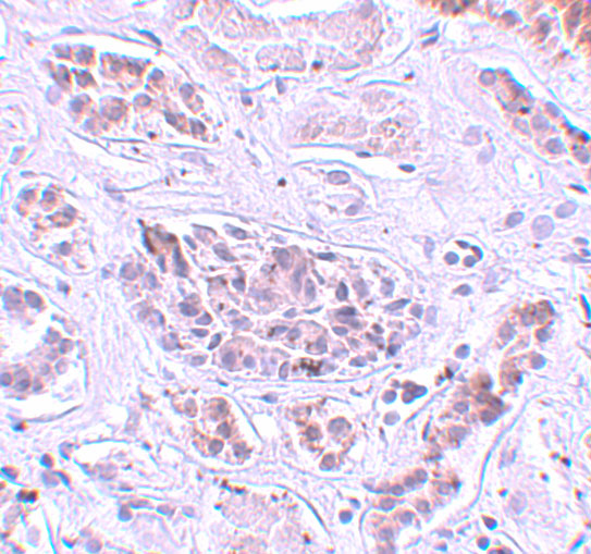
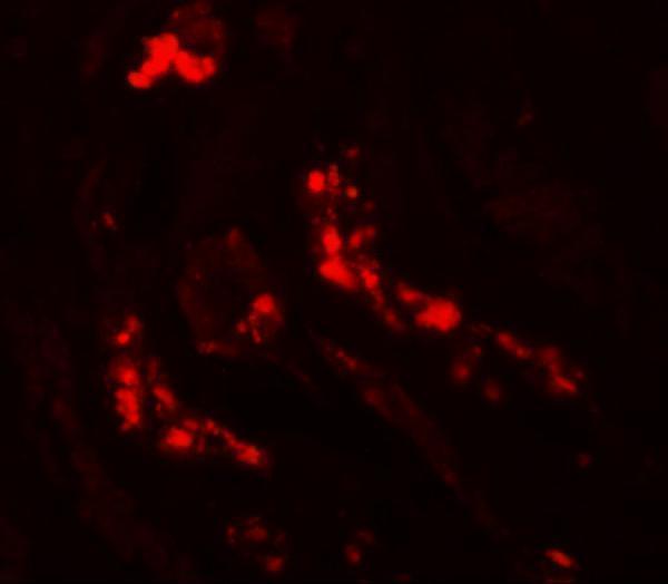
Anti-ATG3 antibodies include IHC and IF images from human kidney; the catalog lists human, mouse and rat reactivity (A01768 image captions; catalog reactivity).

Real IHC data Immunohistochemistry of ATG3 in human kidney tissue with ATG3 antibody at 5 μg/mL.
Anti-ATG3 Antibody
Cat # A01768

A01768 is the only SKU with a rendered card; its IHC image shows human kidney tissue at 5 μg/mL (A01768 IHC image caption). Its IF image shows human kidney at 20 μg/mL, and its listed reactivity is human, mouse and rat (A01768 IF image caption; catalog reactivity).

Which to pick: For paraffin-section IHC, choose A01768: IHC-P is listed and its own IHC image shows human kidney tissue; the fixative is unreported (A01768 applications; A01768 IHC image caption). For IF, A01768 has a human-kidney image; for ICC, M01768 lists ICC/IF but has no supplied image (A01768 IF image caption; M01768 applications and image alts). For work across human, mouse and rat, all three SKUs list reactivity in those species, but the supplied IHC and IF images for A01768 show human kidney only (catalog reactivity; A01768 image captions).

Each figure is that product's own IHC / IF validation image from its datasheet.

References

  1. UniProt Consortium. UniProt entry Q9NT62 (ATG3_HUMAN, Ubiquitin-like-conjugating enzyme ATG3).
  2. Human Protein Atlas. ATG3 tissue IHC expression (reliability: Enhanced).
  3. Human Protein Atlas. ATG3 subcellular location (ICC-IF): Localized to the plasma membrane and cytosol..
  4. Human Protein Atlas. ATG3 antibody validation summary (2 antibodies).
  5. Autophagy-Related Proteins' Immunohistochemical Expression and Their Potential Role as Biomarkers in Thymic Epithelial Tumors. Cancers 2026 — PMC12897376.
  6. Autophagy inhibition elicits emergence from metastatic dormancy by inducing and stabilizing Pfkfb3 expression. Nature communications 2019 — PMC6694140.
  7. Exploring the interplay between EBV and autophagy-related gene expression patterns in nasopharyngeal carcinoma. Frontiers in oncology 2025 — PMC12234470.
  8. ATG3, a Target of miR-431-5p, Promotes Proliferation and Invasion of Colon Cancer via Promoting Autophagy. Cancer management and research 2019 — PMC6911302.
  9. PubMed PMID:11825910 — UniProt-cited evidence.
  10. PubMed PMID:17974005 — UniProt-cited evidence.
  11. PubMed PMID:16641997 — UniProt-cited evidence.