This website uses cookies to ensure you get the best experience on our website.
- Table of Contents
3 Citations 8 Q&As
2 Citations
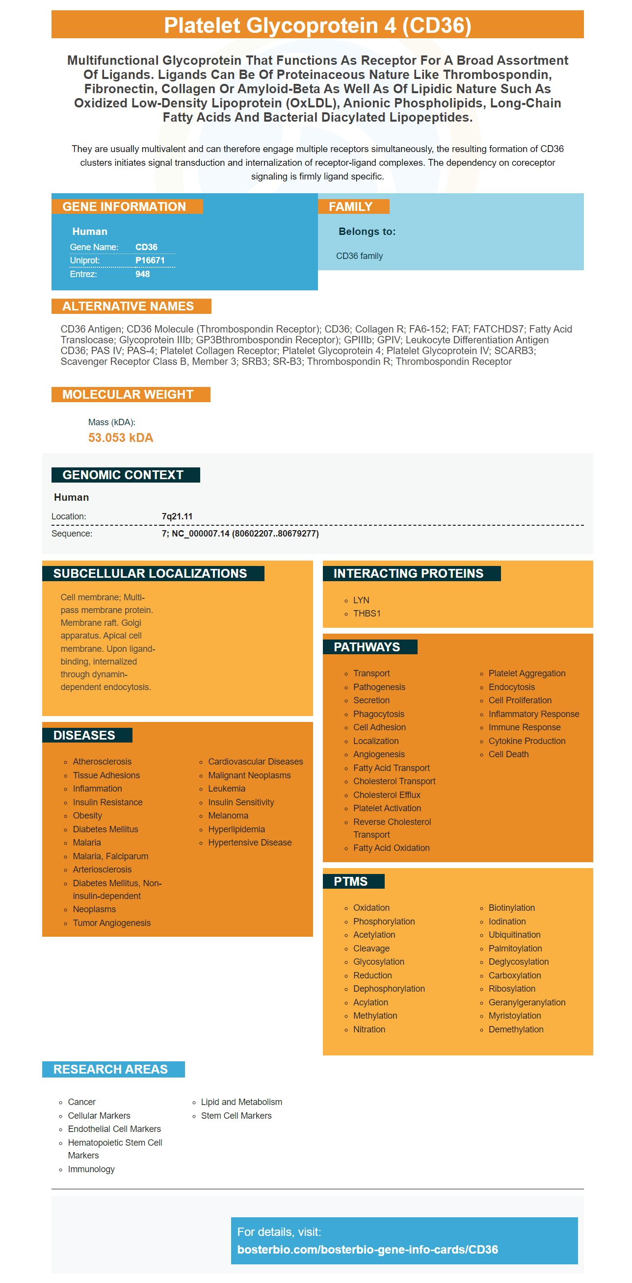
Facts about Platelet glycoprotein 4.
They are usually multivalent and can therefore engage multiple receptors simultaneously, the resulting formation of CD36 clusters initiates signal transduction and internalization of receptor-ligand complexes. The dependency on coreceptor signaling is firmly ligand specific.
Human | |
---|---|
Gene Name: | CD36 |
Uniprot: | P16671 |
Entrez: | 948 |
Belongs to: |
---|
CD36 family |
CD36 antigen; CD36 molecule (thrombospondin receptor); CD36; Collagen R; FA6-152; FAT; FATCHDS7; Fatty acid translocase; Glycoprotein IIIb; GP3Bthrombospondin receptor); GPIIIb; GPIV; Leukocyte differentiation antigen CD36; PAS IV; PAS-4; Platelet collagen receptor; platelet glycoprotein 4; Platelet glycoprotein IV; SCARB3; scavenger receptor class B, member 3; SRB3; SR-B3; Thrombospondin R; Thrombospondin receptor
Mass (kDA):
53.053 kDA
Human | |
---|---|
Location: | 7q21.11 |
Sequence: | 7; NC_000007.14 (80602207..80679277) |
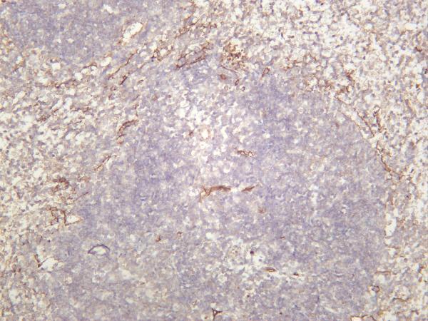
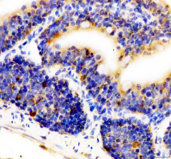
Cell membrane; Multi-pass membrane protein. Membrane raft. Golgi apparatus. Apical cell membrane. Upon ligand-binding, internalized through dynamin-dependent endocytosis.
What Is CD36? What is CD36? How do you test it? Find out in this article the ways Boster Bio can help you. This article will explain how to test your CD36 antibodies with boster bio products. This product is beneficial for many reasons. Boster Bio will confirm the CD36 antibody and provide an antibody that is specific to CD36.
If you're in search of an antibody that recognizes the CD36 marker you're in the right spot. Boster Bio is an antibody manufacturer that specializes in the picogram sensitive ELISA kit and IHC-optimized monoclonal antibody. Boster Bio offers more than 12,000 antibodies that have been validated for WB, Flow Cytometry, and Immunofluorescence. These antibodies are tested on over 250 different samples to ensure high specificity and high affinity. The Boster Bio range of antibodies is specifically designed for IHC, WB, and flow applications. Boster Bio also validates antibodies quantitatively against the known quantities of recombinant proteins.
Researchers have compared the levels of in soluble mediators as well as CD36 protein by using freshly isolated adherent monocytes. The treatment raised CD36 mRNA levels from 6 to 12 fold. Dexamethasone, however, dramatically decreased the levels of CD36's mRNA. The decrease was sustained for up to 24 hours. Multiple soluble mediators responsible for causing CD36 mRNA expression were found. This was reversed when dexamethasone and LPS were not present.
After the incubation period with primary antibodies, cells were stained with a secondary antibody against CD36. After this cell removal was performed using SDS-PAGE. The protein-protein complexes were detected by using the Chemidoc imaging system (Bio-Rad), while intensity of the bands was determined using ImageJ software. A sample of monoclonal antibody to CD36 (Cat# 562702) was used for each analysis.
While a soluble mediator, soluble factor, and cell surface receptor (MHC) were examined to determine the role played by CD36 in lipophagy However, they were unable to establish a connection between these molecules. These studies suggest that soluble mediators may be a key factor when CD36 levels are controlled in cells. In addition, upregulation by force of CD36 inhibits lipophagy.
The antibody inhibited CD36 in microphages from the peritoneal region. It was discovered that BMDM have lower levels of CD36 than macrophages residing in the peritoneal area. However, peritoneal macrophages exhibited higher levels of the anti-CD36 protein , and released more TNF-a. This finding provides a rationale for a new treatment for atherosclerosis.
The CD36 marker is a pleiotropic agent that has many functions, including negative regulation of autophagy. It is also present in cardiac muscle cells as well as platelets. CD36 has also been implicated in platelet activation and cell adhesion. The CD36 molecule is present on many different types of cells, including the hepatocytes.
The CD36 molecule has two transmembrane regions, an extracellular one with multiple binding sites for ligands as well as short cytoplasmic tails that are located on both ends. The extracellular domain is able to recognize and cleaves the ligands, such as fatty acid and oxygenated LDL. It also undergoes many posttranslational modifications. The recognition of b–amyloid peptides and AGEs is done by the CD36 molecule.
The TSP-1 marker is associated with CD36 and inhibits VEGF signaling. Therefore, targeting TSP-1 could be a beneficial therapy option for cancer patients. The TSP-1 ligand also hinders VEGF synthesis and degradation, which is a key regulator of the growth of vascular cells. Hence, anti-LC3 antibodies are an essential part of cancer therapy. The treatment of anti-LC3 can cause irritation and complications.
Although there isn't consensus regarding the role of CD36 as a tumor suppressor, many studies have shown its importance in tumor progression. CD36 is a major scavenger, and is involved in inflammation, lipid homeostasis and recognition by immunological means. The CD36 molecule is also known to interact with multiple ligands. The ligand TSP-1 is known to reduce tumor metastasis through the SR-BI pore, a biomarker that could be a therapeutic target.
In cancer, the CD36 marker plays a crucial part in immune responses. Inflammation stimulates the growth of tumor cells by promoting the infiltration of NK cells and T cells. The CD36 marker regulates the expression of inflammatory mediators in tumor cells. This causes the proliferation, invasion, and metastasis. Therefore, a thorough investigation of CD36 expression and distribution is required to understand its function.
At present, the only way to determine if cancer metastasis-initiating cancer cells are those that have been expressed in cancer tumors is to test their expression. CD36 is expressed in tumor stroma cells. The ability to stop this signaling pathway could hinder the growth of tumors that contain high levels of CD36. This discovery will be an important step forward in our understanding of the process of cancer metastasis. This means that we now have a reliable way to identify cancerous tumors.
The expression of CD36 is a major determinant of the activity of intratumoral Treg cells and accumulation. This gene is also known to affect the Stability of intratumoral Trg cells. Ablation of CD36 in Boster Bio mice eliminates this determinant and allows tumor cells to express more activation markers. However, CD36 ablation does not hinder tumor growth.
CD36 blockade is an anti-tumor medication which can be utilized as an alternative to PD-1 blocking. This means that it can be used in conjunction with PD-1 inhibition to reduce the side effects of T-cell exhaustion. Patients with CD36-deficient tumor lines are significantly less likely than those who do not have it to develop cancer. Furthermore, CD36 deficiency decreases the expression of other PD-1-based cancer-fighting molecules.
In addition, GW501516 treatment enhances NAD/NADH ratios in CD36-deficient Treg cells. This treatment could also result in enhanced anti-tumor immunity. The combination of this treatment with anti-PD-1 therapy could also be the reason for further development of CD36 inhibitors. When a breakthrough is made in the development of an effective treatment, the possibility of a safe, effective, and affordable option for treating cancer is extremely high.
The results of this research show that CD36-mediated metabolic adaptation could instruct Treg cells in the intratumoral area. Boster has actually validated the metabolic adaptation mediated by CD36 in a variety of animal models. Utilizing anti-CD36-based medicines and vaccines, we have shown that CD36-targeted antibodies can be used to treat cancer in mice. The results of the Boster Bio antibody are promising.
The CD36 marker is involved in many biological processes. Many tissues and cells express this marker. It is involved in many developmental, pathological and homeostatic processes. It regulates numerous functions and transmits intracellular signals. It is composed of two transmembrane zones and an extracellular region that contains six Cystineines that are joined by three disulfide bonds. The protein also contains an hydrophobic region that spans amino acids in 184 and 204.
CD36, glycoprotein IV was first identified more than 30 years ago. It was first identified as the fourth major band of gels that contain platelets. Later studies revealed that the protein was identical to an antigen identified by the OKM5 antibody. This is a marker for monocytes and macrophages. CD36 is a ubiquitous membrane protein that is found in wide variety of mammalian cell types including microvascular endothelium, macrophages with epithelia that is specialized, as well as macrophage.
The CD36 marker is a glycoprotein located on the membranes of platelets, monocytes Adipocytes and phagocytes myocytes, liver cells, and some epithelia. It also acts as a receptor for thrombospondin-1 and is involved in the internalization of apoptotic cells and modified low-density lipoproteins. The CD36 molecule contributes to the regulation of angiogenesis, inflammatory reactions and atherothrombotic disorders. Furthermore, it binds long-chain fatty acids, facilitating their transportation.
PMID: 2473841 by Oquendo P., et al. CD36 directly mediates cytoadherence of Plasmodium falciparum parasitized erythrocytes.
PMID: 7693552 by Taylor K.T., et al. Characterization of two alternatively spliced 5'-untranslated exons of the human CD36 gene in different cell types.
*More publications can be found for each product on its corresponding product page