This website uses cookies to ensure you get the best experience on our website.


- Table of Contents
Plan CEL chromogenic IHC in paraffin sections with pancreatic exocrine cells as a high-staining reference and lactating glandular cells as a medium-staining comparison (HPA tissue IHC). This guide covers fixation consistency and the catalog antibody’s 1:100–1:300 IHC dilution (datasheet A01821-1).

Expected localisation, validated protocols, controls and antibodies — the at-a-glance facts below, then the full design guide.
| Expected localisation | CEL is secreted (UniProt); glandular cell staining is observed (HPA tissue IHC). | |
| Staining pattern | Pancreatic exocrine cells: high; lactating glands: medium; subcellular pattern unreported (HPA tissue IHC). | |
| Antigen retrieval | Tris-EDTA pH 9.0 HIER, heat-mediated (datasheet A01821-1) | |
| Positive control | Pancreas+1 more · see all | |
| Negative control | Adipose tissue+4 more · see all |
| Fixation | Keep fixation consistent across paraffin sections. (standard IHC practice; not target-specific) | |
| Caveat | Secreted CEL may stain away from its tissue RNA source (HPA tissue IHC). | |
| Regulation | Lactating glands show medium staining (HPA tissue IHC). | |
| Isoform / epitope | 2 isoforms; mature chain starts at residue 21; epitope differences unspecified (UniProt). |
The catalog antibody’s IHC-P protocol (datasheet A01821-1) is paired with a published CEL protein IHC protocol in mouse pancreas (PMC9117557).
| Sample | Paraffin-embedded human tonsil tissue; fixative not specified (datasheet A01821-1) |
| Fixation | Image fixative and duration unreported (datasheet A01821-1); verify before use. |
| Sectioning | 4–5 µm sections on charged slides (standard) |
| Deparaffinisation | Xylene, graded ethanol series to water (standard) |
| Antigen retrieval | Heat retrieval: Tris-EDTA pH 9.0 (datasheet A01821-1); 20 min, 95–100 °C (standard) |
| Peroxidase block | 3% H2O2, 10 min, room temperature (standard) |
| Blocking | 10% normal serum of the secondary host, 30 min, room temperature (standard) |
| Primary antibody | Rabbit anti-CEL, 1:100-1:300 (datasheet A01821-1) |
| Primary incubation | Overnight at 4 °C (standard) |
| Detection | HRP-polymer secondary, DAB chromogen 5–10 min (standard) |
| Counterstain | Hematoxylin, blue, dehydrate and mount (standard) |
| Expected result | CEL-positive staining in exocrine glandular cells of pancreas (HPA tissue IHC: High). HPA tissue profile: Selective expression in exocrine glandular cells of pancreas and glandular cells in lactating breast. No signal in the no-primary control. |
CEL is a secreted protein with no transmembrane segment (UniProt P19835 topology). In paraffin section IHC, expect the strongest staining in pancreatic exocrine glandular cells and a weaker positive pattern in lactating breast glandular cells (HPA: High in pancreas; Medium in lactating breast). HPA rates the tissue IHC profile Enhanced, while cautioning that a secreted protein’s staining location may differ from its site of RNA expression (HPA: reliability description).
| Strong staining in pancreatic exocrine glandular cells, with weaker staining in lactating breast glandular cells. | This matches the reported cell types and relative levels (HPA: High in pancreatic exocrine cells; Medium in lactating glandular cells). Assess the relevant cells rather than assigning one intensity threshold to both tissues; secretion can also separate protein location from RNA location (HPA: reliability description). |
| Predominantly nuclear staining, or a crisp cell-surface outline without a glandular secretory pattern. | Question target specificity: CEL is annotated as secreted and has no transmembrane segment (UniProt P19835 topology). Those features do not establish a precise intracellular IHC pattern; HPA provides no ICC-IF image-based location for CEL (HPA: subcellular summary). |
| Prominent staining in cell types reported as negative, such as adipocytes in adipose tissue. | HPA reports no detection in those adipocytes (HPA: adipose tissue IHC). If staining persists, consider off-target binding or endogenous detection activity as general IHC possibilities. Compare tissue morphology and appropriate staining controls before calling CEL positive. |
| Diffuse chromogen across glands, stroma, and cells without a coherent cell-type pattern. | Diffuse background cannot establish CEL localisation. The reported tissue pattern is selective for pancreatic exocrine and lactating breast glandular cells (HPA: tissue IHC profile). In general IHC practice, nonspecific reagent binding, detection chemistry, or incomplete washing can obscure a selective signal. |
| No convincing staining in pancreatic exocrine glandular cells. | Treat the run as inconclusive before interpreting another tissue as CEL negative: this is the strongest reported positive population (HPA: High in pancreatic exocrine cells). Check tissue identity and preservation, controls, antibody use, and detection performance using general IHC practice; HPA supplies no CEL-specific fixation sensitivity. |
| Secreted protein and processing | CEL has a signal peptide at residues 1–20 and a mature chain at 21–753, with no transmembrane segment (UniProt P19835). Interpret cellular staining alongside possible secreted protein outside the producing cell; neither source supplies a precise IHC compartment map. |
| Tissue and cell context | Pancreatic exocrine cells are High, lactating glandular cells Medium, and several sampled cell types are Not detected (HPA: tissue IHC). HPA also notes that secreted protein and RNA locations may differ; RNA enrichment alone cannot set the stained-cell boundary. |
| Isoforms and glycosylation | UniProt lists Long and Short isoforms and 11 glycosylation sites (UniProt P19835). Antibody epitopes are not supplied here, so the record cannot predict whether either feature changes recognition or justify an antigen retrieval choice. |
| IHC validation and subcellular limits | Two listed antibodies, HPA008023 and HPA052701, have Enhanced IHC status (HPA: antibodies). HPA reports CEL as secreted but provides no ICC-IF images or main intracellular location (HPA: subcellular). These data support tissue-pattern comparison, not a fixed organelle assignment. |
| Situation | Likely cause | Next action |
|---|---|---|
| Pancreatic positive tissue is blank or faint. | A failed run is possible because exocrine glandular cells are reported High (HPA: pancreas IHC); these sources identify no CEL-specific fixation effect. | Verify the exocrine region and control slides, then review the antibody instructions and routine IHC detection steps. Record any retrieval changes as assay optimisation, not a demonstrated CEL requirement (general IHC practice). |
| Lactating breast looks weaker than pancreas. | Different strength is expected: lactating glandular cells are Medium and pancreatic exocrine cells High (HPA: tissue IHC). | Score each cell population against its own expected level and morphology. A weaker breast result alone does not indicate assay failure when the pancreatic positive control performs as expected. |
| Adipocytes or unrelated tissue cells stain strongly. | Those cells may conflict with reported negatives; adipocytes in adipose tissue are Not detected (HPA: adipose tissue IHC). Off-target binding or endogenous detection activity are general IHC possibilities. | Review cell identity, omit-primary and detection controls, and background distribution. Interpret staining as CEL only when the cell pattern is supported by the tissue evidence. |
| Chromogen coats much of the section. | Broad staining lacks the selectivity reported for CEL (HPA: tissue IHC profile). Reagent background or endogenous activity can produce diffuse colour (general IHC practice). | Inspect control slides and repeat with routine blocking, washing, and detection checks as appropriate (general IHC practice). Avoid assigning a CEL-positive cell type from diffuse colour alone. |
| Only nuclei or sharp plasma membranes stain. | A dominant nuclear or membrane outline does not follow the secreted, non-transmembrane annotation (UniProt P19835 topology); a precise intracellular reference pattern is unavailable (HPA: subcellular). | Check morphology and control staining, then compare the result with pancreatic exocrine and lactating glandular patterns (HPA: tissue IHC). Treat the unusual compartment as unresolved unless independently supported. |
| IF/ICC question: should the IHC pattern define an IF-positive cell? | No CEL ICC-IF images or main intracellular location are provided (HPA: subcellular); the listed antibodies have Enhanced IHC status but no ICC status (HPA: antibodies). | Use the separate IF/ICC guide for that application. The IHC tissue pattern can guide expectations, but it does not validate an IF compartment or substitute for IF-specific controls. |
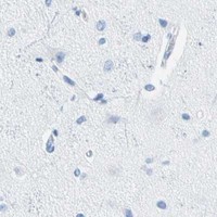


Comprehensive Human Protein Atlas IHC scoring per tissue (reliability: Enhanced — High consistency between antibody staining and RNA expression data. Secreted protein, tissue location of RNA and protein is expected to differ.). Rows are taken directly from the HPA tissue chart — click any row's HPA link to view the source.
| Tissue | Cell type | Level | Evidence | Source |
|---|---|---|---|---|
| Pancreas | Exocrine glandular cells | High | Protein (IHC) | HPA → |
| Lactating breast | Lactating glandular cells | Medium | Protein (IHC) | HPA → |
| Tissue | Cell type | Level | Evidence | Source |
|---|---|---|---|---|
| Adipose tissue | Adipocytes | Not detected | Protein (IHC) | HPA → |
| Adrenal gland | Glandular cells | Not detected | Protein (IHC) | HPA → |
| Appendix | Glandular cells | Not detected | Protein (IHC) | HPA → |
| Bone marrow | Hematopoietic cells | Not detected | Protein (IHC) | HPA → |
| Breast | Adipocytes | Not detected | Protein (IHC) | HPA → |
Troubleshoot CEL staining in paraffin sections by checking retrieval, cellular context, controls, and scoring before interpreting the signal.
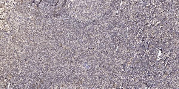
A01821-1 has IHC data from paraffin-embedded human tonsil (catalog IHC image caption). IF is listed, and Human, Mouse and Rat are listed as reactive species (catalog applications; catalog reactivity).
A01821-1 has an IHC image from paraffin-embedded human tonsil, using 1:200 primary antibody and Tris-EDTA retrieval at pH 9.0 (catalog IHC image caption). IF is listed for A01821-1, but no IF image is supplied (catalog applications; catalog IF image alts).
Which to pick: Choose A01821-1 for paraffin-section tissue IHC because its own image documents human tonsil staining; the fixative is unreported (catalog IHC image caption). For IF/ICC, A01821-1 lists IF at 1:50, but the payload supplies no IF image or ICC validation (catalog applications; catalog IF dilution; catalog IF image alts). For cross-species work, A01821-1 lists Human, Mouse and Rat reactivity; it is rabbit-hosted, and its clone is unreported (catalog reactivity; catalog host; catalog clone).