This website uses cookies to ensure you get the best experience on our website.


- Table of Contents
Plan CGN staining in paraffin sections with the IHC-validated antibody at 1:50–1:200 (datasheet: A02373). Use high-staining epithelial tissue as a positive control and assess cytoplasmic and membranous signal in light of CGN’s expected junctional location (HPA tissue IHC; UniProt).

Expected localisation, validated protocols, controls and antibodies — the at-a-glance facts below, then the full design guide.
| Expected localisation | Cytoplasmic and membranous staining (HPA tissue IHC); apical junction expected (UniProt) | |
| Staining pattern | Epithelia show cytoplasmic and membranous staining (HPA tissue IHC) | |
| Antigen retrieval | Citrate pH 6.0 HIER, 95–98 °C, 20 min (rule: cytoplasmic / membrane antigen) | |
| Positive control | Appendix+4 more · see all | |
| Negative control | Adipose tissue+4 more · see all |
| Fixation | Keep formalin fixation consistent across sections. (standard IHC practice; not target-specific) | |
| Caveat | Antibody staining and RNA show medium consistency (HPA tissue IHC) | |
| Regulation | Intestine-enhanced RNA expression (HPA tissue RNA) | |
| Isoform / epitope | 2 isoforms; check epitope coverage (UniProt) |
The catalog antibody’s IHC-P protocol is followed by published CGN staining methods for human lung tissue (PMC10855320), dog skin (PMC13314070), and salivary duct adenocarcinoma tissue (PMC9179926).
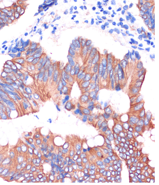
| Sample | Paraffin-embedded Human colon carcinoma tissue; fixative not specified (datasheet A02373) |
| Fixation | Image fixative and duration unreported (datasheet A02373); verify before use. |
| Sectioning | 4–5 µm sections on charged slides (standard) |
| Deparaffinisation | Xylene, graded ethanol series to water (standard) |
| Antigen retrieval | Heat-induced epitope retrieval in citrate buffer, pH 6.0, 20 min at 95–98 °C (standard rule: cytoplasmic / membrane antigen) |
| Peroxidase block | 3% H2O2, 10 min, room temperature (standard) |
| Blocking | 10% normal serum of the secondary host, 30 min, room temperature (standard) |
| Primary antibody | Rabbit anti-CGN, 1:50-1:200 (datasheet A02373) |
| Primary incubation | Overnight at 4 °C (standard) |
| Detection | HRP-polymer secondary, DAB chromogen 5–10 min (standard) |
| Counterstain | Hematoxylin, blue, dehydrate and mount (standard) |
| Expected result | CGN-positive staining in enterocytes - Microvilli of appendix (HPA tissue IHC: High). HPA tissue profile: Cytoplasmic and membranous expression in several tissues, most abundant in gastrointestinal tract. No signal in the no-primary control. |
CGN is a junctional protein on the cytoplasmic face of tight junctions in polarized epithelia and some endothelia; it has no transmembrane segment (UniProt Q9P2M7). In paraffin-section IHC, expect staining in selected epithelial cells, especially gastrointestinal epithelium (HPA tissue IHC). HPA describes cytoplasmic and membranous staining, with Enhanced reliability but only medium consistency between antibody staining and RNA expression (HPA tissue IHC).
| Apical-border staining in colon or duodenal enterocytes, with some cytoplasmic signal. | This fits high enterocyte staining and HPA’s cytoplasmic and membranous profile (HPA tissue IHC). Junction-associated enrichment fits CGN’s location on the cytoplasmic face of epithelial tight junctions (UniProt Q9P2M7). The tissue image alone does not resolve every junction. |
| Strong, isolated nuclear staining dominates otherwise positive epithelium. | A nuclear-dominant pattern is discordant with the reported cell-junction location and HPA membrane/cell-junction ICC-IF localization (UniProt Q9P2M7; HPA subcellular). Treat it as suspect; review controls and detection before assigning it to CGN. |
| Prominent staining appears in an HPA-negative cell population or across unrelated cells. | That distribution needs scrutiny for antibody cross-reactivity or endogenous detection activity (general IHC practice). HPA reports no detection in adipocytes or bone-marrow hematopoietic cells, for example; those findings apply to the specified cells, not every cell in those tissues (HPA tissue IHC). |
| Uniform haze covers cells and surrounding tissue, obscuring cell borders. | The haze does not show the expected cell-linked distribution (UniProt Q9P2M7; HPA tissue IHC). Nonspecific antibody binding, incomplete blocking, or chromogen background are possible general IHC causes; use controls to distinguish them (general IHC practice). |
| No signal appears in colon enterocytes or kidney collecting ducts. | Both are reported as High in the named cell populations (HPA tissue IHC). A blank run therefore warrants checking tissue preservation, primary-antibody use, retrieval, and detection with controls (general IHC practice); it does not alone establish absent CGN. |
| Compartment and topology | CGN is on the cytoplasmic face of tight junctions and has no transmembrane segment (UniProt Q9P2M7). HPA also reports cytoplasmic and membranous tissue staining (HPA tissue IHC). Score the cell-border pattern with that resolution limit in mind; membrane staining alone does not prove precise tight-junction placement. |
| Choice of positive and comparison cells | HPA rates appendix, colon, and duodenal enterocytes, bronchial respiratory epithelium, and kidney collecting ducts High (HPA tissue IHC). Compare the named cell populations, because the HPA calls are cell-specific; a negative call for liver cholangiocytes does not negate UniProt’s broader report of liver expression (HPA tissue IHC; UniProt Q9P2M7). |
| Strength of antibody evidence | Four listed HPA antibodies have Enhanced IHC validation, while HPA reports medium consistency between staining and RNA expression (HPA antibodies; HPA tissue IHC). A matching distribution supports interpretation, but ambiguous staining still needs slide controls and cellular context (general IHC practice). |
| Isoforms and target modifications | UniProt lists two isoforms and multiple modified residues, including phosphoserines (UniProt Q9P2M7). The supplied record gives no antibody epitope or evidence that these features alter IHC staining; do not infer isoform-specific staining or a retrieval requirement from them. |
| Situation | Likely cause | Next action |
|---|---|---|
| Known-positive epithelium is blank. | The run may have a reagent, retrieval, or detection problem (general IHC practice); colon enterocytes are reported High (HPA tissue IHC). | Check a positive control in the same run, then verify primary-antibody application and detection steps (general IHC practice). The supplied sources specify no CGN-specific retrieval condition. |
| The slide shows mostly nuclear signal. | That location conflicts with junctional CGN and supported HPA plasma-membrane/cell-junction localization (UniProt Q9P2M7; HPA subcellular). | Check the no-primary control and examine whether expected epithelial borders stain (general IHC practice); avoid scoring isolated nuclei as CGN-positive. |
| Many unrelated cells stain strongly. | Cross-reactivity or endogenous detection activity is possible (general IHC practice); HPA staining levels are assigned to specific cell populations (HPA tissue IHC). | Review no-primary and detection controls, then compare each stained population with its HPA cell-level call (general IHC practice; HPA tissue IHC). |
| Background obscures cell boundaries. | Nonspecific binding or background from detection may be contributing (general IHC practice). | Use appropriate blocking and a no-primary control; adjust staining conditions only against a positive control so interpretable epithelial signal is retained (general IHC practice). |
| A nominally negative tissue contains focal staining. | The negative call may concern a different cell population: HPA reports liver cholangiocytes as Not detected, while UniProt reports liver expression (HPA tissue IHC; UniProt Q9P2M7). | Identify the stained cells before calling the result discordant; assess their compartment and control staining (general IHC practice). |
| Can ICC-IF help assess localization? | HPA supports plasma-membrane and cell-junction localization and lists ICC-IF images for A-431, U-251MG, and U2OS (HPA subcellular). | Use those observations as localization context, while interpreting the paraffin IHC slide against its tissue and cell-level evidence (HPA subcellular; HPA tissue IHC). |
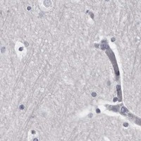


Comprehensive Human Protein Atlas IHC scoring per tissue (reliability: Enhanced — Medium consistency between antibody staining and RNA expression data.). Rows are taken directly from the HPA tissue chart — click any row's HPA link to view the source.
| Tissue | Cell type | Level | Evidence | Source |
|---|---|---|---|---|
| Appendix | Enterocytes - Microvilli | High | Protein (IHC) | HPA → |
| Bronchus | Respiratory epithelial cells | High | Protein (IHC) | HPA → |
| Colon | Enterocytes - Microvilli | High | Protein (IHC) | HPA → |
| Duodenum | Enterocytes - Microvilli | High | Protein (IHC) | HPA → |
| Fallopian tube | Glandular cells | High | Protein (IHC) | HPA → |
| Tissue | Cell type | Level | Evidence | Source |
|---|---|---|---|---|
| Adipose tissue | Adipocytes | Not detected | Protein (IHC) | HPA → |
| Adrenal gland | Glandular cells | Not detected | Protein (IHC) | HPA → |
| Bone marrow | Hematopoietic cells | Not detected | Protein (IHC) | HPA → |
| Cerebral cortex | Endothelial cells | Not detected | Protein (IHC) | HPA → |
| Heart muscle | Cardiomyocytes | Not detected | Protein (IHC) | HPA → |
CGN staining is most useful when junctional signal, tissue context, and processing controls agree (UniProt Q9P2M7; HPA tissue IHC).
A02373 has an IHC image from human paraffin-embedded colon carcinoma (A02373 image caption); A02373-1 lists human, mouse and rat IHC reactivity (catalog applications/reactivity). Neither lists IF validation (catalog applications/images).
The rendered A02373 card shows IHC on paraffin-embedded human colon carcinoma at 1:100 after microwave retrieval in 10 mM PBS, pH 7.2 (A02373 image caption). A02373-1 lists IHC for human, mouse and rat, but has no IHC image in the supplied catalog (A02373-1 applications/reactivity/images).
Which to pick: For tissue IHC, choose A02373 when the documented human paraffin-section example fits your sample; its caption does not report the fixative (A02373 image caption). For mouse or rat tissue, consider the rabbit polyclonal A02373-1, which lists IHC and reactivity with both species but supplies no processing example (A02373-1 catalog host, applications, reactivity and images). Neither SKU is listed for IF/ICC, so there is no catalog-supported IF/ICC pick here (catalog applications/images).