This website uses cookies to ensure you get the best experience on our website.


- Table of Contents
Plan chromogenic IHC on paraffin sections with the catalog antibody at 1:50 (datasheet: IHC 1:50). Interpret the observed cytoplasmic staining, most abundant in immune cells (HPA tissue IHC), alongside CLEC4C’s membrane location (UniProt) and the reported low agreement between staining and RNA expression (HPA tissue IHC).

Expected localisation, validated protocols, controls and antibodies — the at-a-glance facts below, then the full design guide.
| Expected localisation | Cytoplasmic in tissue (HPA tissue IHC); cell membrane (UniProt) | |
| Staining pattern | Cytoplasmic staining in most tissues, strongest in immune cells (HPA tissue IHC) | |
| Antigen retrieval | Citrate pH 6.0 HIER, 95–98 °C, 20 min (rule: cytoplasmic / membrane antigen) | |
| Positive control | Adrenal gland+4 more · see all | |
| Negative control | None in HPA (detected in all 45 tissues); use no-primary + isotype controls |
| Fixation | Keep fixation consistent across paraffin sections. (standard IHC practice; not target-specific) | |
| Caveat | Low agreement between staining and RNA expression (HPA tissue IHC) | |
| Regulation | Downregulated after LPS-induced maturation (UniProt) | |
| Isoform / epitope | 2 isoforms; epitope location requires checking (UniProt) |
Compare the catalog antibody’s IHC-P protocol (datasheet) with the published CLEC4C staining protocol (PMC9045650: methods).
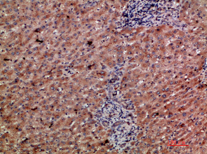
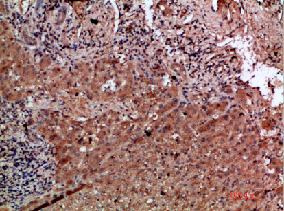
| Sample | Paraffin-embedded human liver-cancer tissue; fixative not specified (datasheet A07313) |
| Fixation | Image fixative and duration unreported (datasheet A07313); verify before use. |
| Sectioning | 4–5 µm sections on charged slides (standard) |
| Deparaffinisation | Xylene, graded ethanol series to water (standard) |
| Antigen retrieval | Heat-induced epitope retrieval in citrate buffer, pH 6.0, 20 min at 95–98 °C (standard rule: cytoplasmic / membrane antigen) |
| Peroxidase block | 3% H2O2, 10 min, room temperature (standard) |
| Blocking | 10% normal serum of the secondary host, 30 min, room temperature (standard) |
| Primary antibody | Rabbit anti-CLEC4C, 1:50 (datasheet A07313) |
| Primary incubation | Overnight at 4 °C (standard) |
| Detection | HRP-polymer secondary, DAB chromogen 5–10 min (standard) |
| Counterstain | Hematoxylin, blue, dehydrate and mount (standard) |
| Expected result | CLEC4C-positive staining in glandular cells of adrenal gland (HPA tissue IHC: High). HPA tissue profile: Cytoplasmic expression in most tissues, most abundant in immune cells. No signal in the no-primary control. |
CLEC4C is a cell membrane receptor with an extracellular domain spanning residues 45–213 (UniProt Q8WTT0 topology). Plasmacytoid dendritic cells are the main biological expectation (UniProt Q8WTT0 tissue specificity). HPA reports predominantly cytoplasmic staining across tissues, most abundant in immune cells; its IHC rating is Approved, with low consistency between staining and RNA expression (HPA tissue IHC).
| Discrete staining in immune cells, with membrane accentuation. | This fits CLEC4C's cell surface location and plasmacytoid dendritic cell expression (UniProt Q8WTT0). Morphology alone cannot establish plasmacytoid dendritic cell identity (general IHC practice); HPA also reports abundant immune cell staining (HPA tissue IHC). |
| Predominantly cytoplasmic staining, including glandular cells. | This resembles HPA's reported tissue pattern, including High staining in several glandular compartments (HPA tissue IHC). It conflicts with a purely surface pattern expected from UniProt topology; interpret it cautiously because HPA reports low staining–RNA consistency (HPA tissue IHC; UniProt Q8WTT0 topology). |
| Dominant nuclear staining or another compartment inconsistent with membrane or reported cytoplasmic staining. | Treat this as a possible artefact: UniProt places CLEC4C at the cell membrane, while HPA reports cytoplasmic tissue staining (UniProt Q8WTT0 subcellular location; HPA tissue IHC). Check controls and staining distribution before assigning target localisation (general IHC practice). |
| Broad staining in cells outside the expected immune population. | Do not automatically call it cross-reactivity: HPA reports High glandular staining in colon, duodenum and gallbladder (HPA tissue IHC). Compare tissue controls and cell morphology; cross-reactivity or endogenous detection activity remains possible if the pattern is inconsistent or persists in a no-primary control (general IHC practice). |
| No signal in a tissue compartment reported as strongly stained. | First check that the expected cells are present: HPA reports High staining in lung macrophages and lymph node non-germinal center cells (HPA tissue IHC). A blank result may reflect a failed staining run or antibody performance; HPA's Approved rating does not guarantee staining in every preparation (HPA tissue IHC; general IHC practice). |
| Epitope position | CLEC4C has a cytoplasmic region at residues 1–21, a transmembrane segment at 22–44 and an extracellular region at 45–213 (UniProt Q8WTT0 topology). The supplied record does not locate the IHC antibody's epitope, so it cannot predict its section staining pattern. |
| Glycosylation and variants | UniProt lists glycosylation sites at 110, 137 and 164 and 2 isoforms (UniProt Q8WTT0). These facts do not establish whether this antibody distinguishes isoforms or whether glycosylation changes IHC staining; do not use them to explain a weak result without antibody-specific evidence. |
| Cell population and maturation | UniProt reports expression in plasmacytoid dendritic cells and immature monocyte-derived dendritic cells, with down-regulation after LPS-driven maturation but not TNF treatment (UniProt Q8WTT0 tissue specificity). Cell composition can therefore affect the biological expectation; the supplied HPA IHC data do not identify every stained immune cell subtype (HPA tissue IHC). |
| Tissue IHC evidence | HPA rates the listed antibody Approved and describes low consistency between antibody staining and RNA expression; it does not list an IHC Enhanced rating (HPA tissue IHC; HPA antibody validation). Use its High and Low calls as observed staining levels, not proof of cell-specific expression or validated negative controls. |
| Antigen retrieval | Retrieval is a general paraffin IHC variable to optimize with appropriate controls (general IHC practice). No supplied source reports a CLEC4C-specific retrieval condition or fixation sensitivity; neither UniProt topology nor HPA staining levels justify predicting a target-specific retrieval effect. |
| IF/ICC Q&A | Q: Should IF/ICC show membrane signal? A: Membrane localisation is the biological expectation (UniProt Q8WTT0; HPA subcellular summary), but HPA supplies no ICC-IF images or main-location assignment for CLEC4C (HPA subcellular). Interpret any IF pattern on its own controls; no IF protocol follows from these records. |
| Situation | Likely cause | Next action |
|---|---|---|
| No staining in lung macrophages or lymph node non-germinal center cells. | The expected cells may be absent from the section, or the IHC run may have failed (HPA tissue IHC; general IHC practice). | Confirm the cell compartment on the counterstained section and inspect a known-performing run control; then review retrieval, primary application and detection steps as general IHC checks (general IHC practice). |
| Diffuse color across tissue and blank areas. | Background from detection reagents, inadequate blocking or washing is possible (general IHC practice). | Compare a no-primary control and reagent-only background, then adjust blocking or washes according to the detection system (general IHC practice); diffuse color is not a documented CLEC4C localisation (UniProt Q8WTT0; HPA tissue IHC). |
| Strong staining of unexpected cell types. | It may reflect HPA's broad cytoplasmic pattern, cross-reactivity or endogenous detection activity; HPA reports low staining–RNA consistency (HPA tissue IHC; general IHC practice). | Check whether the cell type appears in HPA's tissue observations, compare tissue controls and examine a no-primary control before assigning CLEC4C expression (HPA tissue IHC; general IHC practice). |
| Nuclear staining dominates. | This differs from UniProt's membrane location and HPA's reported cytoplasmic IHC profile (UniProt Q8WTT0; HPA tissue IHC). | Treat nuclear signal as unconfirmed; inspect controls and review counterstain and detection background before scoring positive cells (general IHC practice). |
| Only weak staining in oral mucosa or splenic red pulp. | HPA lists oral squamous epithelial cells and splenic red pulp cells as Low, rather than negative (HPA tissue IHC). | Avoid using either compartment as an absolute negative control; compare with an HPA High compartment and score the observed cell type separately (HPA tissue IHC; general IHC practice). |
| Predominantly cytoplasmic signal despite an expected surface receptor. | HPA explicitly describes cytoplasmic tissue staining, while UniProt places CLEC4C at the membrane (HPA tissue IHC; UniProt Q8WTT0). | Record the compartment and cell type separately, compare controls and report the source disagreement; do not infer a CLEC4C-specific fixation or retrieval mechanism from these records. |


Comprehensive Human Protein Atlas IHC scoring per tissue (reliability: Approved — Low consistency between antibody staining and RNA expression data.). Rows are taken directly from the HPA tissue chart — click any row's HPA link to view the source.
| Tissue | Cell type | Level | Evidence | Source |
|---|---|---|---|---|
| Adrenal gland | Glandular cells | High | Protein (IHC) | HPA → |
| Colon | Glandular cells | High | Protein (IHC) | HPA → |
| Duodenum | Glandular cells | High | Protein (IHC) | HPA → |
| Endometrium | Cells in endometrial stroma | Medium | Protein (IHC) | HPA → |
| Gallbladder | Glandular cells | High | Protein (IHC) | HPA → |
| Tissue | Cell type | Level | Evidence | Source |
|---|---|---|---|---|
| None in HPA: CLEC4C is detected in all 45 scored tissues. Use a no-primary (secondary-only) and an isotype control instead. | ||||
These questions focus on CLEC4C staining in paraffin sections, with one entry on IF multiplexing (selected IHC caption; UniProt Q8WTT0).
A07313 is a human-reactive anti-CLEC4C antibody with an IHC image from a paraffin-embedded human liver cancer sample (catalog: reactivity; A07313 IHC caption). IF is listed, but no IF image is supplied (catalog: applications; image alts).
A07313 will render with an IHC figure from paraffin-embedded human liver cancer (A07313 IHC caption). Its listed applications include IHC and IF, with human reactivity; no IF figure is supplied (catalog: applications, reactivity, image alts).
Which to pick: Choose A07313 for human tissue IHC: its own image caption documents staining of a paraffin-embedded human liver cancer sample, but does not report the fixative (A07313 IHC caption). For IF/ICC, A07313 lists IF and human reactivity, although the payload provides no IF image or ICC validation; its host is rabbit and clonality is unspecified (catalog: applications, reactivity, host, clone, image alts). No cross-species option is documented because A07313 lists human reactivity only (catalog: reactivity).