This website uses cookies to ensure you get the best experience on our website.


- Table of Contents
Plan CLUH staining in paraffin sections around the cytoplasmic tissue pattern reported by HPA (HPA tissue IHC). The catalog antibody has an IHC dilution range of 1:50–1:200 (datasheet); interpret staining cautiously because HPA rates the tissue evidence uncertain (HPA tissue IHC).

Expected localisation, validated protocols, controls and antibodies — the at-a-glance facts below, then the full design guide.
| Expected localisation | Cytoplasmic staining in most tissues (HPA tissue IHC) | |
| Staining pattern | Cytoplasmic staining in most tissues, including smooth muscle (HPA tissue IHC) | |
| Antigen retrieval | Citrate pH 6.0 HIER, 95–98 °C, 20 min (rule: cytoplasmic / membrane antigen) | |
| Positive control | Breast+4 more · see all | |
| Negative control | Adipose tissue+4 more · see all |
| Fixation | Keep fixation consistent across paraffin sections. (standard IHC practice; not target-specific) | |
| Caveat | Antibody staining and RNA expression have low consistency (HPA tissue IHC) | |
| Regulation | Low tissue RNA specificity; no regulator established (HPA tissue RNA) | |
| Isoform / epitope | One full-length chain; no isoforms reported (UniProt) |
The catalog antibody IHC-P protocol (datasheet) is accompanied by a published CLUH protocol for human colonic biopsy sections (PMC10393232).
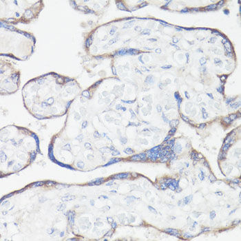
| Sample | Paraffin-embedded human placenta tissue; fixative not specified (datasheet A11184) |
| Fixation | Image fixative and duration unreported (datasheet A11184); verify before use. |
| Sectioning | 4–5 µm sections on charged slides (standard) |
| Deparaffinisation | Xylene, graded ethanol series to water (standard) |
| Antigen retrieval | Heat-induced epitope retrieval in citrate buffer, pH 6.0, 20 min at 95–98 °C (standard rule: cytoplasmic / membrane antigen) |
| Peroxidase block | 3% H2O2, 10 min, room temperature (standard) |
| Blocking | 10% normal serum of the secondary host, 30 min, room temperature (standard) |
| Primary antibody | Rabbit anti-CLUH, 1:50-1:200 (datasheet A11184) |
| Primary incubation | Overnight at 4 °C (standard) |
| Detection | HRP-polymer secondary, DAB chromogen 5–10 min (standard) |
| Counterstain | Hematoxylin, blue, dehydrate and mount (standard) |
| Expected result | CLUH-positive staining in myoepithelial cells of breast (HPA tissue IHC: Medium). HPA tissue profile: Cytoplasmic expression in most tissues, including smooth muscle. No signal in the no-primary control. |
CLUH is a cytoplasmic protein with no transmembrane segment; a fraction occurs in cytoplasmic granules near mitochondria (UniProt O75153). In paraffin section IHC, expect cytoplasmic staining across many tissues, including smooth muscle, with medium staining reported in selected cell populations such as esophageal squamous epithelial cells and cardiomyocytes (HPA tissue IHC). Treat the pattern as provisional: HPA rates tissue IHC reliability Uncertain because staining and RNA expression have low consistency (HPA tissue IHC).
| Cytoplasmic chromogen in esophageal squamous epithelial cells or cardiomyocytes, with recognizable cell boundaries. | This fits the reported medium staining in those populations and the cytoplasmic localization of CLUH (HPA tissue IHC; UniProt O75153). Score intensity within the identified cell population, while allowing for the uncertain tissue IHC reliability (HPA tissue IHC). |
| Predominantly nuclear, membranous, or extracellular staining, with little cytoplasmic signal. | This is discordant with the expected cytoplasmic pattern and the absence of a transmembrane segment (UniProt O75153). Check morphology and detection controls before interpreting it as CLUH; the additional nucleolar signal reported by ICC-IF is uncertain and does not establish a nuclear IHC pattern (HPA subcellular). |
| Strong signal in adipocytes or adrenal glandular cells, especially when expected cytoplasmic populations are unstained. | Those populations are listed as not detected in HPA tissue IHC (HPA tissue IHC). Consider cross-reactivity or endogenous detection activity, then assess a no-primary control; HPA's uncertain reliability means these populations are comparators, not definitive negatives (HPA tissue IHC; general IHC practice). |
| Uniform haze across cells and surrounding tissue, obscuring compartment boundaries. | The pattern cannot support a cytoplasmic CLUH call (UniProt O75153). In chromogenic IHC, incomplete blocking, excess antibody, or detection background can cause diffuse staining; compare a no-primary control and optimize the general IHC workflow before scoring (general IHC practice). |
| No signal in an intact section containing esophageal squamous epithelial cells or cardiomyocytes. | Both populations have reported medium IHC staining, so absence warrants investigation (HPA tissue IHC). Verify tissue identity and assay controls, then check retrieval and detection conditions as general IHC steps; HPA's uncertain reliability prevents treating a single negative section as proof of CLUH absence (HPA tissue IHC; general IHC practice). |
| Cell population and tissue context | HPA reports cytoplasmic expression in most tissues, but staining varies by cell population: esophageal squamous epithelial cells and cardiomyocytes are medium, whereas adipocytes are not detected (HPA tissue IHC). Use the named cells when comparing sections; avoid assigning one intensity to every cell in a tissue (HPA tissue IHC). |
| Localization and protein topology | UniProt places CLUH in the cytoplasm and cytoplasmic granules, with a fraction close to mitochondria, and lists no transmembrane segment (UniProt O75153). Proximity to mitochondria does not establish staining inside mitochondria; compartment calls should follow the visible cytoplasmic pattern (UniProt O75153). |
| Protein processing | UniProt lists one chain spanning residues 1–1309, with no signal peptide or propeptide annotated (UniProt O75153). The supplied record therefore offers no basis for expecting a secreted, shed, or separately processed CLUH staining compartment (UniProt O75153). |
| Strength of IHC evidence | HPA rates tissue IHC Uncertain because antibody staining and RNA expression have low consistency; its IHC entry for HPA044346 is also Uncertain (HPA tissue IHC; HPA antibodies). Treat an unexpected pattern as a finding to check with morphology and controls, not as established CLUH distribution (HPA tissue IHC; general IHC practice). |
| IF/ICC Q: what pattern can be expected? | HPA reports vesicles as the approved main ICC-IF location; its additional nucleoli fibrillar-center location is uncertain, with images listed for A-549, CACO-2, and U2OS (HPA subcellular). HPA031505 is Approved for ICC but has no IHC status listed, so that IF evidence does not validate paraffin-section IHC (HPA antibodies). |
| Situation | Likely cause | Next action |
|---|---|---|
| Expected cytoplasmic cells are blank while the section's detection control also fails. | The assay may have a general detection or reagent problem; this pattern cannot determine whether CLUH is absent (general IHC practice). | Restore the detection control first, then repeat staining and reassess an HPA-reported medium population such as cardiomyocytes (general IHC practice; HPA tissue IHC). |
| Expected cells are blank, but the detection control works. | Possible issues include antibody performance or an unsuitable retrieval condition; no CLUH-specific retrieval or fixation sensitivity is established by the supplied UniProt and HPA records. | Verify the antibody's documented IHC use and optimize retrieval with controlled comparisons as general IHC practice; do not infer CLUH absence from this result alone (general IHC practice; HPA tissue IHC: Uncertain). |
| The no-primary control develops chromogen. | Endogenous enzyme activity or another detection-system background source can produce color without CLUH antibody binding (general IHC practice). | Check the detection chemistry, apply the appropriate endogenous-activity block, and repeat the no-primary control before scoring tissue staining (general IHC practice). |
| Color forms a diffuse haze or obscures cytoplasmic boundaries. | Nonspecific binding or excess primary or detection reagent can obscure the compartment expected for CLUH (general IHC practice; UniProt O75153). | Adjust blocking, washes, and antibody concentration according to the assay controls; score only when cell boundaries and cytoplasmic localization can be assessed (general IHC practice; UniProt O75153). |
| Strong nuclear or cell-surface color dominates an otherwise intact section. | That distribution conflicts with the UniProt cytoplasmic location and lack of a transmembrane segment; HPA's additional nucleolar ICC-IF location is uncertain (UniProt O75153; HPA subcellular). | Compare the no-primary control and cell morphology, then seek independently supported cytoplasmic staining before assigning the signal to CLUH (general IHC practice; UniProt O75153). |
| A reported negative population stains strongly, or reported positive populations vary between sections. | Background or cell-identification error is possible; HPA also rates the tissue pattern Uncertain, so its listed levels are imperfect comparators (HPA tissue IHC; general IHC practice). | Confirm the cell population and section quality, compare positive and negative populations on the same run, and document the discrepancy rather than forcing an HPA-based positive or negative call (HPA tissue IHC; general IHC practice). |


Comprehensive Human Protein Atlas IHC scoring per tissue (reliability: Uncertain — Low consistency between antibody staining and RNA expression data.). Rows are taken directly from the HPA tissue chart — click any row's HPA link to view the source.
| Tissue | Cell type | Level | Evidence | Source |
|---|---|---|---|---|
| Breast | Myoepithelial cells | Medium | Protein (IHC) | HPA → |
| Caudate | Neuronal cells | Medium | Protein (IHC) | HPA → |
| Cerebral cortex | Endothelial cells | Medium | Protein (IHC) | HPA → |
| Colon | Endothelial cells | Medium | Protein (IHC) | HPA → |
| Duodenum | Glandular cells | Medium | Protein (IHC) | HPA → |
| Tissue | Cell type | Level | Evidence | Source |
|---|---|---|---|---|
| Adipose tissue | Adipocytes | Not detected | Protein (IHC) | HPA → |
| Adrenal gland | Glandular cells | Not detected | Protein (IHC) | HPA → |
| Appendix | Glandular cells | Not detected | Protein (IHC) | HPA → |
| Bone marrow | Hematopoietic cells | Not detected | Protein (IHC) | HPA → |
| Cerebellum | Cells in granular layer | Not detected | Protein (IHC) | HPA → |
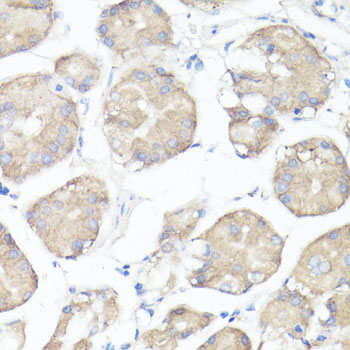
Use the paraffin-section image as a starting reference, and interpret CLUH staining against its reported cytoplasmic distribution and the uncertain tissue-IHC reliability (A11184 caption; UniProt O75153; HPA tissue IHC).
Anti-CLUH A11184 has IHC images from paraffin-embedded human placenta and stomach (catalog image captions); IF/ICC is listed for human and mouse, with no IF image supplied (catalog applications, reactivity, image alts).
A11184 will render with an IHC figure from paraffin-embedded human placenta at 1:100 after microwave retrieval in 10 mM PBS, pH 7.2 (A11184 IHC image caption). A second caption documents human stomach under the same conditions; IF/ICC is listed, but no IF image is supplied (A11184 IHC image captions; catalog applications and IF image alts).
Which to pick: Choose A11184 for paraffin-section tissue IHC because its own captions document staining in human placenta and stomach; the fixative is unreported (A11184 IHC image captions). A11184 is also the listed IF/ICC option, with a 1:50–1:200 dilution range, though no IF image is supplied (catalog applications, dilution, IF image alts). For human or mouse samples, A11184 is the listed rabbit polyclonal option; the pictured IHC samples are human (catalog host, dilution_raw, reactivity; A11184 IHC image captions).