This website uses cookies to ensure you get the best experience on our website.


- Table of Contents
Plan CSNK1A1 staining in paraffin sections using the catalog antibody's IHC protocol (datasheet PB9693). Compare cell types and interpret cytoplasmic staining cautiously: the tissue profile is pending external verification and conflicts with its smooth muscle entry (HPA tissue IHC).

Expected localisation, validated protocols, controls and antibodies — the at-a-glance facts below, then the full design guide.
| Expected localisation | Cytoplasmic in the tissue profile (HPA tissue IHC) | |
| Staining pattern | Cytoplasmic staining in smooth muscle (HPA tissue IHC) | |
| Antigen retrieval | Citrate pH 6 HIER, heat-mediated (datasheet PB9693) | |
| Positive control | Adrenal gland+4 more · see all | |
| Negative control | Liver+3 more · see all |
| Fixation | Keep fixation consistent across sections. (standard IHC practice; not target-specific) | |
| Caveat | Smooth muscle profile conflicts with a 'not detected' entry (HPA tissue IHC) | |
| Regulation | Expression regulation is unannotated (UniProt) | |
| Isoform / epitope | 3 isoforms; check antibody epitope coverage (UniProt) |
The catalog antibody uses citrate pH 6 heat retrieval (datasheet PB9693). The published melanoma and nevus IHC protocol below uses steam retrieval in DIVA buffer (PMC3131429).
| Sample | Paraffin-embedded mouse intestine tissues; fixative not specified (datasheet PB9693) |
| Fixation | Image fixative and duration unreported (datasheet PB9693); verify before use. |
| Sectioning | 4–5 µm sections on charged slides (standard) |
| Deparaffinisation | Xylene, graded ethanol series to water (standard) |
| Antigen retrieval | Heat retrieval: Citrate pH 6, 20 min (datasheet PB9693) |
| Peroxidase block | 3% H2O2, 10 min, room temperature (standard) |
| Blocking | 10% goat serum (datasheet PB9693) |
| Primary antibody | Rabbit anti-CSNK1A1, 0.5-1μg/ml (datasheet PB9693) |
| Primary incubation | Overnight at 4 °C (datasheet PB9693) |
| Detection | Streptavidin-biotin complex (SABC), DAB chromogen (datasheet PB9693) |
| Counterstain | Hematoxylin, blue, dehydrate and mount (standard) |
| Expected result | CSNK1A1-positive staining in glandular cells of adrenal gland (HPA tissue IHC: High). HPA tissue profile: Cytoplasmic expression in smooth muscle. No signal in the no-primary control. |
CSNK1A1 is primarily cytoplasmic, with reported centrosomal, spindle, kinetochore and nuclear-speckle locations (UniProt P48729: subcellular location). On paraffin sections, look for cellular staining in HPA-positive populations, including bronchial respiratory epithelium, placental trophoblasts and testicular seminiferous-duct cells (HPA: High in each). It has no transmembrane segment (UniProt P48729: topology). Treat tissue predictions cautiously: HPA rates its IHC profile Uncertain, pending external verification (HPA: reliability).
| Cytoplasmic staining in bronchial respiratory epithelial cells or placental trophoblasts (HPA: High in both). | This fits the reported tissue pattern and predominant compartment (HPA: tissue IHC; UniProt P48729: cytoplasm). Judge intensity against an appropriate control on the same run; HPA’s Uncertain reliability means agreement is supportive, not independent validation (HPA: reliability; general IHC practice). |
| Strong, uniform nuclear staining dominates while cytoplasm is clear (UniProt P48729: cytoplasm and nuclear speckle). | Question the pattern before calling it CSNK1A1: UniProt lists nuclear speckles and mitotic structures, but those annotations do not predict uniform nuclear IHC staining (UniProt P48729: subcellular location). Compare morphology and controls, then check whether the pattern persists with an independently validated antibody (general IHC practice). |
| Signal appears in a population listed as Not detected, such as liver cholangiocytes (HPA: Not detected in cholangiocytes). | Review cell identification and detection controls; unexpected chromogen could reflect cross-reactivity or endogenous enzyme activity (general IHC practice). A positive call cannot be ruled out solely from the HPA example because its IHC reliability is Uncertain (HPA: reliability). |
| Chromogen blankets cells and extracellular areas with little cellular contrast (general IHC practice). | This is diffuse background rather than a convincing compartmental pattern (general IHC practice; UniProt P48729: subcellular location). Inspect the no-primary control and reagent background before scoring tissue cells; the broad haze does not establish CSNK1A1 expression (general IHC practice). |
| A chosen high-staining reference population has no discernible signal (HPA: High in adrenal glandular cells, for example). | Check that the expected cell population is present, then review retrieval, antibody dilution and detection using run controls (general IHC practice). HPA’s High designation supplies a reference expectation, not a guarantee for every specimen or antibody (HPA: High in adrenal glandular cells; HPA: reliability Uncertain). |
| Compartment and cell state (UniProt P48729: subcellular location). | Cytoplasm is the broad expectation; centrosomes, spindle and kinetochore fibers are reported in specific cell-cycle contexts (UniProt P48729: subcellular location). Routine chromogenic sections may not resolve those fine structures, so do not require visible puncta to score a cellular positive (general IHC practice). |
| Tissue and cell selection (HPA: tissue IHC). | HPA lists High staining in adrenal glandular cells, bronchial respiratory epithelium, placental trophoblasts and testicular seminiferous-duct cells; it lists cholangiocytes as Not detected (HPA: tissue IHC). Use the named cell population when choosing comparators, with HPA’s Uncertain reliability in view (HPA: reliability). |
| Conflicting smooth-muscle entries (HPA: tissue IHC). | The HPA summary says cytoplasmic expression in smooth muscle, yet its smooth-muscle-cell entry says Not detected (HPA: profile; HPA: Not detected in smooth muscle cells). That conflict makes smooth muscle a poor sole positive or negative reference; document the actual cell-level observation (general IHC practice). |
| Antibody evidence (HPA: antibody validation). | The listed IHC antibody CAB016680 is rated Uncertain; HPA051334 is ICC Approved without an IHC rating in this payload (HPA: antibodies). An ICC rating does not establish performance on paraffin sections; interpret an unexpected IHC pattern through tissue and detection controls (general IHC practice). |
| Isoforms and epitope coverage (UniProt P48729: isoforms). | UniProt lists 3 isoforms and a protein-kinase domain at residues 17–285 (UniProt P48729: isoforms and domain). No antibody epitope is supplied, so this record cannot predict which isoforms the IHC reagent detects or whether retrieval exposes its epitope (general IHC practice). |
| IF/ICC interpretation (HPA: subcellular ICC-IF). | For the separate IF/ICC guide: HPA supports cytosolic localization and lists CACO-2, PC-3 and U2OS images, while cautioning that the localization uses antibodies targeting proteins from multiple genes (HPA: subcellular ICC-IF). Use this as a compartment comparison, not as an IHC protocol or paraffin-section validation (general IHC practice). |
| Situation | Likely cause | Next action |
|---|---|---|
| No staining in a high-reference cell population (HPA: High in bronchial respiratory epithelial cells, for example). | The sampled cells may be absent, or the run may have lost detectable signal; the HPA rating alone cannot identify why (HPA: reliability Uncertain; general IHC practice). | Confirm morphology and a positive run control; review the documented retrieval, antibody dilution and detection steps, changing one variable at a time (general IHC practice). |
| Strong color in the no-primary control (general IHC practice). | Background from the detection system or endogenous enzyme activity can mimic chromogenic cellular staining (general IHC practice). | Check reagent-only controls and the appropriate endogenous-activity blocking step before attributing the signal to CSNK1A1 (general IHC practice). |
| Diffuse chromogen obscures cell boundaries (general IHC practice). | Excess detection background or insufficient washing can prevent compartment-level interpretation (general IHC practice). | Compare the no-primary control and review blocking, washes and antibody concentration under the laboratory’s validated IHC workflow (general IHC practice). |
| Only uniform nuclear color is visible (UniProt P48729: subcellular location). | A dominant uniform nuclear pattern is poorly matched to the cytoplasmic expectation, although UniProt also reports nuclear speckles (UniProt P48729: cytoplasm and nuclear speckle). | Check counterstain and no-primary controls, then seek independent antibody or orthogonal evidence before assigning a nuclear CSNK1A1 pattern (general IHC practice). |
| A reported Not detected population stains strongly, such as liver cholangiocytes (HPA: Not detected in cholangiocytes). | Cell misidentification, cross-reactivity or endogenous detection activity are possibilities; HPA’s Uncertain profile does not prove any one of them (HPA: reliability; general IHC practice). | Verify the cell type on morphology, inspect detection controls and compare a reported High population from the same run (HPA: tissue IHC; general IHC practice). |
| Smooth-muscle staining disagrees with the chosen HPA reference (HPA: profile and smooth-muscle entry). | HPA reports both cytoplasmic smooth-muscle expression and a Not detected smooth-muscle-cell entry (HPA: tissue IHC). | Record the compartment and exact cells observed; use another named HPA High population and appropriate controls to assess the run (HPA: tissue IHC; general IHC practice). |


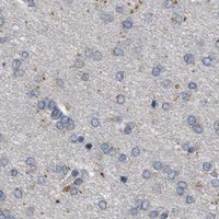
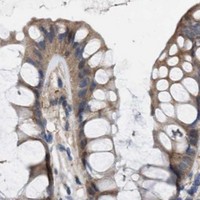

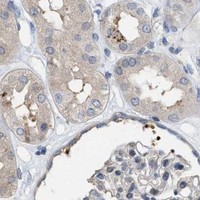


Comprehensive Human Protein Atlas IHC scoring per tissue (reliability: Uncertain — Pending external verification.). Rows are taken directly from the HPA tissue chart — click any row's HPA link to view the source.
| Tissue | Cell type | Level | Evidence | Source |
|---|---|---|---|---|
| Adrenal gland | Glandular cells | High | Protein (IHC) | HPA → |
| Bronchus | Respiratory epithelial cells | High | Protein (IHC) | HPA → |
| Endometrium | Glandular cells | Medium | Protein (IHC) | HPA → |
| Placenta | Trophoblastic cells | High | Protein (IHC) | HPA → |
| Testis | Cells in seminiferous ducts | High | Protein (IHC) | HPA → |
| Tissue | Cell type | Level | Evidence | Source |
|---|---|---|---|---|
| Liver | Cholangiocytes | Not detected | Protein (IHC) | HPA → |
| Ovary | Ovarian stroma cells | Not detected | Protein (IHC) | HPA → |
| Seminal vesicle | Glandular cells | Not detected | Protein (IHC) | HPA → |
| Smooth muscle | Smooth muscle cells | Not detected | Protein (IHC) | HPA → |
Troubleshoot CSNK1A1 staining in paraffin sections by checking retrieval, detection controls, cellular location, and the limits of the antibody evidence.
PB9693 has IHC images from paraffin sections of mouse and rat intestine and human intestinal cancer (PB9693 IHC captions). No IF data are listed (PB9693 catalog).
PB9693 is the only card and lists IHC reactivity with human, mouse, and rat (PB9693 catalog). Its images show paraffin-section staining in mouse and rat intestine and human intestinal cancer (PB9693 IHC captions).
Which to pick: For tissue IHC, choose PB9693: its captions document citrate retrieval at pH 6 for 20 minutes, 1 μg/ml primary antibody overnight at 4°C, and DAB detection; the fixative is unreported (PB9693 IHC captions). For IF/ICC, no listed SKU has an IF/ICC application or image (PB9693 catalog). For cross-species IHC, PB9693 lists human, mouse, and rat reactivity, with paraffin-section images for all three; its clone is unspecified (PB9693 catalog; PB9693 IHC captions).