CUX1 / Homeobox protein cut-like 1 · IHC design guide

Design Immunohistochemistry for CUX1

Plan paraffin CUX1 IHC around the general nuclear tissue pattern (HPA tissue IHC). This guide covers fixation consistency, control selection, antibody dilution, chromogenic detection, and interpretation across isoforms.

Evidence assembled Oct 2026 · For research use; verify linked source records and product datasheet before use
Immunohistochemistry protocol sheet for CUX1 (IHC for CUX1): expected localisation General nuclear staining in tissue (HPA tissue IHC), antibody A01653-4, validated IHC image, and IHC protocol steps
Printable CUX1 IHC protocol sheet — expected localisation General nuclear staining in tissue (HPA tissue IHC), antibody A01653-4, controls and protocol steps. Open the full CUX1 IHC guide →

CUX1 Immunohistochemistry Experimental Design Guide

Expected localisation, validated protocols, controls and antibodies — the at-a-glance facts below, then the full design guide.

Must know before staining
Expected localisation General nuclear staining in tissue (HPA tissue IHC)
Staining pattern Nuclei stain across many tissue cell types (HPA tissue IHC)
Antigen retrieval EDTA pH 8.0 HIER, heat-mediated (datasheet A01653-4)
Positive control ⓘ Appendix+4 more · see all
Negative control ⓘ Liver
Important caveats
Reasons your staining may differ from the expected pattern.
Fixation Keep fixation consistent across samples (standard IHC practice; not target-specific); Matched tissue-IHC evidence does not establish the fixation claim. Validate the specimen-specific method before use. (selected-SKU IHC image A01653-4)
Caveat HPA antibodies target different isoforms; staining may vary (HPA tissue IHC)
Regulation Tissue RNA shows low specificity (HPA tissue RNA)
Isoform / epitope 11 isoforms; p110 spans residues 756–1505, so epitope position matters (UniProt)
Section 1

Recommended CUX1 IHC & IF Protocols

Compare the catalog antibody’s EDTA pH 8.0 heat retrieval protocol (datasheet: A01653-4) with two published CUX1 IHC methods (PMC7409270; PMC8158495).

Recommended immunohistochemistry (IHC-P) protocol parameters
SampleParaffin-embedded human placenta tissue; fixative not specified (datasheet A01653-4)
FixationImage fixative and duration unreported (datasheet A01653-4); verify before use.
Sectioning4–5 µm sections on charged slides (standard)
DeparaffinisationXylene, graded ethanol series to water (standard)
Antigen retrievalHeat retrieval: EDTA pH 8.0 (datasheet A01653-4); 20 min, 95–100 °C (standard)
Peroxidase block3% H2O2, 10 min, room temperature (standard)
Blocking10% goat serum (datasheet A01653-4)
Primary antibodyRabbit anti-CUX1, 2-5 μg/ml (datasheet A01653-4)
Primary incubationOvernight at 4 °C (datasheet A01653-4)
DetectionHRP-conjugated secondary, DAB chromogen (datasheet A01653-4)
CounterstainHematoxylin, blue, dehydrate and mount (standard)
Expected resultCUX1-positive staining in lymphoid tissue of appendix (HPA tissue IHC: High). HPA tissue profile: General nuclear expression. No signal in the no-primary control.
💡Decision noteStart with EDTA pH 8.0 heat retrieval for the catalog antibody (datasheet: A01653-4); the published methods used different retrieval conditions (PMC7409270; PMC8158495).
Section 2

What Is the Expected CUX1 Staining Pattern?

CUX1 is a nuclear transcription factor without a transmembrane segment (UniProt P39880: nucleus, function, topology). In paraffin-section IHC, expect a general nuclear pattern, including high staining in specified epithelial, neuronal, lymphoid and hematopoietic populations (HPA: Approved; general nuclear expression; listed high-staining cells). HPA’s tissue annotation is based on HPA003277; HPA003317 targets different isoforms (HPA: reliability description).

What am I looking at on my slide?
Distinct nuclear staining in bronchial respiratory epithelium, colonic glands or cerebral-cortex neurons.This fits the expected compartment and named high-staining cell populations (HPA: general nuclear expression; High in respiratory epithelial, colonic glandular and cerebral-cortex neuronal cells). Evaluate the named cells, because an organ-wide label would hide differences between its cell types.
Predominantly cytoplasmic, membranous or extracellular chromogen in an IHC section.Treat this as a localization mismatch and investigate artefact or nonspecific binding (UniProt P39880: nucleus; no transmembrane segment; HPA: general nuclear IHC expression). A Golgi signal has separate ICC-IF support and does not, by itself, validate a cytoplasmic IHC pattern (HPA: supported Golgi ICC-IF location).
Strong staining in liver cholangiocytes, or staining that follows deposits rather than cell nuclei.Cholangiocytes are listed as not detected (HPA: Liver, cholangiocytes, Not detected). Check cross-reactivity and endogenous detection activity before calling this CUX1; the HPA observation does not label every liver cell negative.
Diffuse colour across tissue, including spaces without cells, with poor nuclear definition.The distribution does not match the reported nuclear IHC profile (HPA: general nuclear expression). As a general IHC check, compare the no-primary control and review blocking, washes and detection; diffuse background alone cannot identify its cause.
No nuclear signal in a high-staining reference population.An absent signal in bone-marrow hematopoietic cells or appendix lymphoid tissue conflicts with the reported high staining (HPA: Bone marrow, hematopoietic cells, High; Appendix, lymphoid tissue, High). Confirm that the relevant cells are present, then review the assay before interpreting an unknown sample as negative.
💡Expected CUX1 appearanceCall a section positive when the relevant cells show clearly defined, often strong nuclear chromogen—for example, colonic glandular cells (HPA: High in colonic glandular cells; general nuclear expression); diffuse extracellular colour or dominant membrane staining is a false-positive pattern to investigate (UniProt P39880: nucleus; no transmembrane segment).
How each factor affects the staining
Which tissue cells provide useful comparisons?High staining is reported in appendix lymphoid tissue, bone-marrow hematopoietic cells, bronchial respiratory epithelium, cerebellar granular-layer cells, cerebral-cortex neurons, cervical squamous epithelium, colonic glands and endometrial stroma (HPA: listed High populations). Low staining in adrenal, epididymal and prostate glandular cells makes those populations less decisive for an absence check (HPA: listed Low populations).
How should antibody identity affect interpretation?Both HPA003277 and HPA003317 are Approved for IHC, but they target different isoforms; the annotated tissue profile uses HPA003277 (HPA: antibody validation; reliability description). Match an unexpected pattern to the antibody used before treating the HPA tissue annotation as an exact prediction.
Can processing or isoforms change what an antibody detects?UniProt lists 11 isoforms and both a full-length chain and a 756–1505 chain (UniProt P39880: isoforms; processing). Detection of either form depends on an antibody’s epitope, which is not supplied here; neither a stronger nor a weaker tissue signal can be predicted from these annotations alone.
IF/ICC Q&A: is every extranuclear signal unexpected?No. HPA reports supported nucleoplasm and Golgi-apparatus localization in ICC-IF, with images in MCF-7, SH-SY5Y and U2OS (HPA: subcellular ICC-IF). Assess an IF image against that modality-specific observation; the tissue-IHC expectation remains general nuclear expression (HPA: tissue IHC profile).
Why is my staining missing, weak or wrong?
SituationLikely causeNext action
A known high-staining population has little or no nuclear chromogen.The relevant cells may be absent from the section, or the IHC workflow may have failed; the supplied records do not identify a CUX1-specific failure step (HPA: listed High populations).Confirm cell identity and tissue preservation, then check the positive control, antibody dilution, antigen retrieval and detection according to the assay instructions. Treat these as general IHC checks, not documented CUX1 retrieval requirements.
Nuclear staining appears with HPA003277 but differs with HPA003317.The antibodies target different isoforms, and the annotated tissue expression uses HPA003277 (HPA: reliability description). The record does not specify each antibody’s epitope.Record the antibody ID and compare each result with its own validated use; avoid assigning a particular isoform to either staining pattern without epitope evidence (HPA: both antibodies IHC Approved).
Cholangiocytes look strongly positive.This conflicts with the listed not-detected population (HPA: Liver, cholangiocytes, Not detected); cross-reactivity or endogenous detection activity are possible general IHC explanations.Confirm cholangiocyte identity, inspect a no-primary control, and check blocking and detection chemistry. Do not extend the cholangiocyte result to all liver cells (HPA: negative entry specifies cholangiocytes).
Colour is widespread, weakly structured or present outside cells.Diffuse background obscures the expected nuclear pattern (HPA: general nuclear expression); the source data do not identify which workflow step caused it.Compare matched controls, inspect wash and blocking steps, and score only clearly localized nuclear signal. Reassess the section if background prevents cell-level interpretation.
Membrane or broad cytoplasmic staining dominates a tissue IHC section.That pattern conflicts with the nuclear tissue-IHC profile and the absence of a transmembrane segment (HPA: general nuclear expression; UniProt P39880: topology).Check morphology and controls, then repeat or review the assay with its validated antibody conditions. Keep the supported Golgi ICC-IF observation separate from a claim of Golgi staining in tissue IHC (HPA: subcellular ICC-IF).
An IF/ICC image shows nucleoplasmic signal plus a discrete perinuclear structure.Both nucleoplasm and Golgi apparatus are supported ICC-IF locations (HPA: subcellular ICC-IF); the tissue-IHC profile alone cannot adjudicate the perinuclear structure.Use an appropriate Golgi marker and imaging controls to assess that assignment. This is an interpretation check for IF/ICC, whose application has a separate guide, rather than an IHC protocol option.

Sample controls for CUX1 IHC & IF

🧪Run bronchus first: respiratory epithelial cells should stain strongly, with predominantly nuclear signal (HPA: High in respiratory epithelial cells; UniProt P39880: nucleus). Run liver as the negative tissue, focusing on cholangiocytes (HPA: Not detected in cholangiocytes); on the bronchus slide, cells without specific nuclear staining provide an internal background reference, but their negative status should be verified rather than assumed.
Positive control tissue: Appendix (Lymphoid tissue, HPA High)
Negative control tissue: Liver (HPA Not detected)
ICC-IF cell lines (HPA subcellular resource): HPA ICC-IF images show CUX1 in MCF-7, SH-SY5Y, U2OS, with annotated localisation: Nucleoplasm (supported), Golgi apparatus (supported) (HPA subcellular).
Technical controls: Include no-primary (secondary-only) and host- and Ig-class-matched rabbit IgG isotype controls (caption: rabbit anti-CUX1 antibody; standard IHC practice). Use a CUX1 knockout specimen as a biological specificity control and block endogenous peroxidase before chromogenic detection, particularly where blood or inflammatory cells are present (standard IHC practice).
⚠️Feasibility: A target-specific fixation window or fixation effect is unreported, and the selected A01653-4 paraffin-section caption does not state a fixative (caption: fixative not stated). The caption documents heat retrieval in EDTA at pH 8.0, but does not establish that retrieval is required or that frozen sections or IF are easier (caption: EDTA retrieval). In bronchus, interpret chromogen in inflammatory or blood-rich areas against the peroxidase controls (standard IHC practice).

HPA tissue IHC evidence for CUX1

Comprehensive Human Protein Atlas IHC scoring per tissue (reliability: Approved — HPA003317 and HPA003277 target different isoforms. Annotated protein expression is based on HPA003277.). Rows are taken directly from the HPA tissue chart — click any row's HPA link to view the source.

Positive expression · recommended positive controls

TissueCell typeLevelEvidenceSource
Appendix Lymphoid tissue High Protein (IHC) HPA →
Bone marrow Hematopoietic cells High Protein (IHC) HPA →
Bronchus Respiratory epithelial cells High Protein (IHC) HPA →
Cerebellum Cells in granular layer High Protein (IHC) HPA →
Cerebral cortex Neuronal cells High Protein (IHC) HPA →

Undetected expression · recommended negative controls

TissueCell typeLevelEvidenceSource
Liver Cholangiocytes Not detected Protein (IHC) HPA →
Section 3

Advanced CUX1 IHC Tips

Troubleshoot CUX1 staining in paraffin sections by checking retrieval, nuclear localisation, isoform coverage, and cell type before interpreting chromogenic signal.

What retrieval conditions should I start with for CUX1 paraffin-section IHC?
Start with heat-mediated retrieval in EDTA buffer at pH 8.0 (datasheet A01653-4). The catalog antibody was used on a paraffin-embedded human placenta section after that retrieval, followed by 2 μg/ml primary antibody overnight at 4°C (datasheet A01653-4). If nuclear staining is weak, compare a controlled range of heating times on adjacent sections while keeping antibody concentration and detection constant (standard IHC practice). Record tissue damage and background alongside nuclear signal, because harsher heating can reduce section quality (standard IHC practice). Do not infer the caption’s fixation method from its paraffin processing; the fixative was unreported (datasheet A01653-4).
Could fixation explain weak CUX1 staining in my sections?
Target-specific CUX1 sensitivity to fixation is unknown from the supplied IHC evidence; the catalog caption identifies a paraffin section but does not state its fixative (datasheet A01653-4). For new specimens, standardise fixative, tissue thickness, and time in fixative across comparison groups, then document those variables with each staining run (standard IHC practice). If archived blocks vary, compare adjacent sections using the same EDTA pH 8.0 retrieval and antibody conditions before attributing signal differences to biology (datasheet A01653-4; standard IHC practice). Assess morphology and nuclear counterstaining alongside CUX1 signal, and avoid treating staining differences alone as evidence of a CUX1-specific fixation effect (standard IHC practice).
Where should convincing CUX1 signal appear in chromogenic IHC?
Prioritise staining within intact nuclei: CUX1 is annotated as nuclear, and tissue IHC shows general nuclear expression (UniProt P39880 localisation; HPA tissue IHC). Use a nuclear counterstain to determine whether DAB overlaps nuclei rather than merely coating adjacent cytoplasm (standard IHC practice). The HPA subcellular record also supports nucleoplasmic and Golgi localisation in ICC/IF, but that observation does not establish a Golgi staining pattern for this paraffin-section assay (HPA subcellular). Because CUX1 has no annotated transmembrane segment, isolated membrane-rim staining warrants scrutiny with a no-primary control and a review of tissue morphology (UniProt P39880 topology; standard IHC practice).
Could CUX1 isoforms explain discordant IHC results?
Yes: CUX1 has 11 listed isoforms, and its annotated chains include residues 1–1505 and 756–1505 (UniProt P39880 processing and isoforms). An antibody against an epitope before residue 756 would not recognise that shorter annotated chain; the catalog antibody’s epitope is not specified in the supplied evidence (UniProt P39880 processing; datasheet A01653-4). HPA states that HPA003317 and HPA003277 target different isoforms, with its annotated tissue expression based on HPA003277 (HPA tissue IHC). Therefore, compare antibody epitope documentation and the intended isoform before treating disagreement between tissue stains as a retrieval failure (HPA tissue IHC; standard IHC practice).
How should I assess CUX1 in a multiplex IF experiment?
For a separate IF assay, pair CUX1 with a marker identifying the expected cell population, such as neuronal cells in cerebral cortex, and inspect individual nuclei (HPA tissue IHC; UniProt P39880 localisation). Choose a CUX1 fluorophore in a channel with low measured tissue autofluorescence, and include unstained and single-label controls when setting exposures and assessing bleed-through (standard IF practice). Because CUX1 is nuclear and has no transmembrane segment, permeabilise fixed cells sufficiently for antibody access to the nucleus, using the antibody’s documented IF conditions where available (UniProt P39880 localisation and topology; standard IF practice). Treat HPA’s nucleoplasmic and Golgi ICC/IF localisation as context, not as validation of this catalog antibody for IF (HPA subcellular).
How can I reduce diffuse brown background without losing nuclear signal?
Start by matching the demonstrated primary concentration of 2 μg/ml and the 10% goat-serum block, then assess whether the background follows tissue structures or covers the whole section (datasheet A01653-4; standard IHC practice). If background persists, titrate the primary on adjacent sections while keeping retrieval, detection, and DAB development constant (standard IHC practice). Include a no-primary control and apply a peroxidase block before HRP detection to identify endogenous enzyme signal; these are general chromogenic IHC controls (standard IHC practice). The caption used a peroxidase-conjugated secondary and DAB, so interpret brown staining only after checking that the controls remain clear (datasheet A01653-4; standard IHC practice).
How should I quantify CUX1-positive cells across tissue sections? ⚠ ANSWER MARKED FOR VERIFICATION
Define the tissue region and cell population before scoring, then count only interpretable nuclei because CUX1 tissue staining is generally nuclear (HPA tissue IHC; standard IHC practice). Report the percentage of positive viable nuclei and, when intensity matters, an H-score calculated from percentages at intensity grades 0–3, giving a range of 0–300 (standard IHC practice). For spatial comparisons, report positive-nucleus density per mm² of viable tissue as well (standard IHC practice). Normalise each group to its eligible nuclei or viable tissue area, apply one threshold across matched slides, and exclude folds and necrotic regions consistently (standard IHC practice).
What distinguishes true CUX1 staining from an IHC artefact?
A credible result has signal in well-preserved nuclei and a clear no-primary control, consistent with general nuclear CUX1 expression in tissue IHC (HPA tissue IHC; standard IHC practice). Check cell identity: HPA reports high staining in cerebral-cortex neuronal cells and no detected staining in liver cholangiocytes, although those observations are antibody and context dependent (HPA tissue IHC). Be cautious with staining confined to section edges, folds, or necrotic areas, where morphology can compromise interpretation (standard IHC practice). Diffuse cytoplasmic or membrane-rim brown signal also warrants review, given CUX1’s nuclear annotation and lack of an annotated transmembrane segment (UniProt P39880 localisation and topology).
Boster reagents

Best CUX1 / Homeobox protein cut-like 1 IHC Antibodies

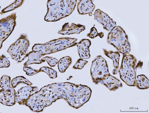
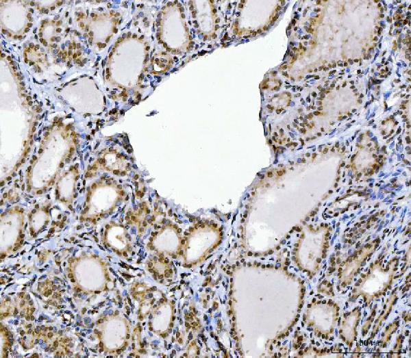
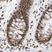
A01653-4 has real IHC images from paraffin-embedded human placenta, thyroiditis, and laryngeal squamous cell carcinoma sections and mouse colon (catalog IHC captions). No IF images are provided (catalog IF image alts).

Real IHC data IHC analysis of CUX1 using anti-CUX1 antibody (A01653-4). CUX1 was detected in a paraffin-embedded section of human placenta tissue. Heat mediated antigen retrieval was performed in EDTA buffer (pH 8.0, epitope retrieval solution). The tissue section was blocked with 10% goat serum. The tissue section was then incubated with 2 μg/ml rabbit anti-CUX1 Antibody (A01653-4) overnight at 4°C. Peroxidase Conjugated Goat Anti-rabbit IgG was used as secondary antibody and incubated for 30 minutes at 37°C. The tissue section was developed using HRP Conjugated Rabbit IgG Super Vision Assay Kit (Catalog # SV0002) with DAB as the chromogen.
Anti-CUX1 Antibody ®
Cat # A01653-4

A01653-4 is listed for IHC in human and mouse samples (catalog applications and reactivity). Its IHC captions document paraffin-embedded human placenta, thyroiditis, and laryngeal squamous cell carcinoma sections and mouse colon (catalog IHC captions).

Which to pick: For tissue IHC, choose A01653-4: this rabbit antibody has paraffin-section IHC images using 2 μg/ml, although the fixative is unreported (catalog IHC captions). No listed SKU has IF/ICC validation or an IF image (catalog applications and IF image alts). For human and mouse IHC, A01653-4 is the cross-species choice (catalog reactivity and IHC captions).

Each figure is that product's own IHC / IF validation image from its datasheet.

References

  1. UniProt Consortium. UniProt entry P39880 (CUX1_HUMAN, Homeobox protein cut-like 1).
  2. Human Protein Atlas. CUX1 tissue IHC expression (reliability: Approved).
  3. Human Protein Atlas. CUX1 subcellular location (ICC-IF): Localized to the nucleoplasm and the Golgi apparatus..
  4. Human Protein Atlas. CUX1 antibody validation summary (2 antibodies).
  5. CUX1-Transcriptional Master Regulator of Tumor Progression in Pancreatic Neuroendocrine Tumors. Cancers 2020 — PMC7409270.
  6. CUX1 Enhances Pancreatic Cancer Formation by Synergizing with KRAS and Inducing MEK/ERK-Dependent Proliferation. Cancers 2021 — PMC8158495.
  7. Altered cortical Cytoarchitecture in the Fmr1 knockout mouse. Molecular brain 2019 — PMC6570929.
  8. Transforming growth factor-β-induced CUX1 isoforms are associated with fibrosis in systemic sclerosis lung fibroblasts. Biochemistry and biophysics reports 2016 — PMC5613511.
  9. PubMed PMID:1301999 — UniProt-cited evidence.
  10. PubMed PMID:14702039 — UniProt-cited evidence.
  11. PubMed PMID:12853948 — UniProt-cited evidence.