This website uses cookies to ensure you get the best experience on our website.
- Table of Contents
16 Citations 7 Q&As
3 Citations 5 Q&As
8 Citations 8 Q&As
3 Citations 17 Q&As
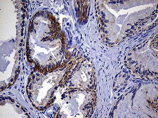
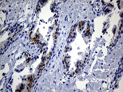
Facts about Pro-epidermal growth factor.
Can induce neurite outgrowth in motoneurons of the pond snail Lymnaea stagnalis in vitro (PubMed:10964941). .
Human | |
---|---|
Gene Name: | EGF |
Uniprot: | P01133 |
Entrez: | 1950 |
Belongs to: |
---|
No superfamily |
beta-urogastrone; EGF; epidermal growth factor (beta-urogastrone); epidermal growth factor; HOMG4; pro-epidermal growth factor; URG; Urogastrone
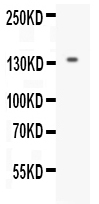
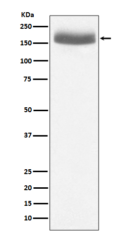
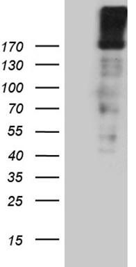
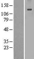
Mass (kDA):
133.994 kDA
Human | |
---|---|
Location: | 4q25 |
Sequence: | 4; NC_000004.12 (109912883..110013766) |
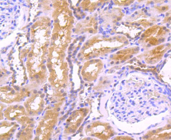
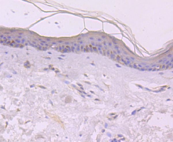
Expressed in kidney, salivary gland, cerebrum and prostate.
Membrane; Single-pass type I membrane protein.
For cancer research An antibody against the EGF marker can aid in the detection of tumor cells. Boster's proprietary technology can also recognize heterodimers of EGFR and ErbB2. Additionally, Boster's antibody can recognize neuroglycan C and neuregulin-1. Here are some examples of best uses of EGF markers in cancer research. Boster has innovative products that can be purchased as antibodies to this marker.
A brand new monoclonal antigen called Anti-EGFR was developed specifically for the treatment of EGFR-expressing tumor cells. The can225IgG chimeric AntiEGFR antibody was made functional with DTPA, a ligand which is able to covalently attach to all lysine residues of monoclonal antibodies. The heavy chain of can225IgG consists of 24 lysine residues while the light chain contains nine. The antibodies were detected in real time when they were bound to cells using a Ligand Tracer (r) method. This allowed to determine their fluorescence intensities and their corresponding intensity of fluorescence. The binding kinetics were calculated using the one-to one ligand-target fitting algorithm.
The monoclonal anti-EGFR antibody does not contain a binding residue for radiometal. Therefore the anti-EGFR Fab is functionalized with diethylene triamine-pentacetic acid (DTPA), which allows it to be radiolabelled with 99mTc. It is a metastable form of technetium-99 , which is extensively used in single-photon emission computed tomography. Further research is needed to determine if anti-EGFR antibodies are compatible with diagnostic nuclides.
EGFR is a well-characterized receptor that tyrosine-kinase. It plays a significant role in cell proliferation, differentiation, and a variety of human cancers are caused by its overexpression. The clinical approach focuses on ligand receptor interactions and tyrosine Kinase activity. EGFR binds with AGR2, a cell-surface protein that allows receptor delivery to the plasma membrane. The effectiveness of EGFR in tackling tumor cells is diminished when the AGR2 protein levels are lower. This can reduce signaling by reducing expression of FOS and AREG and EGR1.
Anti-EGFR Fabs function similarly as monoclonal antibody monoclonal antibodies. However, anti-EGFR mAbs that are high in affinity have higher dissociation rates than monoclonal antibodies , and have less penetration into solid tumors. They also retain large amounts of the anti-EGFR-specific Fabs in normal tissues. Panitumumumab or Cetuximab are highly effective in treating solid tumors.
The epidermal growth factors receptor family (EGFR) comprises HER2, HER3, ErbB4 and HER3. Each member is a tyrosine kinase domain located in the extracellular region, and functions as a transmitter of extracellular signals. This receptor family dimers with other members of the family to trigger intracellular signaling pathways.
The EGFR/ErbB2 gene family regulates normal cell growth, differentiation and survival. While insufficient EGFR signaling has been associated with diseases like Alzheimer's disease, multiple sclerosis, and many other diseases, the excessive expression has been linked with the development of tumors. Specifically, HER2 and EGFR are both overexpressed in breast, ovarian and non-small-cell lung cancers.
It recognizes neuroglycan c (also known as CSPG5) it is a glycoprotein with approximately 120kD in size and a member of the family of neuregulins. This protein is expressed on the surface of neurons as well as other nerve tissues. It has an CS attachment domain and an EGF-like domain. Neuroglycan C can also be observed in two forms. One is recognized by an antibody against the C-terminal component of the protein, whereas the other isoform is not.
CS chain is found on the neuroglycan cell surface, but the mutant form is devoid of the CS chain. Biotinylation permits NGC detection on the cell surface. It recognizes biotinylation. Anti-NGC antibodies recognize neuroglycan C only when it is not biotinylated. This suggests that intracellular trafficking is not dependent on biotinylation.
Neuro 2a cells are a proteoglycan-based version of NGC and are able to attach to brain tissue. They also contain the CS version of NGC. This makes it a valuable model to study the CS glycosylation of NGC. However, it remains unclear how the CS-form of NGC is able to bind to the brain. Next, we need to identify the CS moiety within NGC.
Neuroglycan C is a membrane-type chondroitinsulfate proteoglycan that is crucial for neurite outgrowth. The complexness of dendritic spines increases when NGC/CALEB is overexpressed. Furthermore, it contains an cytoplasmic and transmembrane domain. This study reveals the role of NGC/CALEB in the development and maintenance of the central nervous systems.
This monoclonal antibody has been designed to detect mouse Neuregulin-1. Neuregulins are part of the ErbB/HER receptor family. There are numerous types of neuregulins with different functions. This antibody recognizes NRG1 type III. This antibody is extremely specific and can detect the Neuregulin-1 in mouse, human and rat samples. It recognizes all three forms of Neuregulin-1.
Neuregulin-1 comes in two types that are beta and alpha. The beta form is a direct binding to the receptors on nearby cells, and the alpha form recognizes ErbB4 receptor. Both types of neuregulins are capable of binding to the ErbB4 receptor. The beta form recognizes neuregulin-1 and is able to bind to the downstream ErbB receptor, while the alpha form binds ErbB2.
The axon is a key role in peripheral nerve myelination. The axon Schwann cell unit is involved in controlling the expression of SCIP. The structure of the C-fiber varies based on the location of the peripheral nerve. Additionally, the expression of Krox-20 reveals important steps in the development of peripheral glial cells. Schwann cells require fibroblasts to support basal lamina deposition as well as ensheathment of sympathetic neurites in the culture.
Numerous lines of evidence support the importance of Nrg1 in schizophrenia pathophysiology. Numerous studies from animal and human models support this idea. Many BACE1 inhibitors have entered phase I trials. There are numerous clinical trials that prove the effectiveness of this medication in schizophrenia. This drug is designed to stop the neuroregulin-1 and BACE1 signaling. It also aims to block the effect of BACE1 by blocking the expression of Nrg1.
Tomoregulin is a transmembrane type I protein with two follistatin module modules as well as an extracellular tail, is a transmembrane glycoprotein of the type I variety. It is found in the brain and regulates activin, nodal signaling, through direct binding to Cripto. It is a member of the EGF superfamily. Its structure is similar to the C-type lectin receptors.
Anti-tomoregulin antibody 2H8 was developed to specifically target the tomoregulin proteins in live. The antibody is accumulated in tumor-derived cells, leading to a tumour-specific accumulation. A single i.p. A single i.p. of 150 microCi (163microg) of 90Y-labeled2H8 inhibited the growth rate of mice bearing an LNCaP human prostate cancer xenograft. This was without any apparent toxicities or cross-reactivity to the mousetomoregulin.
PMID: 3491360 by Bell G.I., et al. Human epidermal growth factor precursor: cDNA sequence, expression in vitro and gene organization.
PMID: 300079 by Gregory H., et al. The primary structure of human urogastrone.
*Showing only the more recent 20. More publications can be found for each product on its corresponding product page