This website uses cookies to ensure you get the best experience on our website.
- Table of Contents
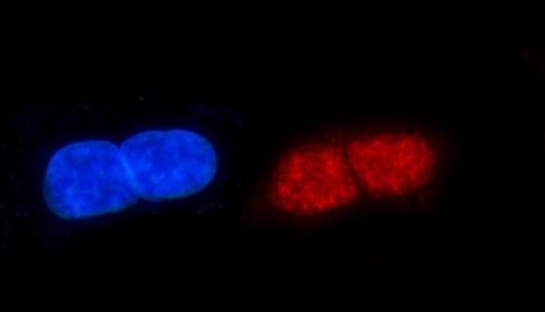
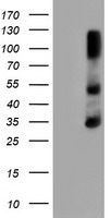
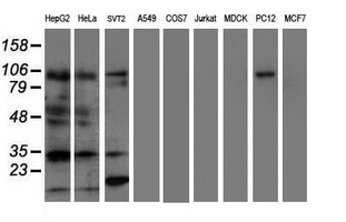
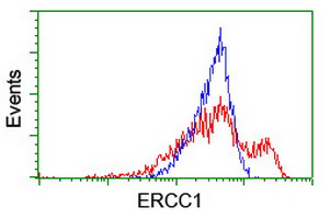
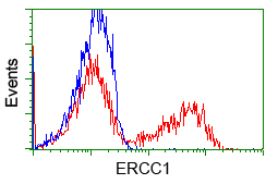
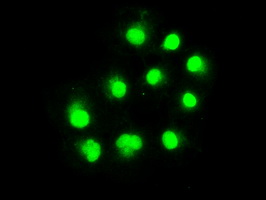
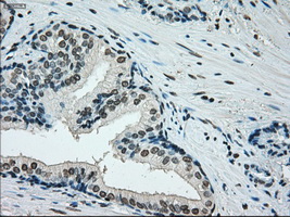
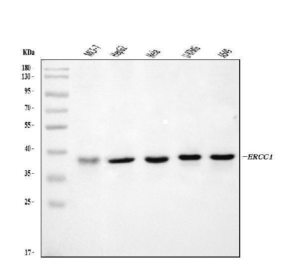
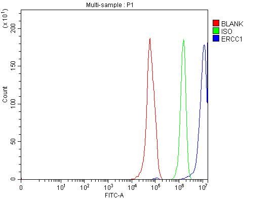
Facts about DNA excision repair protein ERCC-1.
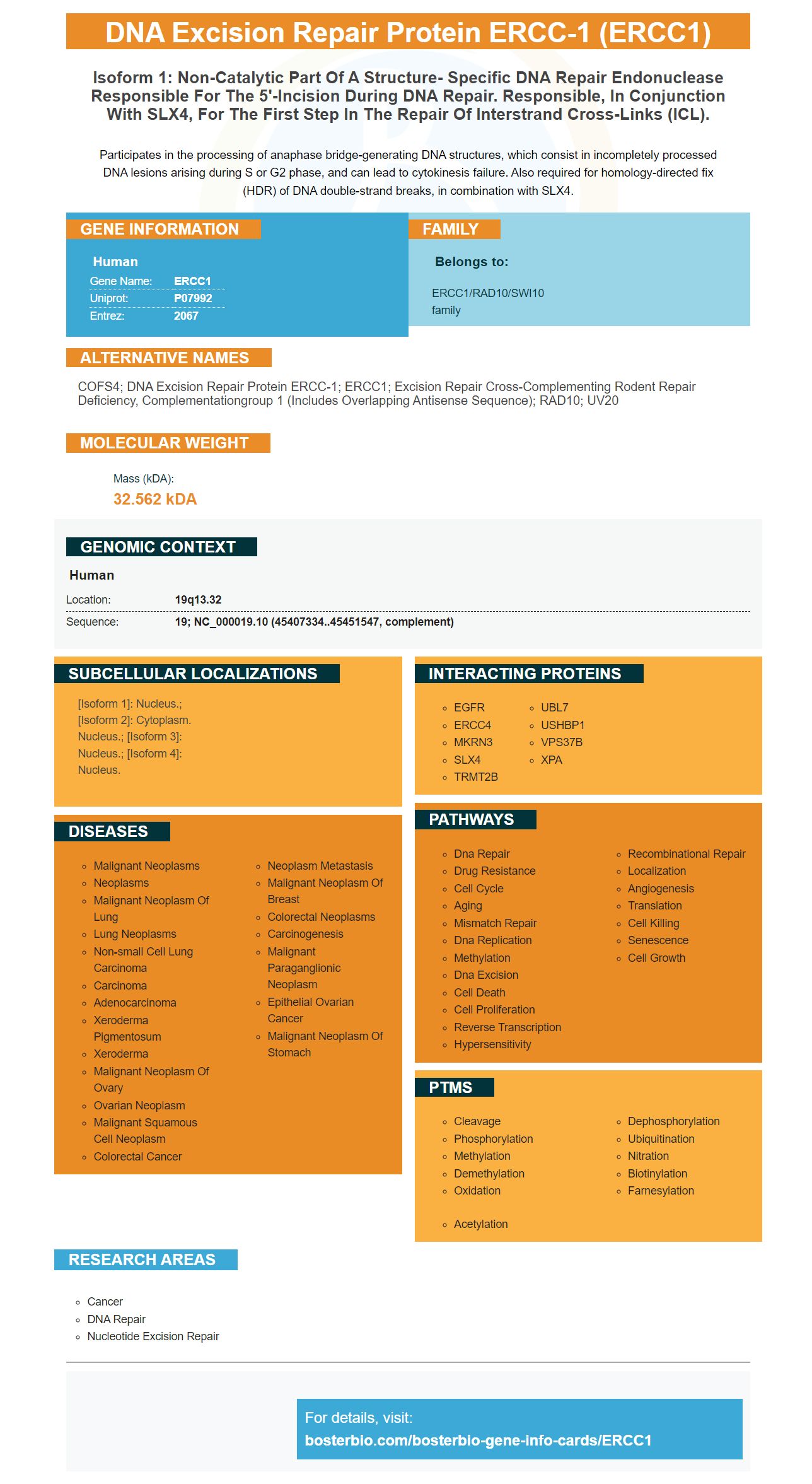
Participates in the processing of anaphase bridge-generating DNA structures, which consist in incompletely processed DNA lesions arising during S or G2 phase, and can lead to cytokinesis failure. Also required for homology-directed fix (HDR) of DNA double-strand breaks, in combination with SLX4.
Human | |
---|---|
Gene Name: | ERCC1 |
Uniprot: | P07992 |
Entrez: | 2067 |
Belongs to: |
---|
ERCC1/RAD10/SWI10 family |
COFS4; DNA excision repair protein ERCC-1; ERCC1; excision repair cross-complementing rodent repair deficiency, complementationgroup 1 (includes overlapping antisense sequence); RAD10; UV20
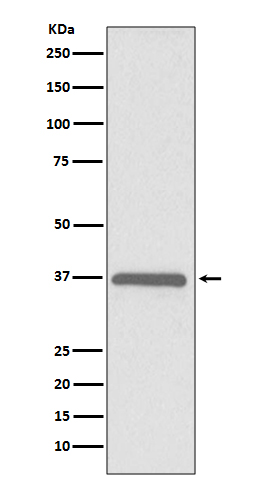
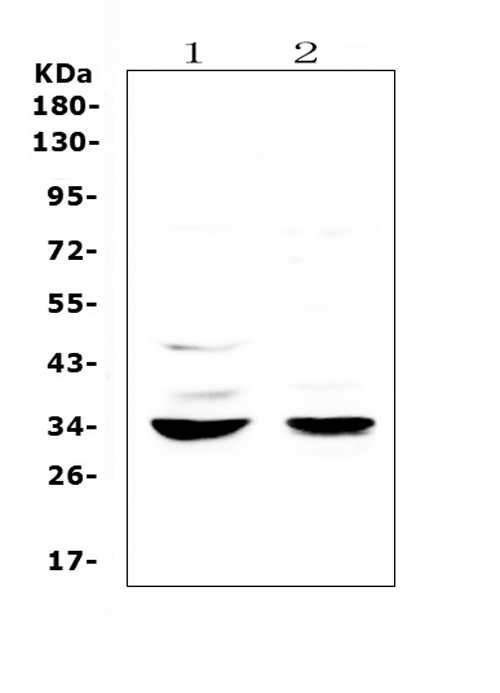
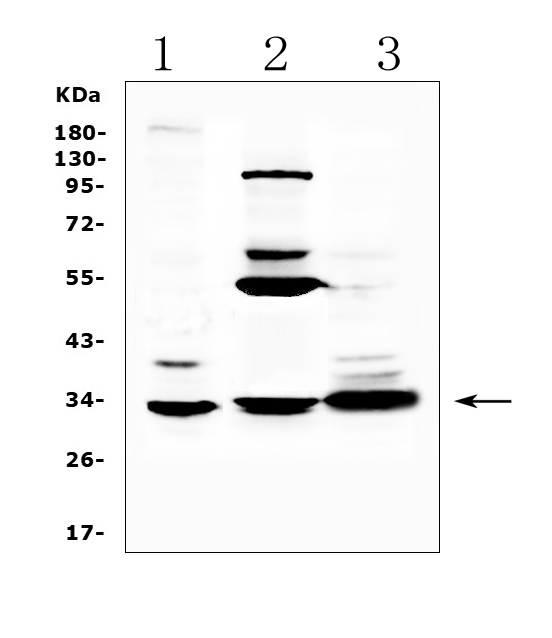
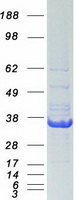
Mass (kDA):
32.562 kDA
Human | |
---|---|
Location: | 19q13.32 |
Sequence: | 19; NC_000019.10 (45407334..45451547, complement) |
[Isoform 1]: Nucleus.; [Isoform 2]: Cytoplasm. Nucleus.; [Isoform 3]: Nucleus.; [Isoform 4]: Nucleus.
The ERCC1 marker is used to determine the expression of the genes ERCC1. This marker has numerous clinical uses. Boster scientists may submit results for species-specific samples, applications and other studies in order to receive product credits. It is available to all scientists around the world. Learn more about it. This article will discuss the clinical and cellular features ERCC1.
The ERCC1 marker has several important functions. It is used to identify cancer cells in tissue sections. It was found in mesenchymal stem cells as well as endothelial and fibroblasts. It can be used to predict the risk of different cancers, including the glioma. However, many questions remain. Here are some of the best uses for this marker.
The ERCC1 Cell-Based ELISA Kit is a convenient and high-throughput tool to measure the expression profile of ERCC1 in human cells. This kit measures relative amounts of ERCC1 in cultured cells. It can also be used to screen the effects of different activators and treatments. However these kits do not contain the individual components. You must therefore think about the application in which you are planning to use markers from ERCC1.
The ERCC1 marker, which is an immunohistochemical marker is used to detect cancerous cells of the colorectal system. In one study the monoclonal antibody (Boster Bio) 4F9 was used to determine the expression of ERCC1 in colorectal cancer tissues. In the research, relevant cell lines were used as controls. To determine the expression of tumors in ERCC1, scoring guidelines were established. The results showed that the antibody 4F9 was specific to all methods and scored 85 percent of colorectal carcinoma specimens.
The current research suggests that ERCC1–TS profiles can aid doctors in managing metastatic colon cancer. These markers could be used to assist doctors in managing advanced stage patients and optimize the treatment. Researchers from UC San Diego and Boster Bio have created the ERCC1-TS test and have published the results. The study has been funded in part by the National Institutes of Health and the Marsha Rivkin Center for Ovarian Cancer Research.
The XPF-ERCC1 protein has been used to verify the marker ERCC1. It cleaves into a simple 3 fluor-labelled substrate. Markers were developed to surround the anticipated position of the incision. The results indicated that XPF-ERCC1 released products were generally 23 nucleotides in length and cleaved at a position of 2 nucleotides 5' to the fork junction.
This study investigated the clinical features of patients suffering from lung cancer. Patients who received platinum chemotherapy demonstrated that ERCC1 expression is a reliable indicator of prognosis. It was also discovered to predict patients' response to immunotherapy. However, further research is needed to establish the validity of ERCC1 expression is a predictor of response to immunotherapy. This is an important step towards clinical practice.
To test the ERCC1 marker, a scoring system was devised. It assessed the expression of ERCC1 within the colorectal crypts. The informed consent of patients was obtained prior to the study. A colorectal tumor was taken from the histopathology department of a hospital. The tissue was frozen with liquid nitrogen and stored at -80 until the time of analysis. Tumor tissue was sourced from this department and embedded in paraffin.
The cellular characteristics of tumor cells were used to determine their score in the evaluation of ERCC1 expression. They were scored from 'low to high' by determining their level of ERCC1 expression. However, tumor cells with homogeneous expression scored high, while those with heterogeneous staining were scored as 'low'. This implies that ERCC1's expression is common in both low and high levels.
The ERCC1 gene encodes a protein that is essential for inter-strand crosslinks and repair of nucleotide excision. A variety of cytotoxic medications have been linked to high levels of ERCC1-XPF's nuclelea. These results suggest that ERCC1 expression could be an important biomarker in predicting the effectiveness of platinum chemotherapy drugs. This suggests that ERCC1 could be used to predict the outcome of non-small-cell lung cancer.
To validate the ERCC1 marker, it was crucial to determine the right substrate. The XPF ERCC1 fluorescent moiety can be enhanced by placing it at different positions in the substrate arm. The fluorophore's position can affect the XPF-ERCC1's function and can alter the site of incision. The XPF–ERCC1 marker is sensitive and scalable, which makes them an ideal tool for high-throughput screening.
A stepwise IHC procedure as well as a workflow illustration are included in the Boster Bio product manual. Optimizing the preparation of samples is crucial for obtaining excellent staining results. BosterBio's comprehensive guide to the retrieval, fixation, and embedding. It also explains the most effective methods for the preparation of various types of tissues. The guide also demonstrates the proper methods for IHC staining different kinds of preserved tissue, such as paraffin wax.
The IHControls were tested against sections of tissue that contained different concentrations of the relevant antibody. To simulate degradation in stain performance the primary antibody was dilute. In the end, the intensity of staining decreased with each dilution. The IHControls were employed as normal controls to determine the intensity of staining. This makes it possible to conduct an accurate and repeatable analysis of data.
The IHControls group showed significantly decreased staining intensity in comparison to the PR group. The stain intensity was statistically significantly lower in tissue controls than in the PR group. Tissue controls also showed a statistically significant decrease in stain intensity at 97°C. These results demonstrate that PR 1294 can recognize a PR antigens without the need for antigen retrieval. In this instance, about 10% of PR+ cells in the "No AgR" group were positive.
For intracellular staining, Boster Bio has several different options to enhance your results. First, you must determine if the target antigen is membrane bound or cytoplasmic. Permeablizing on ice is recommended for antigens that are not surface bound. To restore fluorescence, a fixative could be added. It is best to avoid long-term storage by using alcohol fixatives. When deciding on a fluorochrome, always remember that a weak antigen should be paired with an attractive fluorochrome.
FIGO stage IV is associated with the high expression of the ERCC1 marker. Tumors with this marker displayed high levels of expression in contrast to tumors with unfavorable characteristics, such as an area of greater than four centimeters, an elevated nuclelr grade, or Keratinization. The high expression of the ERCC1 marker also was associated with tumors that displayed an expansive growth pattern, whereas tumors with an infiltrative pattern had a low expression level.
Semi-quantitative immunohistochemical staining for ERCC1 might not be as precise as it should be. This could be due to the aging of tissues enzyme antibodies, interobserver variability and tissue aging. Nevertheless, the findings suggest that a high level of expression of the ERCC1 marker is associated with a favorable OS which suggests that it can be useful in predicting clinical outcomes of gastric cancer patients who are advanced. However, genomic polymorphism analysis is not clinically beneficial.
Although ERCC1 is not a cure for cancer but high expression of the gene could be a sign of a positive outcome, especially in patients who are not treated. It could identify cancers that have little or no molecular progress and have functioning DNA repair mechanisms. The results suggest that the increased expression of the ERCC1 marker is related to clinical resistance to cisplatin-based chemotherapy. However, further research into the mechanism are needed to confirm this link.
The expression of the ERCC1 gene is linked to poor clinical outcomes in patients suffering from locally advanced SCCHN. It is interesting to know that high levels of ERCC1 gene expression are associated to good survival rates in 30% of patients. There are many studies that support this correlation. A high level of ERCC1 can be a predictor for SCCHN clinical outcomes. However, the exact role of ERCC1 in this disease is not yet understood.
The response to chemotherapy was 39.1% in 25 of 64 cases. Table 2 illustrates the relationship between ERCC1 marker expression, and the clinical response. It was interesting to note that there was no correlation between CEA level or age, sex performance status or the number of organs involved. ERCC1 expression was related to clinical response in a significant way, but no other biological marker was of any significance.
PMID: 2420469 by van Duin M., et al. Molecular characterization of the human excision repair gene ERCC-1: cDNA cloning and amino acid homology with the yeast DNA repair gene RAD10.
PMID: 3034490 by Hoeijmakers J.H.J., et al. Identification of DNA repair genes in the human genome.