This website uses cookies to ensure you get the best experience on our website.


- Table of Contents
Plan EXTL3 paraffin IHC using cytoplasmic staining in most tissues as the reference pattern (HPA tissue IHC). Compare high-staining glandular cells with cells reported as unstained, and verify specificity because antibody staining and RNA show low consistency (HPA tissue IHC).

Expected localisation, validated protocols, controls and antibodies — the at-a-glance facts below, then the full design guide.
| Expected localisation | Cytoplasmic staining in most tissues (HPA tissue IHC) | |
| Staining pattern | Cytoplasmic staining in many glandular cells (HPA tissue IHC) | |
| Antigen retrieval | Tris-EDTA pH 9.0 HIER, 95–98 °C, 20 min (rule: nuclear antigen) | |
| Positive control | Cerebellum+4 more · see all | |
| Negative control | Adipose tissue+4 more · see all |
| Fixation | Keep fixation conditions consistent across sections. (standard IHC practice; not target-specific) | |
| Caveat | Antibody staining and RNA show low consistency (HPA tissue IHC) | |
| Regulation | Ubiquitous expression (UniProt) | |
| Isoform / epitope | No annotated isoforms; cytoplasmic vs lumenal epitope matters (UniProt) |
The catalog antibody’s IHC-P protocol is accompanied by one published EXTL3 IHC protocol using mouse pancreas (PMC4741915).
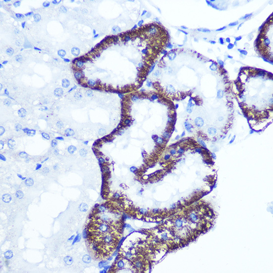
| Sample | Paraffin-embedded rat kidney tissue; fixative not specified (datasheet A07636) |
| Fixation | Image fixative and duration unreported (datasheet A07636); verify before use. |
| Sectioning | 4–5 µm sections on charged slides (standard) |
| Deparaffinisation | Xylene, graded ethanol series to water (standard) |
| Antigen retrieval | Heat-induced epitope retrieval in Tris-EDTA buffer, pH 9.0, 20 min at 95–98 °C (standard rule: nuclear antigen) |
| Peroxidase block | 3% H2O2, 10 min, room temperature (standard) |
| Blocking | 10% normal serum of the secondary host, 30 min, room temperature (standard) |
| Primary antibody | Rabbit anti-EXTL3, 1:50-1:200 (datasheet A07636) |
| Primary incubation | Overnight at 4 °C (standard) |
| Detection | HRP-polymer secondary, DAB chromogen 5–10 min (standard) |
| Counterstain | Hematoxylin, blue, dehydrate and mount (standard) |
| Expected result | EXTL3-positive staining in purkinje cells of cerebellum (HPA tissue IHC: High). HPA tissue profile: Cytoplasmic expression in most tissues. No signal in the no-primary control. |
EXTL3 is a membrane protein reported in the endoplasmic reticulum, Golgi, cell membrane and, under a stated condition, nucleus (UniProt O43909). In tissue IHC, expect predominantly cytoplasmic staining, especially in selected glandular cells, cholangiocytes and Purkinje cells (HPA tissue IHC). HPA rates the tissue pattern Approved but reports low agreement with RNA data and pending external verification (HPA tissue IHC).
| Cytoplasmic staining in pancreatic exocrine glandular cells, liver cholangiocytes or cerebellar Purkinje cells. | These cell populations have High HPA tissue staining and offer useful positive comparators (HPA tissue IHC). Judge the named cells within their tissue context; the HPA profile describes cytoplasmic expression in most tissues, not uniform staining of every cell (HPA tissue IHC). |
| Predominantly nuclear staining, with little cytoplasmic signal, across an ordinary tissue section. | This differs from the reported tissue IHC profile (HPA tissue IHC). UniProt lists the nucleus and specifies that interaction with REG3A induces nuclear translocation (UniProt O43909). Without evidence of that condition, investigate the staining and controls before calling widespread nuclear signal an EXTL3 pattern (standard IHC practice). |
| Strong staining in adipocytes, bone marrow hematopoietic cells or ovarian follicle cells. | HPA reports EXTL3 as not detected in those cell populations (HPA tissue IHC). An isolated strong result raises concern for antibody cross-reactivity or endogenous chromogenic detection activity; examine reagent controls and tissue morphology (standard IHC practice). HPA's negative observations do not establish absolute absence of EXTL3. |
| Diffuse color across multiple compartments or tissue structures, obscuring individual cells. | A broad deposit cannot be assigned confidently to the cytoplasmic cell pattern reported by HPA (HPA tissue IHC). Check a no-primary control, blocking and chromogen development to identify background or endogenous detection activity (standard IHC practice). Interpret any remaining cell signal against morphology. |
| No staining in pancreatic exocrine glandular cells despite an otherwise readable section. | HPA reports High staining in those cells, so the result warrants a technical review (HPA tissue IHC). Verify that the expected cells are present, then assess the IHC-validated antibody, antigen retrieval and detection controls (standard IHC practice). One negative section cannot overturn a tissue-wide expression claim. |
| Membrane topology | EXTL3 has one transmembrane segment at residues 31–51, a cytoplasmic N terminus and a lumenal region spanning residues 52–919 (UniProt O43909 topology). Membrane association is compatible with the HPA cytoplasmic tissue appearance; topology alone cannot specify a chromogenic subcellular pattern. |
| Conditional nuclear location | UniProt lists nuclear localisation and links translocation to interaction with REG3A (UniProt O43909). Treat nuclear staining as context dependent; HPA's tissue summary remains cytoplasmic (HPA tissue IHC). |
| Tissue and antibody evidence | HPA lists CAB025387 as IHC Approved, while the tissue profile has low consistency with RNA expression and awaits external verification (HPA antibodies; HPA tissue IHC). Use the named positive and undetected cell populations as comparators, with that limitation in mind. |
| Annotated processing and glycosylation | UniProt annotates one chain spanning residues 1–919, no signal peptide or propeptide, and glycosylation sites at 290, 592 and 790 (UniProt O43909). These annotations do not establish epitope location, antigen retrieval needs or fixation sensitivity. |
| IF/ICC: is a matching pattern established? | HPA gives a membrane subcellular summary but no main location or ICC/IF image-bearing cell lines; its listed antibody has no ICC status (HPA subcellular; HPA antibodies). UniProt reports several locations (UniProt O43909). This evidence does not define an IF/ICC positive-control image or protocol. |
| Situation | Likely cause | Next action |
|---|---|---|
| The expected positive glandular cells are blank. | The cell population may be missing from the section, or the staining workflow may have failed (standard IHC practice); HPA reports High staining in pancreatic exocrine glandular cells (HPA tissue IHC). | Confirm cell identity on the counterstained section and compare a positive tissue control; review the antibody, antigen retrieval and detection steps using the chosen IHC protocol (standard IHC practice). |
| The no-primary control develops color. | Primary-independent signal indicates background from the detection system or tissue activity (standard IHC practice). | Address endogenous detection activity, blocking and chromogen development before scoring EXTL3; repeat the control alongside the test section (standard IHC practice). |
| Color is strongest in adipocytes or marrow hematopoietic cells. | Those cells are not detected in HPA tissue IHC, so cross-reactivity or detection background is plausible (HPA tissue IHC; standard IHC practice). | Inspect morphology and no-primary controls, then compare a HPA High cell population in the same run before assigning specificity (HPA tissue IHC; standard IHC practice). |
| Nuclear staining dominates the section. | HPA's tissue profile is cytoplasmic, although REG3A interaction can induce nuclear translocation (HPA tissue IHC; UniProt O43909). | Check whether the biological context supports that interaction; inspect controls and accompanying cytoplasmic signal before accepting a nuclear interpretation (UniProt O43909; standard IHC practice). |
| Nearly every structure shows diffuse chromogen. | The deposit obscures the cell-resolved cytoplasmic pattern described by HPA and may reflect nonspecific staining or overdevelopment (HPA tissue IHC; standard IHC practice). | Use the no-primary control to locate background, review blocking and chromogen timing, and score only interpretable cells (standard IHC practice). |
| An IF/ICC result seems inconsistent with the IHC slide. | HPA provides a membrane IF summary without an image-bearing cell line or ICC validation status for the listed antibody (HPA subcellular; HPA antibodies). | Evaluate IF/ICC with its own validated controls and guide; use HPA tissue IHC only for the paraffin-section cell pattern (HPA tissue IHC; standard IF/ICC practice). |


Comprehensive Human Protein Atlas IHC scoring per tissue (reliability: Approved — Low consistency between antibody staining and RNA expression data. Pending external verification.). Rows are taken directly from the HPA tissue chart — click any row's HPA link to view the source.
| Tissue | Cell type | Level | Evidence | Source |
|---|---|---|---|---|
| Cerebellum | Purkinje cells | High | Protein (IHC) | HPA → |
| Gallbladder | Glandular cells | High | Protein (IHC) | HPA → |
| Liver | Cholangiocytes | High | Protein (IHC) | HPA → |
| Pancreas | Exocrine glandular cells | High | Protein (IHC) | HPA → |
| Placenta | Decidual cells | High | Protein (IHC) | HPA → |
| Tissue | Cell type | Level | Evidence | Source |
|---|---|---|---|---|
| Adipose tissue | Adipocytes | Not detected | Protein (IHC) | HPA → |
| Bone marrow | Hematopoietic cells | Not detected | Protein (IHC) | HPA → |
| Ovary | Follicle cells | Not detected | Protein (IHC) | HPA → |
| Soft tissue | Chondrocytes | Not detected | Protein (IHC) | HPA → |
| Spleen | Cells in red pulp | Not detected | Protein (IHC) | HPA → |
Troubleshoot EXTL3 staining in paraffin sections by checking retrieval, compartment, cell identity, and controls before comparing staining intensity.
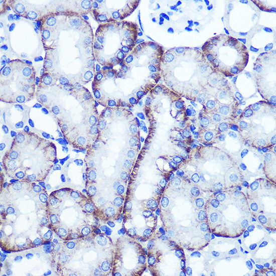
A07636 has real EXTL3 IHC images from paraffin-embedded rat and mouse kidney; catalog reactivity includes human, mouse and rat (catalog IHC image captions; catalog reactivity).
A07636 is listed for IHC and shows staining in paraffin-embedded rat kidney at 1:100 (catalog applications; rat kidney image caption). Its second IHC image shows paraffin-embedded mouse kidney at 1:100; human reactivity is listed without a human IHC image (mouse kidney image caption; catalog reactivity; catalog IHC image captions).
Which to pick: Choose A07636 for paraffin-section IHC: its rat and mouse kidney captions specify microwave antigen retrieval in 10 mM PBS, pH 7.2, and the listed IHC dilution range is 1:50–1:200 (catalog IHC image captions; catalog IHC dilution). The captions do not report a fixative (catalog IHC image captions). For cross-species work, its listed reactivity covers human, mouse and rat, though the IHC images document only mouse and rat; no IF/ICC application or image is listed for this SKU (catalog reactivity; catalog applications; catalog IHC and IF image captions).