This website uses cookies to ensure you get the best experience on our website.


- Table of Contents
Plan FUT2 staining in paraffin sections with the catalog antibody’s 1:100–1:300 IHC dilution range (datasheet A02679). Expect cytoplasmic tissue staining (HPA tissue IHC), but interpret intensity cautiously because staining and RNA expression show low consistency (HPA tissue IHC).

Expected localisation, validated protocols, controls and antibodies — the at-a-glance facts below, then the full design guide.
| Expected localisation | Observed cytoplasmic staining (HPA tissue IHC); expected Golgi membrane location (UniProt) | |
| Staining pattern | Cytoplasmic staining across tissues (HPA tissue IHC) | |
| Antigen retrieval | Tris-EDTA pH 9.0 HIER, heat-mediated (datasheet A02679) | |
| Positive control | Appendix+4 more · see all | |
| Negative control | Vagina |
| Fixation | Keep fixation consistent between sections (standard IHC practice; not target-specific) | |
| Caveat | Low consistency between staining and RNA expression (HPA tissue IHC) | |
| Regulation | Tissue-enhanced RNA in intestine (HPA tissue RNA) | |
| Isoform / epitope | No isoforms annotated; cytosolic versus lumenal epitope matters (UniProt) |
The catalog antibody’s IHC-P protocol (datasheet A02679) is accompanied by published FUT2 IHC methods for lung adenocarcinoma (PMC5722559) and colorectal cancer (PMC9911090).
| Sample | Paraffin-embedded human liver cancer tissue; fixative not specified (datasheet A02679) |
| Fixation | Image fixative and duration unreported (datasheet A02679); verify before use. |
| Sectioning | 4–5 µm sections on charged slides (standard) |
| Deparaffinisation | Xylene, graded ethanol series to water (standard) |
| Antigen retrieval | Heat retrieval: Tris-EDTA pH 9.0 (datasheet A02679); 20 min, 95–100 °C (standard) |
| Peroxidase block | 3% H2O2, 10 min, room temperature (standard) |
| Blocking | 10% normal serum of the secondary host, 30 min, room temperature (standard) |
| Primary antibody | Rabbit anti-FUT2, 1:100-1:300 (datasheet A02679) |
| Primary incubation | Overnight at 4 °C (standard) |
| Detection | HRP-polymer secondary, DAB chromogen 5–10 min (standard) |
| Counterstain | Hematoxylin, blue, dehydrate and mount (standard) |
| Expected result | FUT2-positive staining in glandular cells of appendix (HPA tissue IHC: Medium). HPA tissue profile: Cytoplasmic expression in all tissues. No signal in the no-primary control. |
FUT2 is a Golgi stack membrane enzyme with a cytoplasmic N terminus and a lumenal catalytic region (UniProt Q10981 topology). In paraffin IHC, assess cytoplasmic staining in glandular or respiratory epithelial cells; appendix glandular and bronchial epithelial cells show medium staining (HPA tissue IHC). Treat the pattern as provisional: HPA rates tissue staining reliability Uncertain because staining and RNA expression have low consistency (HPA tissue IHC).
| Localized cytoplasmic staining in appendix glandular cells or bronchial epithelium. | This agrees with medium staining reported in those cells (HPA tissue IHC). A compact perinuclear component is compatible with Golgi localization (UniProt Q10981 subcellular location). Compare the signal with adjacent tissue and a no-primary control before calling cells positive (general IHC practice). |
| Predominantly nuclear staining, with little cytoplasmic signal. | A nuclear pattern is unsupported by the Golgi membrane annotation (UniProt Q10981 subcellular location). Check the no-primary control and review whether chromogen precipitate or counterstain is being read as signal (general IHC practice). Membrane staining alone needs separate assessment because HPA reports an uncertain plasma membrane ICC-IF location (HPA ICC-IF). |
| Strong staining in vaginal squamous epithelium, especially if the expected control is weak. | HPA reports FUT2 as not detected in vaginal squamous cells (HPA tissue IHC). Consider antibody cross-reactivity or endogenous detection activity as possible IHC explanations (general IHC practice); this comparison cannot prove either mechanism because HPA rates tissue staining Uncertain (HPA tissue IHC). |
| Even haze across tissue, stroma, and empty spaces. | A slide-wide haze lacks the cell-specific pattern needed to interpret FUT2 staining (HPA tissue IHC; general IHC practice). Inspect no-primary and reagent controls, then optimize blocking, washes, antibody concentration, and detection conditions one at a time (general IHC practice). |
| No visible signal in appendix glandular cells or bronchial epithelium. | These are reported medium-staining populations, making them useful comparison tissues, although the HPA tissue result is Uncertain (HPA tissue IHC). Check tissue preservation, retrieval conditions, reagent activity, and a run-level positive control before treating the absence of stain as biological (general IHC practice). |
| Compartment and epitope orientation | FUT2 spans the membrane at residues 15–28; residues 1–14 are cytoplasmic and 29–343 are lumenal (UniProt Q10981 topology). The supplied record gives no antibody epitope, so orientation cannot establish a required retrieval method or staining intensity. |
| Tissue and antibody evidence | HPA calls tissue staining Uncertain because antibody staining and RNA expression have low consistency (HPA tissue IHC). Its listed antibody HPA014402 has Uncertain IHC validation (HPA antibody validation). Use reported tissue levels as comparisons, not definitive specificity proof. |
| IF/ICC Q&A: Should membrane signal match IHC? | HPA places FUT2 mainly at the plasma membrane, with additional cytosol, in ICC-IF; both locations are uncertain (HPA ICC-IF). Tissue IHC instead reports cytoplasmic expression (HPA tissue IHC), while UniProt places FUT2 at Golgi membranes (UniProt Q10981 subcellular location). |
| Processing and glycosylation | UniProt lists no signal peptide or propeptide and annotates glycosylation at residues 188, 282, and 308 (UniProt Q10981 processing and glycosylation). Neither annotation establishes a fixation effect, retrieval requirement, or cause of a negative IHC result. |
| Situation | Likely cause | Next action |
|---|---|---|
| Expected epithelial control is blank. | The run may have a tissue, retrieval, antibody, or detection problem (general IHC practice); appendix and bronchus are only provisional medium-positive references (HPA tissue IHC). | Confirm tissue identity and a working run-level control; review retrieval conditions, primary antibody use, and detection reagents in sequence (general IHC practice). |
| Nuclear staining dominates. | Nuclear localization is unsupported for FUT2 (UniProt Q10981 subcellular location); counterstain or deposited chromogen may be misread as target signal (general IHC practice). | Compare the no-primary section and inspect signal separately from counterstain; reassess the result against the expected cytoplasmic distribution (HPA tissue IHC; general IHC practice). |
| Vaginal squamous cells appear strongly positive. | This conflicts with HPA's not-detected observation (HPA tissue IHC). Cross-reactivity or endogenous detection activity are possibilities, not established causes (general IHC practice). | Check a no-primary control and the detection system's endogenous-activity control; compare with a reported medium-positive tissue (HPA tissue IHC; general IHC practice). |
| Diffuse staining obscures cell boundaries. | Excess primary or detection reagent, insufficient blocking, or inadequate washing can raise background (general IHC practice). | Compare reagent controls, then adjust blocking, primary concentration, detection exposure, and washes separately while retaining the same tissue control (general IHC practice). |
| Only surface membrane outlines are visible. | HPA's plasma membrane ICC-IF assignment is uncertain and does not establish the expected paraffin IHC appearance (HPA ICC-IF; HPA tissue IHC). | Review cytoplasmic signal in the same cells and compare controls; record membrane-only staining as unresolved unless independently supported (UniProt Q10981 subcellular location; general IHC practice). |
| Staining varies sharply between sections. | Section condition or run handling may differ (general IHC practice); HPA's Uncertain tissue reliability also limits confidence in a biological explanation (HPA tissue IHC). | Compare matched sections with the same control tissue and document retrieval, antibody application, detection time, and counterstain for each run (general IHC practice). |
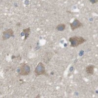
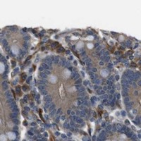


Comprehensive Human Protein Atlas IHC scoring per tissue (reliability: Uncertain — Low consistency between antibody staining and RNA expression data.). Rows are taken directly from the HPA tissue chart — click any row's HPA link to view the source.
| Tissue | Cell type | Level | Evidence | Source |
|---|---|---|---|---|
| Appendix | Glandular cells | Medium | Protein (IHC) | HPA → |
| Bone marrow | Hematopoietic cells | Medium | Protein (IHC) | HPA → |
| Breast | Glandular cells | Medium | Protein (IHC) | HPA → |
| Bronchus | Respiratory epithelial cells | Medium | Protein (IHC) | HPA → |
| Caudate | Neuronal cells | Medium | Protein (IHC) | HPA → |
| Tissue | Cell type | Level | Evidence | Source |
|---|---|---|---|---|
| Vagina | Squamous epithelial cells | Not detected | Protein (IHC) | HPA → |
Use the paraffin-section IHC conditions reported for A02679 as a starting point, and assess staining against FUT2 localisation and tissue context.
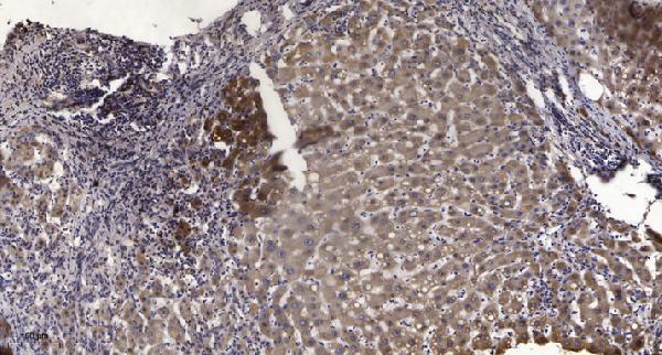
A02679 has real IHC data from paraffin-embedded human liver cancer (IHC image caption). IF is listed, with human, mouse, and rat reactivity (catalog: applications/reactivity).
A02679 shows IHC staining of paraffin-embedded human liver cancer using Tris-EDTA retrieval at pH 9.0 and a 1:200 primary dilution (IHC image caption). The catalog lists IHC and IF applications and human, mouse, and rat reactivity, but provides no IF image (catalog: applications/reactivity/IF image alts).
Which to pick: Choose A02679 for paraffin-section tissue IHC based on its human liver cancer image; the fixative is unreported (A02679 IHC image caption). For IF, A02679 is listed at 1:50; ICC validation and IF images are unreported (catalog: applications/IF dilution/IF image alts). For mouse or rat samples, A02679 lists reactivity with both species, although its IHC image shows human tissue only; the antibody is rabbit-hosted and its clonality is unreported (catalog: reactivity/host/clone; A02679 IHC image caption).