This website uses cookies to ensure you get the best experience on our website.


- Table of Contents
Plan chromogenic GSTA4 IHC in paraffin sections using the catalog antibody at 2–5 μg/ml (datasheet A03699-1). Compare cytoplasmic staining in Leydig and adrenal glandular cells, while interpreting signal cautiously because the tissue antibody may detect protein from more than one gene (HPA tissue IHC).

Expected localisation, validated protocols, controls and antibodies — the at-a-glance facts below, then the full design guide.
| Expected localisation | Cytoplasmic tissue staining (HPA tissue IHC) | |
| Staining pattern | Cytoplasmic staining in Leydig and adrenal glandular cells (HPA tissue IHC) | |
| Antigen retrieval | EDTA pH 8.0 HIER, heat-mediated (datasheet A03699-1) | |
| Positive control | Adrenal gland+4 more · see all | |
| Negative control | Appendix+4 more · see all |
| Fixation | Keep fixation consistent across sections (standard IHC practice; not target-specific) | |
| Caveat | Staining may reflect protein from more than one gene (HPA tissue IHC) | |
| Regulation | No specific regulator reported (UniProt) | |
| Isoform / epitope | 2 isoforms; epitope differences are unspecified (UniProt) |
The catalog antibody uses EDTA pH 8.0 heat retrieval (datasheet A03699-1). The published protocols below describe chromogenic GSTA4 IHC in mouse colon and human tissues (PMC10559223; PMC9619338).
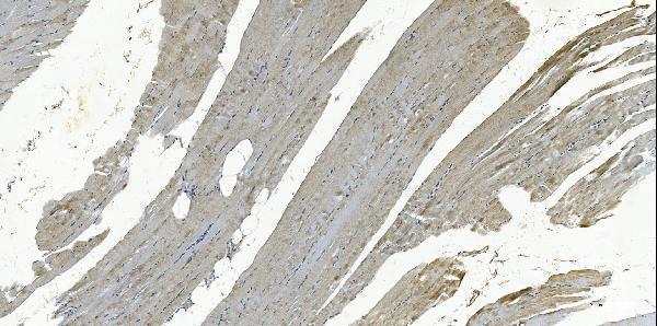
| Sample | Paraffin-embedded human skeletal muscle tissue; fixative not specified (datasheet A03699-1) |
| Fixation | Image fixative and duration unreported (datasheet A03699-1); verify before use. |
| Sectioning | 4–5 µm sections on charged slides (standard) |
| Deparaffinisation | Xylene, graded ethanol series to water (standard) |
| Antigen retrieval | Heat retrieval: EDTA pH 8.0 (datasheet A03699-1); 20 min, 95–100 °C (standard) |
| Peroxidase block | 3% H2O2, 10 min, room temperature (standard) |
| Blocking | 10% goat serum (datasheet A03699-1) |
| Primary antibody | Rabbit anti-GSTA4, 2-5μg/ml (datasheet A03699-1) |
| Primary incubation | Overnight at 4 °C (datasheet A03699-1) |
| Detection | Streptavidin-biotin complex (SABC), DAB chromogen (datasheet A03699-1) |
| Counterstain | Hematoxylin, blue, dehydrate and mount (standard) |
| Expected result | GSTA4-positive staining in glandular cells of adrenal gland (HPA tissue IHC: High). HPA tissue profile: Cytoplasmic expression in several tissues, mainly in Leydig cells in testis, and glandular cells in adrenal gland and fallopian tube. No signal in the no-primary control. |
GSTA4 is a cytoplasmic protein with no transmembrane segment (UniProt O15217). In paraffin-section IHC, look for cytoplasmic staining in Leydig cells of testis and glandular cells of adrenal gland; HPA also reports high staining in several other defined cell populations (HPA tissue IHC). Treat these as reference patterns rather than definitive specificity controls: HPA rates the tissue staining Approved, reports low agreement with RNA data, and cautions that the antibody targets protein from more than one gene (HPA tissue IHC).
| Cytoplasmic staining in adrenal glandular cells or testicular Leydig cells, with adjacent cells less prominent. | This matches the principal tissue profile and the High cell-level staining reported by HPA (HPA tissue IHC), as well as cytoplasmic localisation (UniProt O15217). Score the named cells and their compartment separately from whole-section colour; HPA’s Approved rating carries a low RNA–staining consistency caveat (HPA tissue IHC). |
| Predominantly nuclear, membrane-rim, or extracellular staining, without a convincing cytoplasmic component. | The compartment conflicts with the annotated cytoplasmic location and lack of a transmembrane segment (UniProt O15217). Suspect staining artefact or nonspecific detection first. This appearance alone cannot identify the cause; compare a known-positive cell population and the detection controls before calling it GSTA4. |
| Strong staining in an unexpected cell population, including one HPA lists as Not detected. | Unexpected distribution raises possible cross-reactivity or endogenous chromogen-generating activity, not proof of either. HPA specifically cautions that its tissue antibody targets protein from more than one gene (HPA tissue IHC). Interpret the named cell type: for example, HPA reports High in lung macrophages, while its fallopian-tube entries are internally inconsistent (HPA tissue IHC). |
| Diffuse colour across tissue, stroma, and slide background with little cell-specific contrast. | A uniform haze does not reproduce the cell-restricted HPA examples (HPA tissue IHC). In chromogenic IHC, excessive primary or detection reagent, inadequate blocking or washing, and endogenous enzyme activity are general possible causes (general IHC practice). Review the no-primary control and whether the expected cytoplasmic cells remain distinguishable. |
| No staining in the expected cells of an adrenal-gland or testis reference section. | An absent signal disagrees with HPA’s High glandular-cell or Leydig-cell observations (HPA tissue IHC), but a negative run does not establish absent GSTA4. Check that those cells are present and assess the IHC-validated antibody, retrieval, detection, and positive-control performance using general IHC practice. |
| Compartment and protein architecture (UniProt O15217). | GSTA4 is cytoplasmic, lacks a transmembrane segment, and has one annotated chain spanning residues 1–222 (UniProt O15217). These facts support a cytoplasmic reading of positive cells; they do not specify an antibody epitope or predict which retrieval condition will work. |
| Cell-level reference distribution (HPA tissue IHC). | HPA reports High staining in adrenal glandular cells, testicular Leydig cells, breast myoepithelial cells, duodenal endocrine cells, hepatocytes, and lung macrophages (HPA tissue IHC). The cell identity matters: HPA also lists Not detected populations in other tissues, so whole-organ labels alone are weak comparators. |
| Reference-pattern limitations (HPA tissue IHC). | HPA calls the tissue result Approved but flags low agreement between staining and RNA expression and protein targeting from more than one gene (HPA tissue IHC). Its profile mentions fallopian-tube glandular staining while its cell-level entry says Not detected; avoid treating that tissue as an unambiguous positive control. |
| Isoforms and assay interpretation (UniProt O15217; HPA antibodies). | UniProt lists 2 isoforms (UniProt O15217). The supplied antibody record gives IHC status as Approved and no epitope or isoform coverage (HPA antibodies). Thus, a tissue staining result cannot establish which isoform was detected or whether both are recognized. |
| Situation | Likely cause | Next action |
|---|---|---|
| Reference adrenal gland or testis shows no expected cytoplasmic cells stained. | The selected field may lack the named cells, or the IHC run may have failed; HPA reports them as High reference populations (HPA tissue IHC). | Verify cell identity on the section, then check the run’s positive control, retrieval, primary-antibody use, and detection reagents (general IHC practice). Avoid interpreting an empty or unsuitable field as a biological negative. |
| Colour concentrates in nuclei or outlines cell membranes. | That distribution conflicts with GSTA4’s cytoplasmic annotation and absence of a transmembrane segment (UniProt O15217); nonspecific or detection-related staining is possible. | Compare the expected cytoplasm in HPA-positive cell populations, examine a no-primary control, and review staining conditions (HPA tissue IHC; general IHC practice). Do not score compartment-mismatched colour as a clean positive. |
| Many unrelated cells or extracellular areas carry similar chromogen. | Diffuse background or endogenous enzyme activity can obscure cell-specific signal in chromogenic IHC (general IHC practice). The HPA examples identify particular cell populations (HPA tissue IHC). | Inspect the no-primary control; review blocking, washing, reagent concentration, and endogenous-enzyme quenching as appropriate to the detection system (general IHC practice). Reassess only after the expected cells stand out from background. |
| Lung macrophages stain strongly, but surrounding respiratory cells differ. | HPA reports High staining in lung macrophages and Medium staining in bronchial respiratory epithelial cells (HPA tissue IHC); these are distinct tissue and cell entries. | Record the compartment and identity of each stained cell population. Compare like with like in the HPA tissue entries, and avoid treating every lung or airway cell as an equally strong control (HPA tissue IHC). |
| Fallopian-tube staining appears inconsistent with the HPA overview. | The HPA profile names fallopian-tube glandular cells, yet its detailed negative entry says those cells are Not detected (HPA tissue IHC). The supplied evidence does not resolve the discrepancy. | Document the observed cell type and cytoplasmic pattern, then use a less ambiguous HPA-positive reference such as adrenal glandular cells or testicular Leydig cells for run interpretation (HPA tissue IHC). |
| Can an IF/ICC image be used to confirm the IHC pattern? | HPA supplies no main ICC-IF location and no cell-line ICC-IF images for this record (HPA subcellular). Its tissue observations come from IHC (HPA tissue IHC). | Use the UniProt cytoplasmic annotation as an IF localisation expectation (UniProt O15217), while treating IF/ICC validation and controls as a separate guide question. Do not claim that HPA independently confirmed this IF pattern. |


Comprehensive Human Protein Atlas IHC scoring per tissue (reliability: Approved — Low consistency between antibody staining and RNA expression data. Caution, targets protein from more than one gene.). Rows are taken directly from the HPA tissue chart — click any row's HPA link to view the source.
| Tissue | Cell type | Level | Evidence | Source |
|---|---|---|---|---|
| Adrenal gland | Glandular cells | High | Protein (IHC) | HPA → |
| Breast | Myoepithelial cells | High | Protein (IHC) | HPA → |
| Duodenum | Endocrine cells | High | Protein (IHC) | HPA → |
| Liver | Hepatocytes | High | Protein (IHC) | HPA → |
| Lung | Macrophages | High | Protein (IHC) | HPA → |
| Tissue | Cell type | Level | Evidence | Source |
|---|---|---|---|---|
| Appendix | Endocrine cells | Not detected | Protein (IHC) | HPA → |
| Caudate | Glial cells | Not detected | Protein (IHC) | HPA → |
| Colon | Endocrine cells | Not detected | Protein (IHC) | HPA → |
| Endometrium | Cells in endometrial stroma | Not detected | Protein (IHC) | HPA → |
| Epididymis | Glandular cells | Not detected | Protein (IHC) | HPA → |
Troubleshoot GSTA4 staining in paraffin sections by checking retrieval, cellular localisation, antibody specificity, and cell resolved scoring before interpreting chromogenic signal.
The catalog antibody has real IHC data from paraffin-embedded human skeletal muscle (IHC figure caption). Human and monkey reactivity are listed; IF/ICC validation is absent (catalog applications and images).
A03699-1 is listed for IHC and has a figure showing chromogenic detection in a paraffin-embedded human skeletal muscle section (catalog applications; IHC figure caption). Its listed reactivity is human and monkey, with no IF/ICC application or image supplied (catalog reactivity, applications and images).
Which to pick: Choose A03699-1 for paraffin-section tissue IHC: its own figure shows staining of human skeletal muscle, and the catalog specifies 2–5 μg/ml for IHC (IHC figure caption; datasheet: IHC dilution). The caption does not report the fixative (IHC figure caption). No SKU in this payload has IF/ICC validation; A03699-1 lists monkey reactivity, but its IHC figure documents human tissue only (catalog applications, images and reactivity; IHC figure caption).