This website uses cookies to ensure you get the best experience on our website.


- Table of Contents
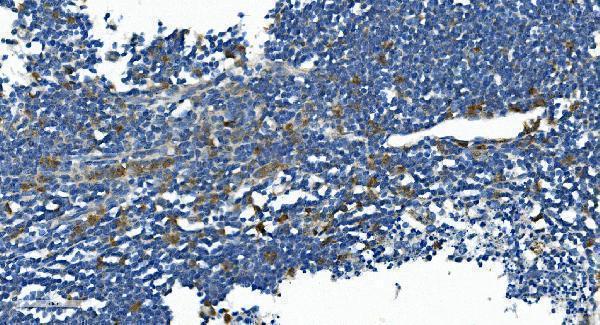
Plan paraffin-section IL2RB staining around selective cytoplasmic signal in immune cells (HPA tissue IHC). The catalog antibody’s IHC example uses 2 μg/mL primary antibody with chromogenic detection (datasheet A03401-3).

Expected localisation, validated protocols, controls and antibodies — the at-a-glance facts below, then the full design guide.
| Expected localisation | Cytoplasmic staining in a subset of immune cells (HPA tissue IHC) | |
| Staining pattern | Selective cytoplasmic signal in some immune cells (HPA tissue IHC) | |
| Antigen retrieval | EDTA pH 8.0 HIER, heat-mediated (datasheet A03401-3) | |
| Positive control | Bone marrow+4 more · see all | |
| Negative control | Adipose tissue+4 more · see all |
| Fixation | Keep fixation consistent across sections. (standard IHC practice; not target-specific) | |
| Caveat | Sparse positive cells may be missed during scoring (HPA tissue IHC) | |
| Regulation | Expression regulation is not annotated (UniProt) | |
| Isoform / epitope | No isoforms annotated; extracellular versus cytoplasmic epitope matters (UniProt) |
Compare the catalog antibody’s IHC-P protocol (datasheet A03401-3) with the published MSI-H colon cancer tissue microarray protocol (PMC8057496).
| Sample | Paraffin-embedded human appendicitis tissue; fixative not specified (datasheet A03401-3) |
| Fixation | Image fixative and duration unreported (datasheet A03401-3); verify before use. |
| Sectioning | 4–5 µm sections on charged slides (standard) |
| Deparaffinisation | Xylene, graded ethanol series to water (standard) |
| Antigen retrieval | Heat retrieval: EDTA pH 8.0 (datasheet A03401-3); 20 min, 95–100 °C (standard) |
| Peroxidase block | 3% H2O2, 10 min, room temperature (standard) |
| Blocking | 10% goat serum (datasheet A03401-3) |
| Primary antibody | Rabbit anti-IL2RB, 2-5μg/ml (datasheet A03401-3) |
| Primary incubation | Overnight at 4 °C (datasheet A03401-3) |
| Detection | Streptavidin-biotin complex (SABC), DAB chromogen (datasheet A03401-3) |
| Counterstain | Hematoxylin, blue, dehydrate and mount (standard) |
| Expected result | IL2RB-positive staining in hematopoietic cells of bone marrow (HPA tissue IHC: Medium). HPA tissue profile: Selective cytoplasmic expression in a subset of immune cells. No signal in the no-primary control. |
IL2RB is a cell-membrane receptor with an extracellular segment at residues 27–240 and a cytoplasmic segment at 266–551 (UniProt P14784 topology). In tissue IHC, expect selective cytoplasmic staining in a subset of immune cells (HPA tissue IHC: Enhanced reliability). Interpret that observed IHC pattern alongside the membrane assignment; do not require a crisp membrane rim in every positive paraffin-section cell (HPA tissue IHC; UniProt P14784 subcellular location).
| Subset of immune cells stain at medium intensity in lymphoid tissue or bone marrow. | This fits the reported selective cytoplasmic IHC profile (HPA tissue IHC: Enhanced reliability). Medium staining is reported in bone-marrow hematopoietic cells and non-germinal-center cells of lymph node and tonsil (HPA tissue IHC: Medium). Judge the labelled cells, since the HPA profile describes a subset rather than uniform staining throughout those tissues (HPA tissue IHC). |
| Signal appears mainly nuclear, or concentrates in structures outside the expected immune-cell subset. | A nuclear pattern does not match the reported selective cytoplasmic tissue staining or cell-membrane assignment (HPA tissue IHC; UniProt P14784 subcellular location). Treat it as suspect until controls and cell identity are checked (standard IHC practice). HPA notes that some stained cells or structures are unannotated, so morphology alone may not identify every positive cell (HPA tissue IHC: reliability description). |
| Adipocytes or respiratory epithelial cells stain as strongly as the proposed positives. | Those cell types are reported as not detected in adipose tissue and bronchus, respectively (HPA tissue IHC: Not detected). Their staining raises possible cross-reactivity or endogenous detection activity; it does not by itself establish which cause applies (standard IHC practice). Compare the suspect signal with an omitted-primary control and inspect cell morphology before scoring it as IL2RB (standard IHC practice). |
| Diffuse colour covers cells, stroma, and empty areas without a discrete positive subset. | That distribution conflicts with selective staining of immune cells (HPA tissue IHC). General IHC causes include nonspecific antibody binding, incomplete blocking, or detection-reagent background (standard IHC practice). Check whether the same haze appears in the omitted-primary control, then assess the primary-antibody and detection steps separately (standard IHC practice). |
| No labelled cells appear in a bone-marrow or lymph-node section. | HPA reports medium staining in bone-marrow hematopoietic cells and lymph-node non-germinal-center cells (HPA tissue IHC: Medium). A blank section therefore warrants a control review, although positivity is confined to particular cells rather than every cell in either tissue (HPA tissue IHC). Confirm that relevant cells are present before adjusting retrieval or detection conditions (standard IHC practice). |
| Observed compartment versus receptor topology | HPA describes selective cytoplasmic staining in tissue IHC, while UniProt assigns IL2RB to the cell membrane (HPA tissue IHC; UniProt P14784 subcellular location). Use the reported IHC appearance to assess the slide without treating a missing sharp membrane rim as an automatic failure (HPA tissue IHC; standard IHC interpretation). |
| Cell and tissue selection | HPA reports medium signal in bone-marrow hematopoietic cells, endometrial lymphocytes, splenic red-pulp cells, and non-germinal-center cells in lymph node and tonsil (HPA tissue IHC: Medium). HPA reports several other specified cell populations as not detected; these are cell-level comparisons, not whole-tissue absence claims (HPA tissue IHC: Not detected). |
| Antibody evidence | The listed antibody HPA062657 has Enhanced IHC validation, and the tissue profile has Enhanced reliability based on staining and RNA-expression consistency (HPA antibodies: HPA062657; HPA tissue IHC: reliability description). This supports the reported pattern but does not establish that every stained structure has been identified (HPA tissue IHC: reliability description). |
| Epitope and processing limits | UniProt places the mature chain at residues 27–551 and lists four glycosylation sites in its extracellular region (UniProt P14784 processing, topology, glycosylation). The supplied record does not locate the IHC antibody epitope, so these annotations alone cannot predict how a particular retrieval condition will change staining (UniProt P14784; standard IHC interpretation). |
| IF/ICC: what can this record establish? | HPA summarizes IL2RB as membrane-localized for ICC-IF but supplies no main-location assignment or cell-line images here (HPA subcellular ICC-IF). That summary is consistent with the UniProt membrane assignment; it does not establish an IF/ICC staining pattern for a particular cell line or override the observed tissue-IHC profile (HPA subcellular ICC-IF; UniProt P14784). |
| Situation | Likely cause | Next action |
|---|---|---|
| Positive tissue is entirely blank. | The relevant positive-cell subset may be absent from the section, or the IHC workflow may have failed (HPA tissue IHC: selective profile; standard IHC practice). | First verify hematopoietic cells in bone marrow or non-germinal-center cells in lymph node; both have reported medium staining (HPA tissue IHC: Medium). Then check the run control, primary-antibody step, retrieval, and chromogenic detection using the validated IHC procedure (standard IHC practice). |
| Only a few cells stain, making the section look mostly negative. | Sparse signal can reflect the reported selective immune-cell pattern rather than assay failure (HPA tissue IHC: profile). | Score the specific cell population and its compartment instead of averaging across the section (HPA tissue IHC: selective cytoplasmic profile; standard IHC interpretation). Compare a second reported positive cell population if the candidate cells are hard to identify (HPA tissue IHC: positive list). |
| Adipocytes or bronchial respiratory epithelium stain. | These cells are reported as not detected; cross-reactivity or endogenous chromogenic activity is possible (HPA tissue IHC: Not detected; standard IHC practice). | Compare an omitted-primary control, check the relevant endogenous-activity block, and reassess primary-antibody specificity before calling the signal IL2RB (standard IHC practice). Keep the comparison at cell level rather than labelling the entire tissue negative (HPA tissue IHC). |
| Background is diffuse across the section. | Broad staining departs from HPA's selective immune-cell profile and may arise from nonspecific binding or detection background (HPA tissue IHC; standard IHC practice). | Inspect the omitted-primary control, confirm blocking and washes, and adjust the primary-antibody or detection conditions only after locating the background source (standard IHC practice). Recheck whether a discrete immune-cell subset remains visible (HPA tissue IHC: profile). |
| Staining is predominantly nuclear. | A nuclear-dominant result disagrees with the reported cytoplasmic IHC profile and membrane assignment (HPA tissue IHC; UniProt P14784 subcellular location). | Review counterstain and chromogen separation, compare controls, and check whether the nuclear colour tracks with the primary antibody before interpreting it as target signal (standard IHC practice). |
| Changing retrieval seems to alter the signal. | Target-specific fixation effects are not established by the supplied assay evidence. Verify with a matched IHC source before attributing a result to fixation. | Use the catalog antibody's validated IHC-P procedure as the starting condition, then compare conditions with the same reported positive-cell population and appropriate run controls (standard IHC practice; HPA tissue IHC: positive list). Avoid inferring an IL2RB-specific fixation mechanism from signal changes alone (standard IHC interpretation). |


Comprehensive Human Protein Atlas IHC scoring per tissue (reliability: Enhanced — High consistency between antibody staining and RNA expression data. Antibody staining in cells/structures not annotated, view images.). Rows are taken directly from the HPA tissue chart — click any row's HPA link to view the source.
| Tissue | Cell type | Level | Evidence | Source |
|---|---|---|---|---|
| Bone marrow | Hematopoietic cells | Medium | Protein (IHC) | HPA → |
| Endometrium | Lymphocytes | Medium | Protein (IHC) | HPA → |
| Lymph node | Non-germinal center cells | Medium | Protein (IHC) | HPA → |
| Spleen | Cells in red pulp | Medium | Protein (IHC) | HPA → |
| Tonsil | Non-germinal center cells | Medium | Protein (IHC) | HPA → |
| Tissue | Cell type | Level | Evidence | Source |
|---|---|---|---|---|
| Adipose tissue | Adipocytes | Not detected | Protein (IHC) | HPA → |
| Adrenal gland | Glandular cells | Not detected | Protein (IHC) | HPA → |
| Appendix | Glandular cells | Not detected | Protein (IHC) | HPA → |
| Breast | Adipocytes | Not detected | Protein (IHC) | HPA → |
| Bronchus | Respiratory epithelial cells | Not detected | Protein (IHC) | HPA → |
Use the catalog antibody’s paraffin-section evidence and IL2RB’s membrane topology to guide staining, controls, and interpretation (caption A03401-3; UniProt P14784 topology).
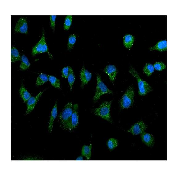
A03401-3 has human paraffin-section IHC and A549-cell IF images (catalog image captions). The catalog lists human, mouse and rat reactivity (catalog: reactivity).
A03401-3 was demonstrated by IHC on a paraffin-embedded human appendicitis section (A03401-3 IHC caption). The same SKU was demonstrated by IF in A549 cells and lists IHC, IF and ICC applications (A03401-3 IF caption; catalog: applications).
Which to pick: For tissue IHC, choose A03401-3: its own image shows a human paraffin section with EDTA pH 8.0 retrieval and 2 μg/ml primary antibody; the fixative is unreported (A03401-3 IHC caption). For IF/ICC, A03401-3 lists both applications and has an A549-cell IF image using 5 μg/ml primary antibody (catalog: applications; A03401-3 IF caption). For mouse or rat samples, A03401-3 lists reactivity to both species, although its supplied IHC and IF images show human samples; clonality is unreported (catalog: reactivity and clone; A03401-3 image captions).