This website uses cookies to ensure you get the best experience on our website.


- Table of Contents
Plan MEF2A paraffin IHC around nuclear staining (HPA tissue IHC). Use high-staining cardiomyocytes as a positive control and liver cholangiocytes, where staining was not detected, as a comparison (HPA tissue IHC).

Expected localisation, validated protocols, controls and antibodies — the at-a-glance facts below, then the full design guide.
| Expected localisation | Nuclear staining in tissue (HPA tissue IHC) | |
| Staining pattern | General nuclear staining across cell types (HPA tissue IHC) | |
| Antigen retrieval | EDTA pH 8.0 HIER, heat-mediated (datasheet PB9953) | |
| Positive control | Adipose tissue+4 more · see all | |
| Negative control | Liver+1 more · see all |
| Fixation | Keep fixation consistent between sections (standard IHC practice; not target-specific) | |
| Caveat | Staining shows medium agreement with RNA (HPA tissue IHC) | |
| Regulation | MEF2/MEFA: skeletal muscle, heart, brain (UniProt) | |
| Isoform / epitope | 8 isoforms; antibody epitope coverage is unreported (UniProt; datasheet PB9953) |
The catalog antibody’s IHC-P protocol (datasheet: PB9953) is accompanied by four published MEF2A IHC protocols (PMC5415962; PMC11654142; PMC9715834; PMC5759946).
| Sample | Paraffin-embedded mouse brain tissue; fixative not specified (datasheet PB9953) |
| Fixation | Image fixative and duration unreported (datasheet PB9953); verify before use. |
| Sectioning | 4–5 µm sections on charged slides (standard) |
| Deparaffinisation | Xylene, graded ethanol series to water (standard) |
| Antigen retrieval | Heat retrieval: EDTA pH 8.0 (datasheet PB9953); 20 min, 95–100 °C (standard) |
| Peroxidase block | 3% H2O2, 10 min, room temperature (standard) |
| Blocking | 10% goat serum (datasheet PB9953) |
| Primary antibody | Rabbit anti-MEF2A, 0.5-1μg/ml (datasheet PB9953) |
| Primary incubation | Overnight at 4 °C (datasheet PB9953) |
| Detection | Streptavidin-biotin complex (SABC), DAB chromogen (datasheet PB9953) |
| Counterstain | Hematoxylin, blue, dehydrate and mount (standard) |
| Expected result | MEF2A-positive staining in adipocytes of adipose tissue (HPA tissue IHC: High). HPA tissue profile: General nuclear expression. No signal in the no-primary control. |
MEF2A is a nuclear transcription factor with no transmembrane segment (UniProt Q02078: location, topology). In paraffin section IHC, expect nuclear staining in cells reported as high by HPA, including cardiomyocytes, cerebellar granular layer cells, and cerebral cortical neurons (HPA: High in each). HPA describes general nuclear expression, but rates its tissue IHC evidence Approved with medium consistency between staining and RNA data (HPA: tissue IHC).
| Distinct nuclear staining in cardiomyocytes or cerebellar granular layer cells (HPA: High in both). | This matches the expected compartment and reported positive cell types (UniProt Q02078: nucleus; HPA: general nuclear expression). Compare nuclei with adjacent tissue structures and the counterstain; judge the pattern within each cell type rather than treating every brown area as positive (general IHC practice). |
| Predominantly cytoplasmic chromogen with little nuclear signal in an otherwise positive tissue (HPA: general nuclear expression). | That is discordant with the IHC pattern and UniProt location (HPA: tissue IHC; UniProt Q02078: nucleus). Investigate staining artefact and antibody specificity. HPA also reports uncertain cytosolic localisation by ICC-IF, so cytoplasmic colour alone cannot establish a true IHC signal (HPA: subcellular). |
| Strong staining in liver cholangiocytes or stomach glandular cells (HPA: Not detected in both). | The result conflicts with those HPA cell-level observations and warrants checks for cross-reactivity or endogenous chromogenic activity (HPA: tissue IHC; general IHC practice). “Not detected” describes the reported samples and assay; it is not proof that every such cell must always be negative. |
| Broad colour across cells, spaces, or slide areas without discernible nuclei (HPA: general nuclear expression). | Treat this as background until a nuclear pattern survives comparison with the negative control (general IHC practice). Uneven blocking, excess antibody, or detection background are possible workflow causes (general IHC practice); the supplied HPA and UniProt records do not assign a MEF2A-specific cause. |
| No nuclear signal in heart cardiomyocytes or cerebral cortical neurons (HPA: High in both). | A missing signal in these reported positive cells raises a failed-assay or low-sensitivity question (HPA: tissue IHC; general IHC practice). Confirm tissue identity and control performance before interpreting the absence biologically; HPA’s Approved rating has only medium staining–RNA consistency (HPA: tissue IHC). |
| Cell and tissue choice (HPA: tissue IHC). | HPA reports High staining in adipocytes, adrenal glandular cells, cerebellar granular layer cells, cortical neurons, esophageal squamous cells, cardiomyocytes, and lung alveolar cells (HPA: High in each). It reports Not detected in liver cholangiocytes and stomach glandular cells (HPA: tissue IHC). These are useful comparison contexts, not universal controls. |
| Isoform distribution (UniProt Q02078: tissue specificity). | UniProt reports MEF2 and MEFA isoforms only in skeletal and cardiac muscle and brain, while RSRFC4 and RSRFC9 were found in all tissues examined (UniProt Q02078: isoforms). Thus, broad HPA staining does not by itself identify which isoform an antibody detects; that requires epitope or isoform-specific evidence. |
| Antibody evidence (HPA: antibody validation). | CAB004499 has Approved IHC status, while HPA046597 and HPA056563 have Supported ICC status with no IHC status supplied (HPA: antibodies). HPA calls the tissue pattern Approved with medium staining–RNA consistency (HPA: tissue IHC). Review the IHC result in that evidence context rather than treating an ICC validation as IHC validation. |
| IF/ICC Q&A: where should MEF2A appear? | Mainly in the nucleoplasm (HPA: supported ICC-IF localisation). HPA additionally lists cytosol as uncertain (HPA: subcellular), so a cytosolic IF signal needs cautious interpretation. The supplied IF evidence informs localisation; it supplies no IF/ICC protocol option for this IHC page. |
| Retrieval, detection, and modifications (general IHC practice; UniProt Q02078: modified residues). | Antigen retrieval and chromogenic detection settings can affect an IHC assay in general (general IHC practice). UniProt lists phosphorylation and acetylation sites, but the supplied records do not map an antibody epitope to them or report MEF2A-specific retrieval or fixation sensitivity (UniProt Q02078: modified residues). |
| Situation | Likely cause | Next action |
|---|---|---|
| The known-positive section lacks nuclear staining (HPA: High in heart cardiomyocytes). | The assay may have insufficient signal, or its detection steps may have failed (general IHC practice); this observation alone does not identify the failed step. | Check that the positive-control tissue and expected cells are present, then review antibody use, retrieval, detection reagents, and control performance (general IHC practice). Avoid calling the test sample negative until the positive control works. |
| Most colour lies outside nuclei (HPA: general nuclear expression). | Non-specific staining or detection background is possible (general IHC practice). HPA’s uncertain cytosolic ICC-IF observation does not establish a predominantly cytoplasmic paraffin IHC pattern (HPA: subcellular). | Compare cell outlines with the counterstain and a negative control; review blocking and detection conditions if colour persists without matching nuclei (general IHC practice). |
| Liver cholangiocytes or stomach glandular cells stain strongly (HPA: Not detected in both). | Cross-reactivity or endogenous detection activity may explain an unexpected chromogenic signal (general IHC practice). HPA’s reported absence is an assay observation, not an absolute biological exclusion (HPA: tissue IHC). | Check a negative detection control and compare nuclear localisation with a reported positive tissue before attributing that staining to MEF2A (general IHC practice; HPA: general nuclear expression). |
| The whole section looks hazy, obscuring nuclei (HPA: general nuclear expression). | Excess primary antibody, incomplete blocking, or detection background can obscure cell-level interpretation (general IHC practice). The supplied sources give no MEF2A-specific background mechanism. | Review the antibody dilution against its own IHC instructions, blocking, washes, and negative control; reassess whether discrete nuclear staining remains (general IHC practice). No dilution is specified in the supplied evidence. |
| Some cells in one tissue stain while nearby cells do not (HPA: cell-level tissue IHC). | HPA reports staining by cell type: for example, adipocytes are High in adipose tissue, whereas colon endothelial cells are Low (HPA: tissue IHC). Mixed staining need not mean that every cell failed. | Identify the stained cell type and score nuclear signal by cell population; compare each population with the corresponding HPA observation rather than scoring the whole section as one unit (HPA: tissue IHC; general IHC practice). |
| An ICC-supported antibody gives an ambiguous paraffin section result (HPA: antibodies). | Supported ICC status for HPA046597 or HPA056563 does not supply an IHC validation status (HPA: antibodies). Assay formats can yield different results (general IHC practice). | Review IHC-specific antibody validation and use an appropriate positive tissue and negative control for the paraffin assay (general IHC practice; HPA: High in heart cardiomyocytes). |



Comprehensive Human Protein Atlas IHC scoring per tissue (reliability: Approved — Medium consistency between antibody staining and RNA expression data.). Rows are taken directly from the HPA tissue chart — click any row's HPA link to view the source.
| Tissue | Cell type | Level | Evidence | Source |
|---|---|---|---|---|
| Adipose tissue | Adipocytes | High | Protein (IHC) | HPA → |
| Adrenal gland | Glandular cells | High | Protein (IHC) | HPA → |
| Breast | Adipocytes | High | Protein (IHC) | HPA → |
| Cerebellum | Cells in granular layer | High | Protein (IHC) | HPA → |
| Cerebral cortex | Neuronal cells | High | Protein (IHC) | HPA → |
Troubleshoot MEF2A staining in paraffin sections by checking retrieval, nuclear localisation, cell identity and matched controls before interpreting intensity.
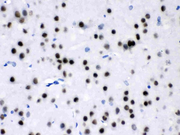
PB9953 has IHC images from paraffin sections of mouse and rat brain and human glioma and meningioma, plus an ICC/IF image of U20S cells (PB9953 image captions).
PB9953 is the SKU that renders, with its own mouse brain paraffin-section IHC figure (PB9953 card figure). Additional captions document rat brain, human glioma and human meningioma IHC, and U20S cell ICC/IF (PB9953 image captions).
Which to pick: Choose PB9953 for tissue IHC: its paraffin-section captions describe EDTA retrieval at pH 8.0 and 1 μg/ml primary antibody; the fixative is unreported (PB9953 IHC image captions). For IF/ICC, PB9953 has a U20S cell image, while rabbit monoclonal M01398-1 lists ICC/IF but has no supplied image (PB9953 IF image caption; M01398-1 catalog). For work across human, mouse and rat, both list those species, but PB9953 also lists IHC and has paraffin-section images from all three; M01398-1 does not list IHC (PB9953 catalog and IHC image captions; M01398-1 catalog).