This website uses cookies to ensure you get the best experience on our website.


- Table of Contents
Plan chromogenic MYH4 IHC in paraffin sections around cytoplasmic staining in skeletal and heart muscle (HPA tissue IHC). Keep fixation consistent (standard IHC practice) and interpret staining with the reported multi-gene targeting caution (HPA tissue IHC).

Expected localisation, validated protocols, controls and antibodies — the at-a-glance facts below, then the full design guide.
| Expected localisation | Cytoplasm in skeletal and heart muscle (HPA tissue IHC) | |
| Staining pattern | Myocyte and cardiomyocyte cytoplasm; medium staining (HPA tissue IHC) | |
| Antigen retrieval | EDTA pH 8.0 HIER, heat-mediated (datasheet A09143-1) | |
| Positive control | Heart muscle+1 more · see all | |
| Negative control | Adipose tissue+4 more · see all |
| Fixation | Keep fixation consistent across paraffin sections. (standard IHC practice; not target-specific) | |
| Caveat | Antibody may target proteins from more than one gene (HPA tissue IHC) | |
| Regulation | No expression regulators annotated (UniProt) | |
| Isoform / epitope | No annotated isoforms or chain cleavage; full-length protein (UniProt) |
The catalog antibody uses EDTA retrieval (datasheet: A09143-1). Published IHC protocols cover dog frozen muscle sections (PMC11672843) and human eyelid FFPE sections (PMC11802858).
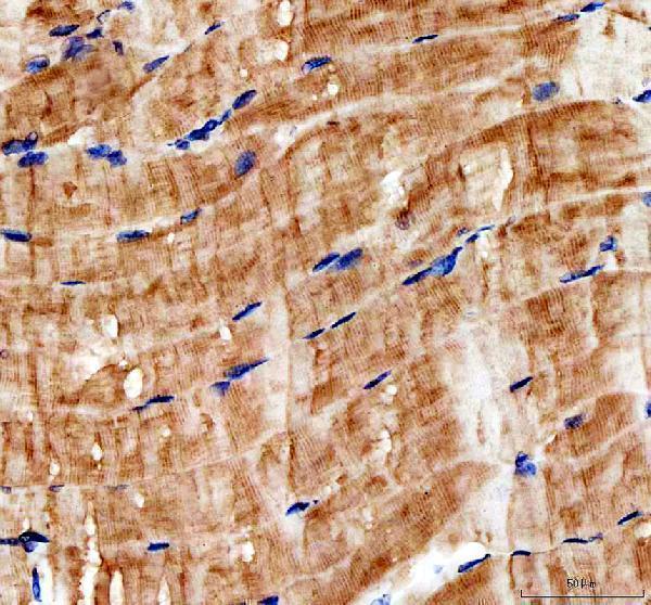
| Sample | Paraffin-embedded mouse skeletal muscle tissue; fixative not specified (datasheet A09143-1) |
| Fixation | Image fixative and duration unreported (datasheet A09143-1); verify before use. |
| Sectioning | 4–5 µm sections on charged slides (standard) |
| Deparaffinisation | Xylene, graded ethanol series to water (standard) |
| Antigen retrieval | Heat retrieval: EDTA pH 8.0 (datasheet A09143-1); 20 min, 95–100 °C (standard) |
| Peroxidase block | 3% H2O2, 10 min, room temperature (standard) |
| Blocking | 10% goat serum (datasheet A09143-1) |
| Primary antibody | Rabbit anti-MYH4, 1:50 recommended; image 1:100 (datasheet A09143-1) |
| Primary incubation | Overnight at 4 °C (datasheet A09143-1) |
| Detection | HRP-conjugated secondary, DAB chromogen (datasheet A09143-1) |
| Counterstain | Hematoxylin, blue, dehydrate and mount (standard) |
| Expected result | MYH4-positive staining in cardiomyocytes of heart muscle (HPA tissue IHC: Medium). HPA tissue profile: Cytoplasmic expression in skeletal muscle and heart muscle. No signal in the no-primary control. |
MYH4 is a cytoplasmic myofibrillar protein of muscle thick filaments and has no transmembrane segment (UniProt Q9Y623). In paraffin section IHC, expect staining in skeletal myocytes and cardiomyocytes (HPA: Medium in both). Interpret that pattern cautiously: HPA rates tissue IHC as Supported, reports medium consistency with RNA data, and warns that the antibody staining may represent proteins from more than one gene.
| Cytoplasmic staining within skeletal myocytes or cardiomyocytes, with surrounding structures comparatively clear. | This fits the reported Medium staining in both cell types (HPA tissue IHC) and MYH4's location in myofibrillar thick filaments (UniProt Q9Y623). The observed pattern supports a muscle associated signal; it does not by itself resolve the antibody's multi gene specificity warning (HPA tissue IHC). |
| Predominantly nuclear or sharply cell surface staining, without a convincing cytoplasmic muscle signal. | Treat this as discordant with the reported cytoplasmic pattern (HPA tissue IHC) and myofibrillar location without a transmembrane segment (UniProt Q9Y623). Review morphology and controls before scoring it as MYH4; the compartment alone cannot identify which staining step caused the discrepancy. |
| Prominent staining in cells reported as negative, such as adipocytes or adrenal glandular cells. | HPA reports these cells as Not detected (HPA tissue IHC). Check for antibody cross reaction and detection related signal using suitable controls (general IHC practice). HPA's warning that staining may represent proteins from more than one gene limits any claim that unexpected staining is MYH4 specific. |
| Widespread, low contrast color across muscle and nonmuscle areas, with little cellular definition. | Diffuse background is difficult to reconcile with the cell associated cytoplasmic pattern (HPA tissue IHC). Uneven blocking, excessive detection signal, or inadequate washing can produce background in chromogenic IHC (general IHC practice); appearance alone cannot distinguish these causes. |
| No staining in a skeletal muscle or heart muscle section that serves as the positive tissue. | Both tissues have reported Medium staining, so an absent signal calls for a run check (HPA tissue IHC). Inspect tissue preservation and assay controls, then review retrieval, antibody conditions, and detection in that order as appropriate (general IHC practice). Do not infer a MYH4 specific fixation effect. |
| Compartment and muscle structure | MYH4 resides in cytoplasmic myofibrillar thick filaments and lacks a transmembrane segment (UniProt Q9Y623). Use muscle cell cytoplasm as the anatomical reference when scoring IHC; a membrane or nuclear dominant pattern needs independent scrutiny. |
| Reference tissue distribution | HPA reports Medium staining in skeletal myocytes and cardiomyocytes and Not detected staining in the listed negative cell types (HPA tissue IHC). Its RNA summary is tissue enriched in skeletal muscle (HPA tissue IHC); that summary does not negate the reported heart IHC signal. |
| Antibody level confidence | The listed antibody HPA001349 is IHC Supported, while HPA describes medium agreement with RNA and cautions that staining may represent more than one gene (HPA antibody record; HPA tissue IHC). Score a compatible pattern as supportive evidence, with specificity interpreted cautiously. |
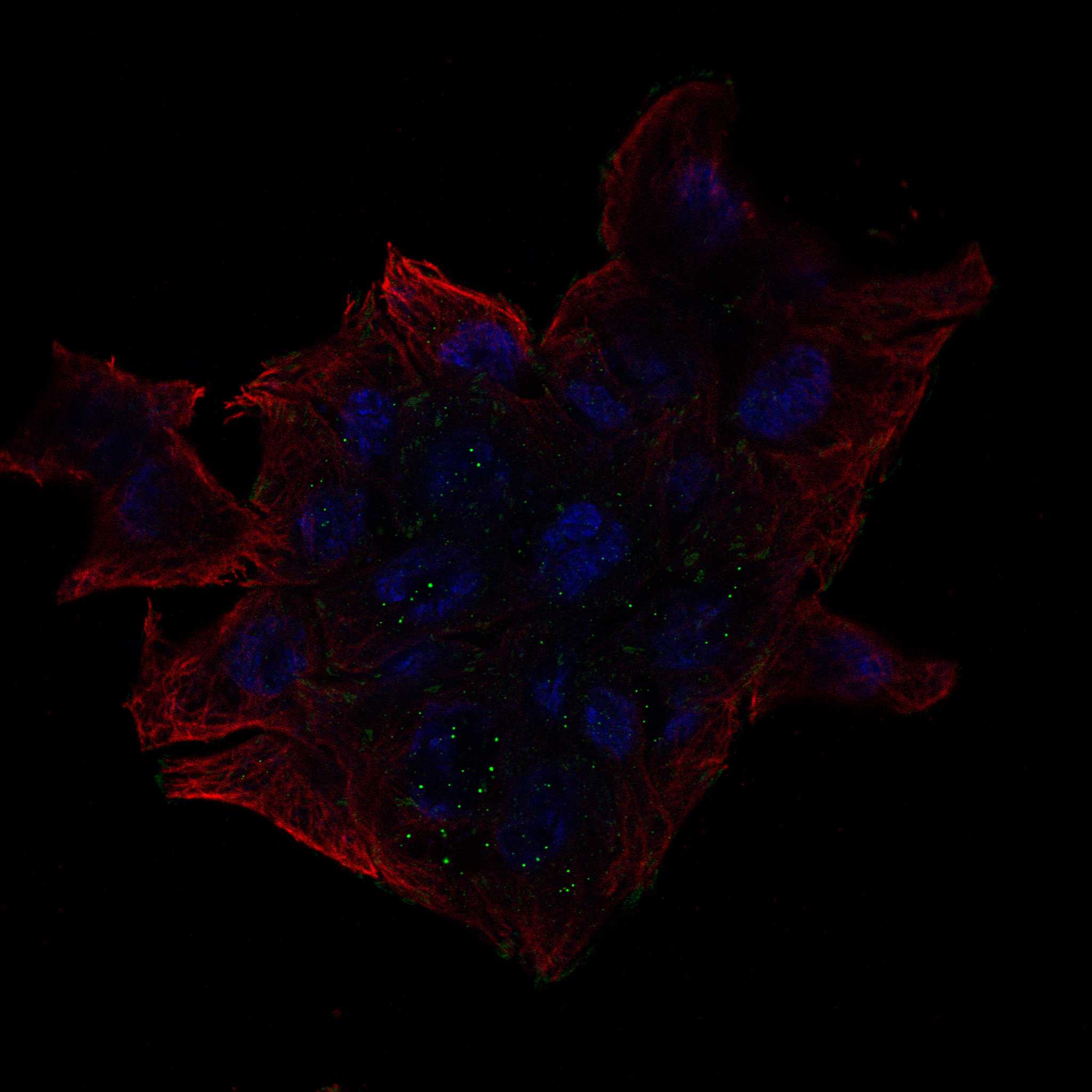
| IF or ICC interpretation | For the separate IF or ICC guide: is focal adhesion staining the expected MYH4 readout? HPA lists focal adhesion sites as uncertain and the antibody's ICC status as Uncertain (HPA subcellular; HPA antibody record). That observation cannot replace the tissue IHC cytoplasmic reference. |
| Situation | Likely cause | Next action |
|---|---|---|
| The positive muscle control is completely blank. | An assay or sample problem is plausible because HPA reports Medium staining in skeletal myocytes and cardiomyocytes (HPA tissue IHC); the blank section does not identify a target specific cause. | Confirm tissue identity and morphology, then check reagent delivery, retrieval record, antibody application, and detection controls (general IHC practice). Record the failed control before interpreting test sections. |
| Muscle staining is faint or uneven despite an intact positive control. | Section variation or uneven assay exposure can change visible chromogenic signal (general IHC practice). HPA's Medium reference is a qualitative observation, not a required intensity threshold (HPA tissue IHC). | Compare matched section areas and assay controls; review section quality, coverage, and documented retrieval and detection settings (general IHC practice). Avoid labeling a faint area negative solely by comparison with the darkest field. |
| Nuclei or cell outlines dominate the staining. | That distribution conflicts with cytoplasmic muscle staining (HPA tissue IHC) and MYH4's myofibrillar, nontransmembrane location (UniProt Q9Y623). A particular procedural cause cannot be assigned from the pattern alone. | Recheck cell boundaries against the counterstain and inspect negative controls (general IHC practice). Score the dominant discordant pattern separately from any convincing myocyte cytoplasmic signal. |
| Color appears in reported negative cells or without primary antibody. | HPA lists adipocytes and adrenal glandular cells as Not detected (HPA tissue IHC). Signal without primary antibody suggests detection background; staining confined to the full assay may also involve antibody cross reaction (general IHC practice). | Compare the no primary control, inspect endogenous chromogen or enzyme activity, and review blocking and detection conditions (general IHC practice). Keep HPA's multi gene antibody warning in the specificity assessment. |
| A uniform haze obscures otherwise plausible muscle staining. | Low contrast background can arise from nonspecific reagent binding or excessive detection development (general IHC practice); HPA's reference pattern is cellular and cytoplasmic (HPA tissue IHC). | Compare negative controls, review antibody concentration and detection time, and assess wash and blocking steps (general IHC practice). Reassess cell localization only after the background is controlled. |
| ICC or IF shows focal adhesion staining instead of the tissue IHC pattern. | HPA calls the focal adhesion assignment uncertain, notes antibodies may target more than one gene, and rates the listed antibody ICC Uncertain (HPA subcellular; HPA antibody record). | Interpret the IF or ICC result under its separate guide and validation controls; do not use it to redefine the cytoplasmic muscle IHC reference (HPA tissue IHC; HPA subcellular). |
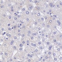
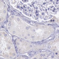

Comprehensive Human Protein Atlas IHC scoring per tissue (reliability: Supported — Medium consistency between antibody staining and RNA expression data. Caution, targets protein from more than one gene.). Rows are taken directly from the HPA tissue chart — click any row's HPA link to view the source.
| Tissue | Cell type | Level | Evidence | Source |
|---|---|---|---|---|
| Heart muscle | Cardiomyocytes | Medium | Protein (IHC) | HPA → |
| Skeletal muscle | Myocytes | Medium | Protein (IHC) | HPA → |
| Tissue | Cell type | Level | Evidence | Source |
|---|---|---|---|---|
| Adipose tissue | Adipocytes | Not detected | Protein (IHC) | HPA → |
| Adrenal gland | Glandular cells | Not detected | Protein (IHC) | HPA → |
| Appendix | Glandular cells | Not detected | Protein (IHC) | HPA → |
| Bone marrow | Hematopoietic cells | Not detected | Protein (IHC) | HPA → |
| Breast | Adipocytes | Not detected | Protein (IHC) | HPA → |
Troubleshoot MYH4 staining in paraffin sections by checking retrieval, intracellular localisation, controls and scoring against the reported skeletal muscle IHC result (datasheet A09143-1).
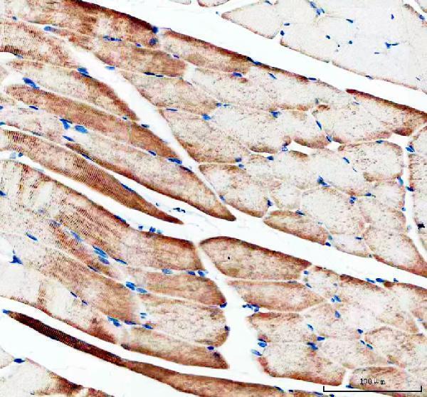
A09143-1 has IHC images from paraffin-embedded mouse and rat skeletal muscle sections (A09143-1 IHC captions). Human reactivity is listed, but no IF image is supplied (A09143-1 catalog reactivity; IF image alts).
A09143-1 is listed for IHC and has a mouse skeletal muscle paraffin-section image (A09143-1 catalog applications; mouse IHC caption). Its second image shows rat skeletal muscle paraffin sections; human reactivity is listed without a human IHC image (A09143-1 rat IHC caption; catalog reactivity; IHC image alts).
Which to pick: For tissue IHC, choose A09143-1 based on its mouse and rat paraffin-section images; the fixative is unreported (A09143-1 IHC captions). For IF/ICC, this catalog provides no validated option: A09143-1 has no listed IF/ICC application or IF image (A09143-1 catalog applications; IF image alts). For cross-species IHC, A09143-1 is a rabbit polyclonal listed as reactive with human, mouse, and rat, with direct IHC images for mouse and rat only (A09143-1 catalog host/dilution_raw/reactivity; IHC captions).