PMCH / Pro-MCH · IHC design guide

Design Immunohistochemistry for PMCH

Plan chromogenic IHC on paraffin sections around the hypothalamic neuronal pattern (HPA tissue IHC). The catalog antibody lists an IHC dilution of 1:50–1:200 (datasheet); account for PMCH secretion when interpreting staining (UniProt).

Evidence assembled Oct 2026 · For research use; verify linked source records and product datasheet before use
Immunohistochemistry protocol sheet for PMCH (IHC for PMCH): expected localisation Hypothalamic neuronal projections (HPA tissue IHC), antibody A07730, validated IHC image, and IHC protocol steps
Printable PMCH IHC protocol sheet — expected localisation Hypothalamic neuronal projections (HPA tissue IHC), antibody A07730, controls and protocol steps. Open the full PMCH IHC guide →

PMCH Immunohistochemistry Experimental Design Guide

Expected localisation, validated protocols, controls and antibodies — the at-a-glance facts below, then the full design guide.

Must know before staining
Expected localisation Hypothalamic neuronal projections (HPA tissue IHC)
Staining pattern High staining in hypothalamic neurons and neural processes (HPA tissue IHC)
Antigen retrieval Citrate pH 6.0 HIER, 95–98 °C, 20 min (rule: cytoplasmic / membrane antigen)
Positive control ⓘ Hypothalamus
Negative control ⓘ Adipose tissue+4 more · see all
Important caveats
Reasons your staining may differ from the expected pattern.
Fixation Keep fixation consistent across paraffin sections (standard IHC practice; not target-specific); Matched tissue-IHC evidence does not establish the fixation claim. Validate the specimen-specific method before use. (selected-SKU IHC image A07730)
Caveat Secretion can separate protein staining from PMCH RNA location (HPA tissue IHC)
Regulation No staining regulator specified (UniProt)
Isoform / epitope No isoforms annotated; Pro-MCH chain starts at residue 22 (UniProt)
Section 1

Recommended PMCH IHC & IF Protocols

Compare the catalog antibody’s IHC-P protocol with the published Pro-MCH IHC protocol for human lateral hypothalamus (PMC10175407).

Recommended immunohistochemistry (IHC-P) protocol parameters
SampleParaffin-embedded human testis tissue; fixative not specified (datasheet A07730)
FixationImage fixative and duration unreported (datasheet A07730); verify before use.
Sectioning4–5 µm sections on charged slides (standard)
DeparaffinisationXylene, graded ethanol series to water (standard)
Antigen retrievalHeat-induced epitope retrieval in citrate buffer, pH 6.0, 20 min at 95–98 °C (standard rule: cytoplasmic / membrane antigen)
Peroxidase block3% H2O2, 10 min, room temperature (standard)
Blocking10% normal serum of the secondary host, 30 min, room temperature (standard)
Primary antibodyRabbit anti-PMCH, 1:50 recommended; image 1:200 (datasheet A07730)
Primary incubationOvernight at 4 °C (standard)
DetectionHRP-polymer secondary, DAB chromogen 5–10 min (standard)
CounterstainHematoxylin, blue, dehydrate and mount (standard)
Expected resultPMCH-positive staining in neuronal projections of hypothalamus (HPA tissue IHC: High). HPA tissue profile: Highly expressed in neurons and neural processes in hypothalamus. No signal in the no-primary control.
💡Decision noteStart with citrate pH 6.0 retrieval at 95–98 °C for 20 min (page retrieval); the published protocol used 95 °C for 10–15 min (PMC10175407).
Section 2

What Is the Expected PMCH Staining Pattern?

PMCH is a secreted neuropeptide precursor with no transmembrane segment (UniProt P20382). In paraffin sections, expect staining in hypothalamic neurons and their neural processes, especially neuronal projections; HPA rates hypothalamic projection staining High and the tissue IHC pattern Enhanced (HPA tissue IHC). UniProt also reports expression in the lateral hypothalamus (UniProt P20382).

What am I looking at on my slide?
Strong staining follows neuronal projections in a hypothalamic section, with staining also present in neurons.This matches the reported High projection signal and expression in neurons and neural processes (HPA tissue IHC). Evaluate the pattern against the section’s anatomy; secreted PMCH need not appear as a crisp cell membrane outline (UniProt P20382 topology).
Staining is predominantly nuclear, or forms a uniform membrane rim across many cells.That distribution is discordant with the reported neuronal and neural process pattern (HPA tissue IHC) and PMCH’s secreted, non-transmembrane annotation (UniProt P20382). Treat it as a possible staining artefact and review controls before scoring it as PMCH.
Prominent signal appears in adipocytes, adrenal glandular cells, caudate glia, or cerebellar granular-layer cells.HPA reports PMCH as Not detected in those respective cells (HPA tissue IHC). Such staining merits a cross-reactivity or endogenous detection activity check, particularly if the hypothalamic neuronal pattern is absent; an unexpected field alone does not establish its cause.
A diffuse chromogen haze covers neuronal and non-neuronal areas alike.The broad haze lacks the cell and process contrast expected from the hypothalamic profile (HPA tissue IHC). Consider nonspecific background from the IHC workflow (general IHC practice); PMCH being secreted does not, by itself, validate uniform staining (UniProt P20382).
A well-preserved hypothalamic section has no convincing neuronal or projection signal.This conflicts with the reported High staining in hypothalamic neuronal projections (HPA tissue IHC). Check that the sampled section includes the relevant hypothalamic region, then review the positive control and IHC workflow before interpreting absence as biological loss (general IHC practice; UniProt P20382 tissue specificity).
💡Expected PMCH appearanceCall a section positive when hypothalamic neurons and especially their projections show distinct, strong staining (HPA tissue IHC: High); widespread nuclear staining or signal in HPA-negative cell types is a suspect positive (HPA tissue IHC; UniProt P20382 topology).
How each factor affects the staining
Anatomic samplingHPA reports High staining in hypothalamic neuronal projections, while UniProt specifies lateral hypothalamic expression; a section that misses the relevant area can weaken interpretation (HPA tissue IHC; UniProt P20382).
Secretion and processingPMCH has a signal peptide at residues 1–21 and a Pro-MCH chain at 22–165 (UniProt P20382). Its secreted, non-transmembrane annotation supports attention to neuronal processes but does not identify which processed form an antibody detects (UniProt P20382; HPA tissue IHC).
Antibody validation and epitopeHPA lists Enhanced IHC validation for HPA046055 and HPA061884 (HPA antibodies). The supplied evidence does not locate their epitopes, so staining alone cannot distinguish precursor from processed PMCH species.
IHC versus IF evidenceHPA reports Secreted for subcellular summary but provides no ICC-IF images or main ICC-IF location (HPA subcellular). Its Enhanced tissue IHC assessment does not establish an IF staining pattern (HPA tissue IHC; HPA subcellular).
Why is my staining missing, weak or wrong?
SituationLikely causeNext action
Hypothalamic projections are unstained.The sampled field may miss the relevant anatomy, or an IHC workflow step may have failed (UniProt P20382 tissue specificity; general IHC practice).Review section location and tissue integrity; inspect a known-positive hypothalamic control and the antibody’s IHC-P workflow before calling the result negative (HPA tissue IHC; general IHC practice).
Every compartment shows similar brown signal.Uniform signal conflicts with the reported neuronal and projection profile (HPA tissue IHC); nonspecific background or endogenous detection activity is possible (general IHC practice).Compare a no-primary control, inspect blocking and detection steps, and reassess whether processes remain distinguishable from surrounding tissue (general IHC practice; HPA tissue IHC).
Nuclei dominate the signal.Predominantly nuclear staining is discordant with secreted PMCH and the reported neural process pattern (UniProt P20382; HPA tissue IHC).Check the no-primary control and compare localization with a known-positive hypothalamic section before scoring nuclear signal as specific (general IHC practice; HPA tissue IHC).
Adipose or adrenal cells stain strongly.HPA reports Not detected in adipocytes and adrenal glandular cells (HPA tissue IHC); cross-reactivity or endogenous detection activity warrants investigation (general IHC practice).Compare those fields with no-primary controls and the hypothalamic positive pattern; document the unexpected cell type separately from confirmed neuronal staining (general IHC practice; HPA tissue IHC).
The positive control is faint or patchy.Uneven tissue sampling or workflow variation may obscure the reported High hypothalamic projection pattern (HPA tissue IHC; general IHC practice).Confirm the section contains relevant hypothalamic neurons and processes; review retrieval, antibody incubation, detection, and counterstain using the applicable IHC-P instructions (UniProt P20382; general IHC practice).
Can IF/ICC confirm an ambiguous IHC pattern?HPA supplies no ICC-IF images or main ICC-IF location for PMCH (HPA subcellular).Use the separate IF/ICC guide for that application; here, judge the paraffin-section result against HPA’s hypothalamic neuronal and projection IHC pattern (HPA tissue IHC).

Sample controls for PMCH IHC & IF

🧪Run hypothalamus first; neuronal projections should stain (HPA: High in hypothalamic neuronal projections). Run adipose tissue as a negative (HPA: Not detected in adipocytes), and expect cells outside the stained projections on the hypothalamus slide to show only background signal.
Positive control tissue: Hypothalamus (Neuronal projections, HPA High)
Negative control tissue: Adipose tissue (HPA Not detected)
ICC-IF cell lines (HPA subcellular resource): HPA carries no ICC-IF cell line for PMCH; derive a cell-line control from the positive tissue's cell type (Neuronal projections) and confirm it by RNA or western blot first.
Technical controls: Include a no-primary, secondary-only control; use host-matched nonimmune IgG for a polyclonal antibody or a matched isotype for a monoclonal antibody; and confirm specificity with PMCH knockout tissue or a cognate peptide block when available (standard IHC practice). For chromogenic hypothalamus IHC, block endogenous peroxidase and, if using biotin-based detection, check for endogenous biotin; assess tissue autofluorescence if examining IF (standard IHC/IF practice).
⚠️Feasibility: A target-specific fixation window or fixation effect is unreported, and the selected paraffin-section caption does not report its fixative (caption: A07730, human testis, 1:200). Retrieval dependency and any advantage of frozen sections or IF over paraffin IHC are unreported in the supplied evidence; optimize retrieval empirically (standard IHC practice). In hypothalamus, distinguish staining of neuronal projections from background and tissue pigment when scoring (HPA: High in hypothalamic neuronal projections; standard IHC practice).

HPA tissue IHC evidence for PMCH

Comprehensive Human Protein Atlas IHC scoring per tissue (reliability: Enhanced — Secreted protein, tissue location of RNA and protein is expected to differ. Antibody staining consistent with external characterization data and GTEx RNA expression data.). Rows are taken directly from the HPA tissue chart — click any row's HPA link to view the source.

Positive expression · recommended positive controls

TissueCell typeLevelEvidenceSource
Hypothalamus Neuronal projections High Protein (IHC) HPA →

Undetected expression · recommended negative controls

TissueCell typeLevelEvidenceSource
Adipose tissue Adipocytes Not detected Protein (IHC) HPA →
Adrenal gland Glandular cells Not detected Protein (IHC) HPA →
Appendix Glandular cells Not detected Protein (IHC) HPA →
Bone marrow Hematopoietic cells Not detected Protein (IHC) HPA →
Breast Adipocytes Not detected Protein (IHC) HPA →
Section 3

Advanced PMCH IHC Tips

Troubleshoot PMCH staining in paraffin sections using the catalog image, tissue expression evidence, and controls that distinguish neuronal signal from artefact.

What retrieval conditions should I start with for PMCH in paraffin sections?
Start with citrate buffer at pH 6.0 for heat-induced retrieval at 95–98 °C for 20 min (page retrieval setting). Cool sections consistently, then compare staining with a no-primary control and hypothalamic neuronal projections, where PMCH staining is high (HPA: High in hypothalamic neuronal projections; standard IHC practice). The catalog image used antibody A07730 at 1:200 in paraffin-embedded human testis, but its caption does not report retrieval conditions (A07730 tissue-IHC caption). If staining remains weak, vary retrieval time on matched sections before considering another buffer, and check whether tissue morphology and background remain acceptable (standard IHC practice).
Could fixation explain weak or uneven PMCH staining?
PMCH-specific fixation sensitivity is unknown from the supplied evidence; the A07730 paraffin-section caption does not state a fixative or fixation duration (A07730 tissue-IHC caption). Record the fixative, processing history, and section age, then compare matched sections processed with a consistent schedule (standard IHC practice). If weak staining tracks with a processing batch, check section attachment, retrieval uniformity, and antibody incubation before assigning the difference to fixation (standard IHC practice). Keep a no-primary control alongside each comparison, and judge signal in an appropriate positive region such as hypothalamic neuronal projections when available (HPA: High in hypothalamic neuronal projections; standard IHC practice).
Where should convincing PMCH staining appear in tissue?
Look for staining in hypothalamic neurons and their projections, with high signal reported in neuronal projections (HPA: hypothalamus tissue IHC). PMCH is annotated as secreted and has no transmembrane segment, so do not require a crisp plasma-membrane rim to call staining plausible (UniProt P20382: subcellular location and topology). Use tissue architecture to distinguish neuronal processes from diffuse chromogen deposited across the section, and compare the pattern with a no-primary control (HPA: hypothalamus tissue IHC; standard IHC practice). The A07730 image documents staining in paraffin-embedded human testis at 1:200, but it does not establish a cell-specific localisation there (A07730 tissue-IHC caption).
How should PMCH processing affect antibody and epitope selection?
PMCH is a 165-aa precursor with a signal sequence at residues 1–21 and a Pro-MCH chain at 22–165 (UniProt P20382: sequence processing). Check the antibody's stated immunogen or mapped epitope against those regions before interpreting a missing signal, because a cleaved sequence may not remain in every processed product (UniProt P20382: sequence processing; standard IHC practice). No isoforms or glycosylation sites are annotated, while an amidated residue is annotated at position 143 (UniProt P20382: isoforms and modifications). If the epitope is undisclosed, describe staining as antibody-detected PMCH material and avoid assigning it to a specific processed peptide (standard IHC practice).
How can I adapt PMCH detection to a multiplex IF experiment?
Treat IF as a separate validation: the supplied A07730 image documents paraffin-section IHC, and no ICC/IF cell-line images are listed (A07730 tissue-IHC caption; HPA: subcellular record). Multiplex PMCH with a validated neuronal marker to test whether candidate signal follows the expected hypothalamic neurons and projections (HPA: hypothalamus tissue IHC; standard IF practice). Choose a fluorophore channel after measuring tissue autofluorescence, and include single-stain and no-primary controls to assess bleed-through and background (standard IF practice). Because PMCH is secreted with no transmembrane segment, use permeabilisation when the antibody epitope requires access to intracellular precursor; establish that requirement from the epitope and matched conditions (UniProt P20382: topology and processing; standard IF practice).
What should I check when PMCH chromogenic staining is widespread?
Start with a no-primary control and inspect whether chromogen persists in the same compartments, which would point toward detection-system background (standard IHC practice). For peroxidase-based detection, verify the endogenous peroxidase block, then compare matched sections with a shorter chromogen development time (standard IHC practice). Titrate the primary antibody around the catalog image's 1:200 dilution while keeping retrieval and detection conditions fixed (A07730 tissue-IHC caption; standard IHC practice). Widespread staining in adipocytes would conflict with the reported absence of PMCH detection there, whereas hypothalamic neuronal projections provide a reported positive pattern for comparison (HPA: adipose tissue and hypothalamus tissue IHC).
How should I quantify PMCH staining across sections? ⚠ ANSWER MARKED FOR VERIFICATION
Define regions before scoring and quantify the same anatomical compartment across matched sections, using hypothalamic neuronal projections when that is the study region (HPA: hypothalamus tissue IHC; standard IHC practice). For neuronal processes, report chromogen-positive area or process density per mm²; for identifiable cell bodies, report percent positive cells or an H-score (standard IHC practice). Normalize area measurements to analyzable tissue area and cell scores to the number of eligible cells, excluding folds, edges, and damaged regions by a fixed rule (standard IHC practice). Keep section thickness, retrieval, antibody dilution, imaging, and threshold settings consistent, and report how background from no-primary controls was handled (standard IHC practice).
How can I distinguish true PMCH staining from artefact?
Favor a reproducible pattern in hypothalamic neurons and projections, where protein staining is reported as high (HPA: hypothalamus tissue IHC). Interpret isolated staining in an unexpected cell type cautiously, particularly when it follows section edges, folds, or necrotic material rather than recognizable neuronal structures (HPA: hypothalamus tissue IHC; standard IHC practice). Check no-primary and peroxidase-block controls when a diffuse or granular signal could reflect detection background or endogenous enzyme activity (standard IHC practice). PMCH is secreted, so RNA and protein locations can differ; an extracellular or projection-associated signal needs anatomical and control support before assignment to a producing cell (UniProt P20382: secreted; HPA: reliability description; standard IHC practice).
Boster reagents

Best PMCH / Pro-MCH IHC Antibodies

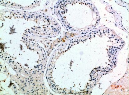
A07730 has a human paraffin-section IHC image and lists IF as an application; catalog reactivity covers human, mouse, and rat (A07730 catalog; A07730 IHC caption).

Real IHC data Immunohistochemical analysis of paraffin-embedded human-testis, antibody was diluted at 1:200
Anti-Pro-MCH PMCH Antibody
Cat # A07730

A07730 will render with an IHC image of paraffin-embedded human testis (A07730 IHC caption). Its listed applications are IHC, IF, and ELISA, with human, mouse, and rat reactivity; no IF image is supplied (A07730 catalog).

Which to pick: Choose A07730 for paraffin-section IHC because its own caption documents that sample type; the fixative is unreported (A07730 IHC caption). For IF, A07730 is listed at 1:50, but the payload supplies no IF image or ICC validation (A07730 catalog). For human, mouse, or rat samples, A07730 is the listed polyclonal option based on catalog reactivity; its IHC image documents human tissue only (A07730 catalog; A07730 IHC caption).

Each figure is that product's own IHC / IF validation image from its datasheet.

References

  1. UniProt Consortium. UniProt entry P20382 (MCH_HUMAN, Pro-MCH).
  2. Human Protein Atlas. PMCH tissue IHC expression (reliability: Enhanced).
  3. Human Protein Atlas. PMCH subcellular location (ICC-IF): Secreted.
  4. Human Protein Atlas. PMCH antibody validation summary (2 antibodies).
  5. Loss of hypothalamic MCH decreases food intake in amyotrophic lateral sclerosis. Acta neuropathologica 2023 — PMC10175407.
  6. Dissociated Pmch and Cre Expression in Lactating Pmch-Cre BAC Transgenic Mice. Frontiers in neuroanatomy 2020 — PMC7475711.
  7. Methylation-based epigenetic studies and gene integration analysis of preeclampsia. Annals of translational medicine 2022 — PMC9843334.
  8. Development of posterior hypothalamic neurons enlightens a switch in the prosencephalic basic plan. PloS one 2011 — PMC3241628.
  9. PubMed PMID:2149166 — UniProt-cited evidence.
  10. PubMed PMID:8326825 — UniProt-cited evidence.
  11. PubMed PMID:15489334 — UniProt-cited evidence.