This website uses cookies to ensure you get the best experience on our website.


- Table of Contents
Plan chromogenic IHC for RGS10 using the cytoplasmic and nuclear tissue pattern (HPA tissue IHC). This guide identifies lymphoid tissues as high-staining references (HPA tissue IHC) and covers fixation, retrieval, and staining controls.

Expected localisation, validated protocols, controls and antibodies — the at-a-glance facts below, then the full design guide.
| Expected localisation | Cytoplasmic and nuclear staining (HPA tissue IHC) | |
| Staining pattern | Cytoplasmic and nuclear; most abundant in lymphoid tissues (HPA tissue IHC) | |
| Antigen retrieval | Tris-EDTA pH 9.0 HIER, heat-mediated (datasheet A05197-1) | |
| Positive control | Appendix+4 more · see all | |
| Negative control | Epididymis+4 more · see all |
| Fixation | Keep paraffin-section fixation consistent across samples. (standard IHC practice; not target-specific) | |
| Caveat | Antibody staining and RNA show medium consistency (HPA tissue IHC) | |
| Regulation | Forskolin promotes nuclear translocation (UniProt) | |
| Isoform / epitope | 3 isoforms; epitope coverage is unspecified (UniProt) |
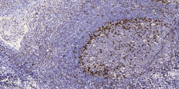
The catalog antibody’s IHC-P protocol (datasheet: A05197-1) and one published RGS10 paraffin-section protocol (PMC11326775) provide starting conditions.
| Sample | Paraffin-embedded human tonsil tissue; fixative not specified (datasheet A05197-1) |
| Fixation | Image fixative and duration unreported (datasheet A05197-1); verify before use. |
| Sectioning | 4–5 µm sections on charged slides (standard) |
| Deparaffinisation | Xylene, graded ethanol series to water (standard) |
| Antigen retrieval | Heat retrieval: Tris-EDTA pH 9.0 (datasheet A05197-1); 20 min, 95–100 °C (standard) |
| Peroxidase block | 3% H2O2, 10 min, room temperature (standard) |
| Blocking | 10% normal serum of the secondary host, 30 min, room temperature (standard) |
| Primary antibody | Rabbit anti-RGS10, 1:100-1:300 (datasheet A05197-1) |
| Primary incubation | Overnight at 4 °C (standard) |
| Detection | HRP-polymer secondary, DAB chromogen 5–10 min (standard) |
| Counterstain | Hematoxylin, blue, dehydrate and mount (standard) |
| Expected result | RGS10-positive staining in lymphoid tissue of appendix (HPA tissue IHC: High). HPA tissue profile: General cytoplasmic and nuclear expression, most abundant in lymphoid tissues. No signal in the no-primary control. |
RGS10 can stain the cytoplasm and nucleus, consistent with a protein lacking a transmembrane segment (UniProt O43665: localization and topology). In paraffin sections, expect the strongest representative staining in lymphoid tissue, including lymph node germinal center cells, alongside other HPA high populations (HPA: tissue IHC). HPA rates its tissue IHC evidence Enhanced, while noting only medium consistency between antibody staining and RNA expression (HPA: reliability description).
| Cytoplasmic and nuclear stain appears in lymph node germinal center cells or appendix lymphoid tissue. | This fits the reported compartments and high staining in these populations (UniProt O43665: localization; HPA: tissue IHC). Score the named cell population and its compartment rather than calling the entire section uniformly positive (standard IHC practice). |
| Stain appears only on cell borders or in extracellular material, without a convincing intracellular pattern. | Recheck localization: UniProt lists cytosol and nucleus and no transmembrane segment (UniProt O43665: localization and topology). Border staining alone does not establish RGS10; compare morphology, controls and the specific population before accepting it (standard IHC practice). |
| Strong stain dominates a population listed as not detected, such as skeletal muscle myocytes. | That conflicts with the reported IHC observation for that population (HPA: not detected in skeletal muscle myocytes). Consider antibody cross-reactivity or endogenous detection activity, then inspect a reagent control (standard IHC practice). HPA not detected is not proof that expression is impossible. |
| Color spreads diffusely across cells and surrounding tissue, obscuring nuclear and cytoplasmic boundaries. | The distribution cannot be scored reliably against the reported intracellular pattern (HPA: tissue IHC; UniProt O43665: localization). Review reagent controls, blocking and detection background before interpreting intensity (standard IHC practice). |
| No convincing stain appears in lymph node germinal center cells or bone marrow hematopoietic cells. | Both populations are reported High, so an absent result calls for an assay check (HPA: tissue IHC). Confirm that the chosen population is present, then review the antibody, retrieval and detection controls (standard IHC practice); tissue positivity is not guaranteed in every specimen. |
| Tissue and cell selection | Appendix lymphoid tissue and lymph node germinal center cells are High; several glandular populations are Low or not detected (HPA: tissue IHC). Identify the cell population before comparing signal intensity across sections (standard IHC practice). |
| Localization and stimulation | UniProt lists cytosol and nucleus and says forskolin promotes phosphorylation and nuclear translocation (UniProt O43665: localization). Nuclear predominance may depend on treatment; it should not be required of untreated tissue. |
| Antibody validation | HPA021305 and CAB004559 each have Enhanced IHC status (HPA: antibody validation). HPA describes the tissue profile as having medium antibody–RNA consistency (HPA: reliability description); neither label establishes specificity in every specimen. |
| Isoform interpretation | UniProt lists three RGS10 isoforms (UniProt O43665: isoforms). The supplied evidence gives no epitope mapping, so staining alone cannot distinguish isoforms or establish that every isoform is detected. |
| IF/ICC Q: What localization is reported? | A: HPA reports supported nucleoplasmic localization and additional nuclear bodies in ICC-IF (HPA: subcellular). This cell-image observation can inform interpretation; it is not an IHC protocol or a requirement that every paraffin section show nuclear bodies. |
| Detection background | Endogenous enzyme activity or nonspecific reagent binding can produce chromogenic signal (standard IHC practice). A reagent control helps separate that background from cell-associated staining; the supplied sources do not quantify either effect for RGS10. |
| Situation | Likely cause | Next action |
|---|---|---|
| The high-expression reference tissue looks blank. | The expected population may be absent from the field, or the IHC assay may have failed (HPA: High in lymph node germinal center and bone marrow hematopoietic cells; standard IHC practice). | Locate the named cells on a counterstained section, then check antibody, retrieval and detection steps with appropriate controls (standard IHC practice). Do not infer biological absence from one blank field. |
| A reported not-detected population stains strongly. | Cross-reactivity or endogenous chromogenic activity is possible (standard IHC practice); HPA reports no detection in skeletal muscle myocytes and ovarian stroma cells (HPA: tissue IHC). | Check tissue identity and morphology, compare a reagent control, and assess staining with an independently validated antibody if available (standard IHC practice). Treat the HPA category as a comparison, not an absolute exclusion. |
| Color is diffuse and compartment boundaries cannot be scored. | Background from blocking, washing or detection may obscure the intracellular pattern (standard IHC practice; UniProt O43665: localization). | Inspect reagent controls and background outside cells; adjust blocking, washing or detection conditions using standard IHC practice before assigning an RGS10 score. |
| Stain is confined to an apparently extracellular deposit. | That pattern does not match the cytosolic and nuclear localization or lack of a transmembrane segment (UniProt O43665: localization and topology). | Check morphology and reagent controls, then seek reproducible intracellular staining in an HPA High cell population before calling the deposit RGS10 (HPA: tissue IHC; standard IHC practice). |
| A nucleus-positive field differs from a mainly cytoplasmic field. | Both compartments are reported, and forskolin can promote nuclear translocation (UniProt O43665: localization). Differences alone cannot establish treatment history or a technical fault. | Record nuclear and cytoplasmic scores separately and compare like cell populations and known treatment conditions (standard IHC practice). Avoid inferring stimulation solely from stain location. |
| Nuclear bodies are absent in paraffin IHC. | HPA's supported nuclear-body observation comes from ICC-IF images, while tissue IHC is summarized as general cytoplasmic and nuclear expression (HPA: subcellular; HPA: tissue IHC). | Judge the paraffin result against its tissue IHC pattern and controls (HPA: tissue IHC; standard IHC practice). Do not require ICC-IF nuclear bodies to validate chromogenic IHC. |
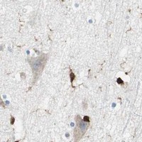
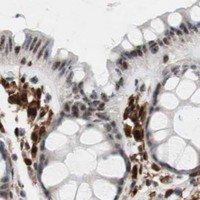

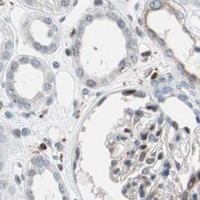


Comprehensive Human Protein Atlas IHC scoring per tissue (reliability: Enhanced — Medium consistency between antibody staining and RNA expression data.). Rows are taken directly from the HPA tissue chart — click any row's HPA link to view the source.
| Tissue | Cell type | Level | Evidence | Source |
|---|---|---|---|---|
| Appendix | Lymphoid tissue | High | Protein (IHC) | HPA → |
| Bone marrow | Hematopoietic cells | High | Protein (IHC) | HPA → |
| Bronchus | Goblet cells | High | Protein (IHC) | HPA → |
| Cerebellum | Granular cells - cytoplasm/membrane | High | Protein (IHC) | HPA → |
| Cervix | Glandular cells | High | Protein (IHC) | HPA → |
| Tissue | Cell type | Level | Evidence | Source |
|---|---|---|---|---|
| Epididymis | Glandular cells | Not detected | Protein (IHC) | HPA → |
| Ovary | Ovarian stroma cells | Not detected | Protein (IHC) | HPA → |
| Parathyroid gland | Glandular cells | Not detected | Protein (IHC) | HPA → |
| Prostate | Glandular cells | Not detected | Protein (IHC) | HPA → |
| Salivary gland | Glandular cells | Not detected | Protein (IHC) | HPA → |
Troubleshoot RGS10 staining in paraffin-section chromogenic IHC using the catalog antibody’s tonsil image, reported tissue patterns, and appropriate section controls.
Two anti-RGS10 antibodies have IHC figures from human paraffin sections (catalog IHC image captions); one also lists IF and human, mouse, and rat reactivity (A05197-1 applications/reactivity).
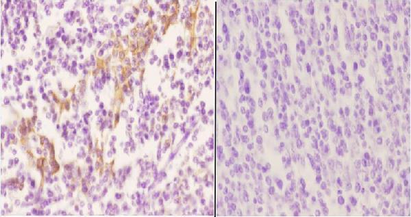
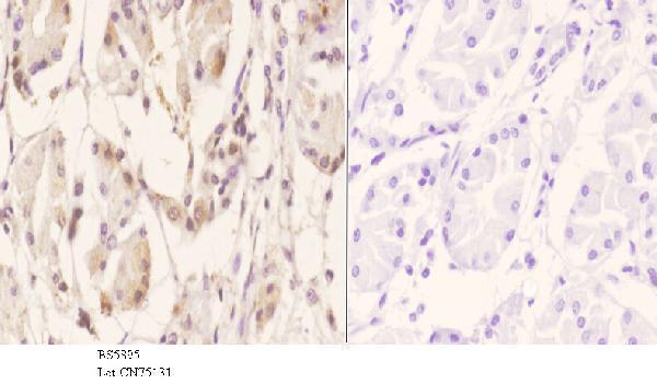
A05197-1 shows IHC in paraffin-embedded human tonsil (A05197-1 IHC image caption) and lists IF plus human, mouse, and rat reactivity (A05197-1 applications/reactivity). A05197 shows IHC in paraffin-embedded human tonsil carcinoma and stomach carcinoma (A05197 IHC image captions) and lists human reactivity (A05197 reactivity).
Which to pick: For tissue IHC, choose A05197-1 for its human tonsil example or A05197 for its human carcinoma examples (catalog IHC image captions); both captions show paraffin sections, but neither reports the fixative (catalog IHC image captions). For IF/ICC planning, choose polyclonal A05197-1 because it lists IF; ICC validation and an IF figure are unreported (A05197-1 dilution_raw; applications; if_image_alts). For mouse or rat samples, choose A05197-1 based on its listed reactivity (A05197-1 reactivity).